- Dentin

Содержание
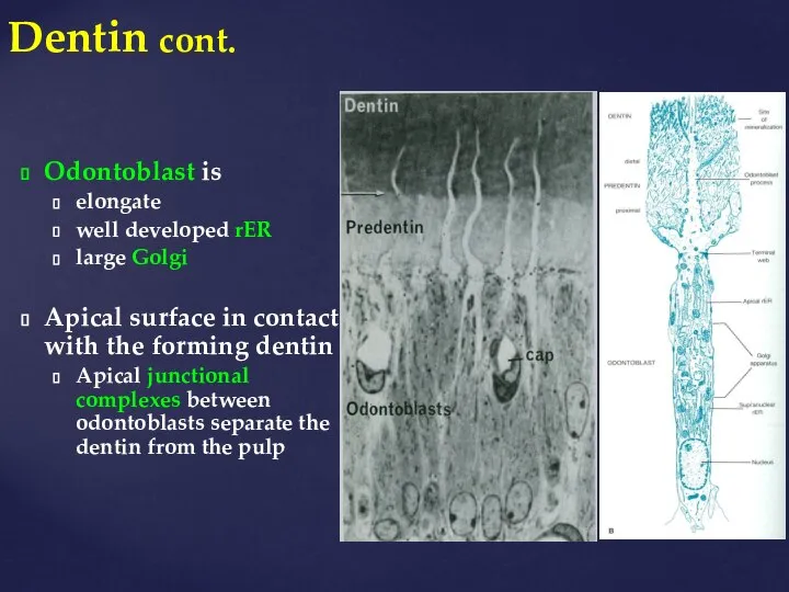
- 2. Dentin cont. Odontoblast is elongate well developed rER large Golgi Apical surface in contact with the
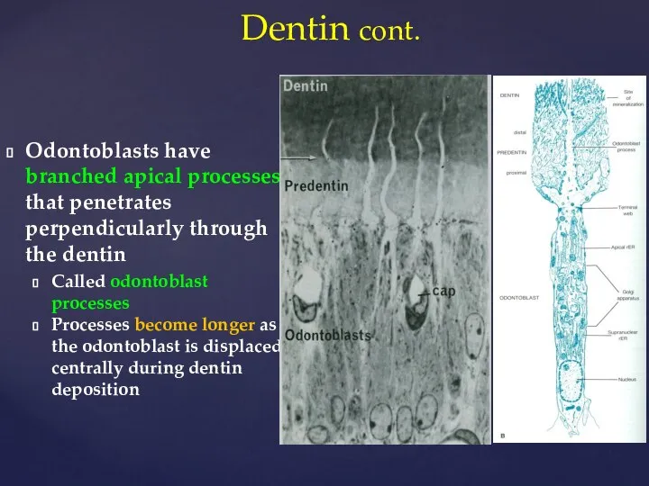
- 3. Odontoblasts have branched apical processes that penetrates perpendicularly through the dentin Called odontoblast processes Processes become
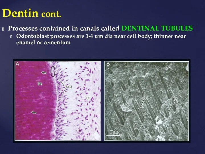
- 4. Dentin cont. Processes contained in canals called DENTINAL TUBULES Odontoblast processes are 3-4 um dia near
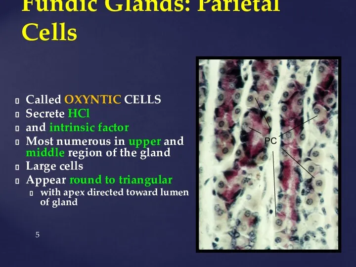
- 5. Fundic Glands: Parietal Cells Called OXYNTIC CELLS Secrete HCl and intrinsic factor Most numerous in upper
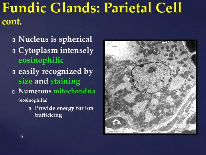
- 6. Fundic Glands: Parietal Cell cont. Nucleus is spherical Cytoplasm intensely eosinophilic easily recognized by size and
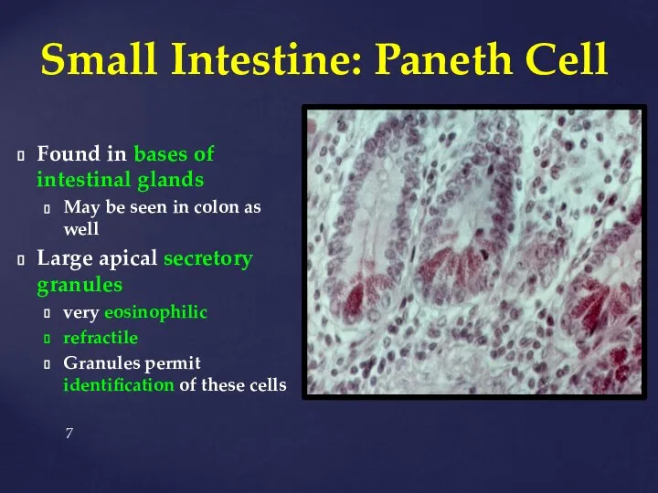
- 7. Small Intestine: Paneth Cell Found in bases of intestinal glands May be seen in colon as
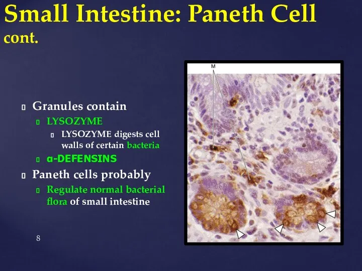
- 8. Small Intestine: Paneth Cell cont. Granules contain LYSOZYME LYSOZYME digests cell walls of certain bacteria α-DEFENSINS
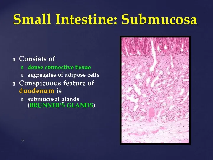
- 9. Small Intestine: Submucosa Consists of dense connective tissue aggregates of adipose cells Conspicuous feature of duodenum
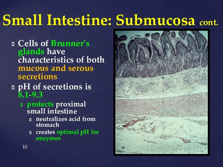
- 10. Small Intestine: Submucosa cont. Cells of Brunner’s glands have characteristics of both mucous and serous secretions
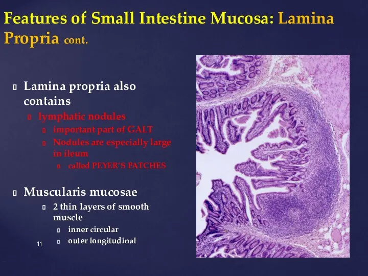
- 11. Features of Small Intestine Mucosa: Lamina Propria cont. Lamina propria also contains lymphatic nodules important part
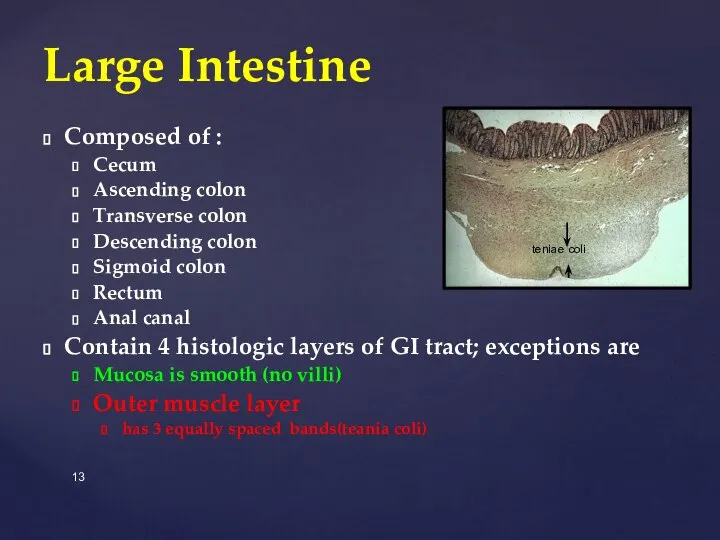
- 13. Composed of : Cecum Ascending colon Transverse colon Descending colon Sigmoid colon Rectum Anal canal Contain
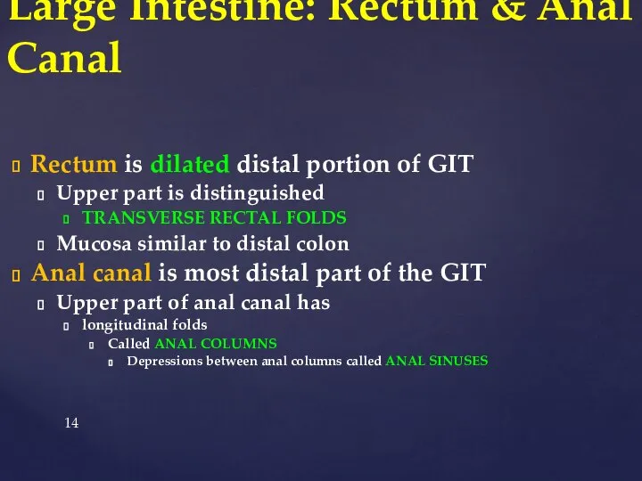
- 14. Rectum is dilated distal portion of GIT Upper part is distinguished TRANSVERSE RECTAL FOLDS Mucosa similar
- 16. Скачать презентацию
Слайд 3Odontoblasts have branched apical processes that penetrates perpendicularly through the dentin
Called odontoblast
Odontoblasts have branched apical processes that penetrates perpendicularly through the dentin
Called odontoblast
Слайд 4Dentin cont.
Processes contained in canals called DENTINAL TUBULES
Odontoblast processes are 3-4 um
Dentin cont.
Processes contained in canals called DENTINAL TUBULES
Odontoblast processes are 3-4 um
Слайд 5Fundic Glands: Parietal Cells
Called OXYNTIC CELLS
Secrete HCl
and intrinsic factor
Most numerous in
Fundic Glands: Parietal Cells
Called OXYNTIC CELLS
Secrete HCl
and intrinsic factor
Most numerous in
Слайд 6Fundic Glands: Parietal Cell cont.
Nucleus is spherical
Cytoplasm intensely eosinophilic
easily recognized by size
Fundic Glands: Parietal Cell cont.
Nucleus is spherical
Cytoplasm intensely eosinophilic
easily recognized by size
Слайд 7Small Intestine: Paneth Cell
Found in bases of intestinal glands
May be seen in
Small Intestine: Paneth Cell
Found in bases of intestinal glands
May be seen in
Слайд 8Small Intestine: Paneth Cell cont.
Granules contain
LYSOZYME
LYSOZYME digests cell walls of certain
Small Intestine: Paneth Cell cont.
Granules contain
LYSOZYME
LYSOZYME digests cell walls of certain
Слайд 9Small Intestine: Submucosa
Consists of
dense connective tissue
aggregates of adipose cells
Conspicuous feature of
Small Intestine: Submucosa
Consists of
dense connective tissue
aggregates of adipose cells
Conspicuous feature of
Слайд 10Small Intestine: Submucosa cont.
Cells of Brunner’s glands have characteristics of both mucous
Small Intestine: Submucosa cont.
Cells of Brunner’s glands have characteristics of both mucous
Слайд 11Features of Small Intestine Mucosa: Lamina Propria cont.
Lamina propria also contains
lymphatic nodules
important
Features of Small Intestine Mucosa: Lamina Propria cont.
Lamina propria also contains
lymphatic nodules
important

Слайд 13Composed of :
Cecum
Ascending colon
Transverse colon
Descending colon
Sigmoid colon
Rectum
Anal canal
Contain 4 histologic
Composed of :
Cecum
Ascending colon
Transverse colon
Descending colon
Sigmoid colon
Rectum
Anal canal
Contain 4 histologic
Слайд 14Rectum is dilated distal portion of GIT
Upper part is distinguished
TRANSVERSE RECTAL
Rectum is dilated distal portion of GIT
Upper part is distinguished
TRANSVERSE RECTAL
 What’s the capital city?
 Animal - Zoo
 Molle's House
 September 11 attacks
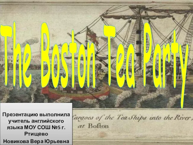 Презентация на тему The Boston Tea Party
  Verb to be
 Health Unit 8
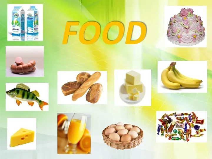 Food
Food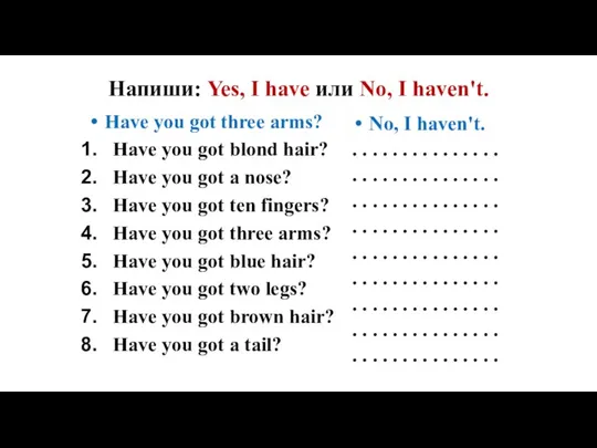 Напиши: Yes, I have или No, I haven't. (задание)
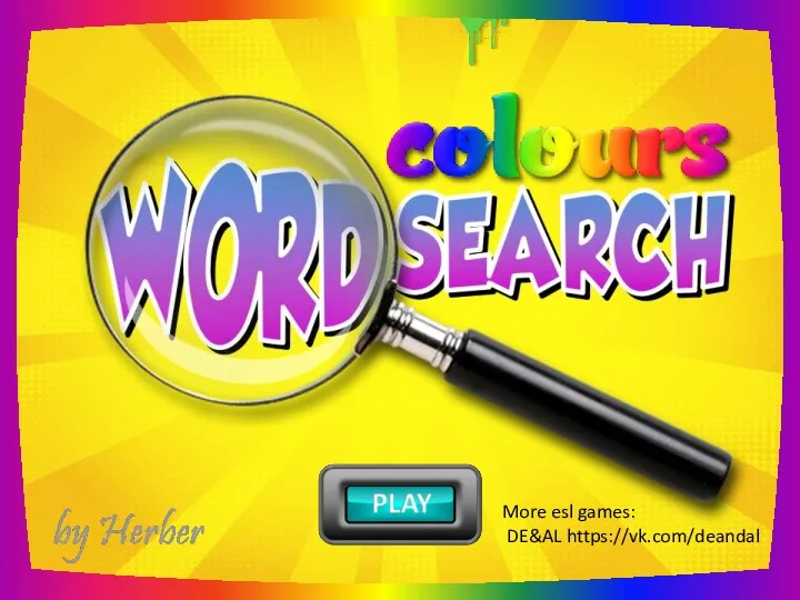 Colors word search
 Smack
Smack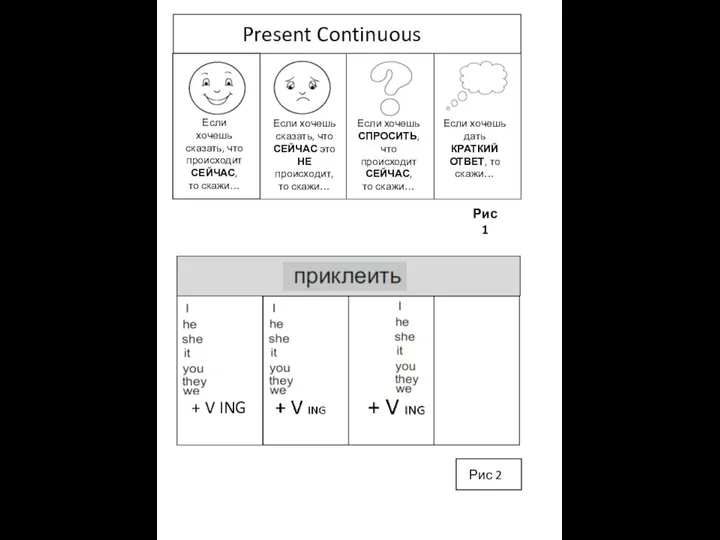 Present continuous
 Types of business letters
 Понимание английского языка через образное восприятие
 What season is it?
 We explain the way. Dialogue speech
 Презентация на тему Prince Andrew
  ABC holday
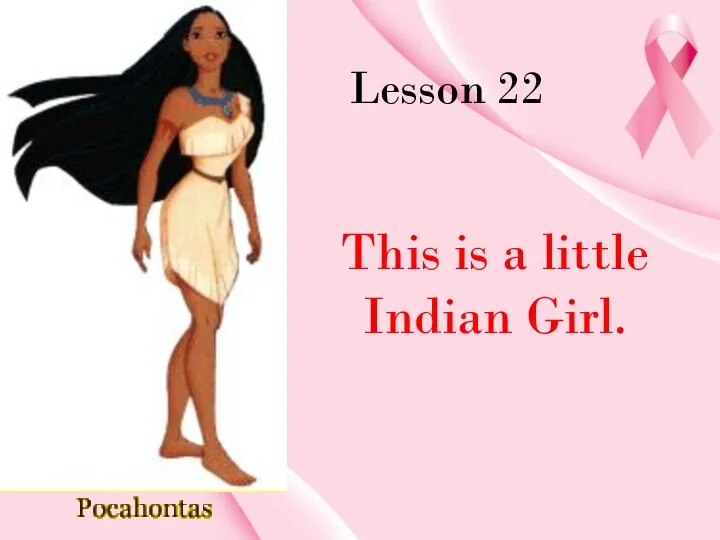 This is a little Indian Girl
 Self Introduction
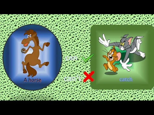 Can-cant
 Short u
 Презентация на тему Welcome the United Kingdom of Great Britain and Northern Ireland
  A historical tour round old streets of Mikhaylovka
 Games. The vowels (a, e, i, o, u) have been removed from these words. Try to guess what each word is. Buildings & Places
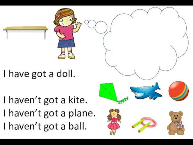 Reading toys
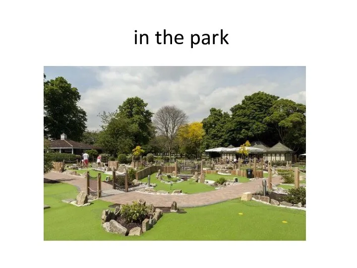 In the, on the
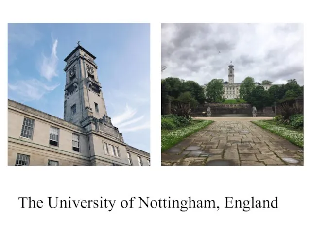 The University of Nottingham, England