- Tissue parasites

Содержание
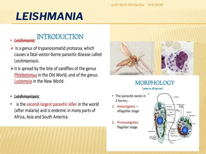
- 3. LEISHMANIA 4/9/2020 prof. Mahi Ghobashy
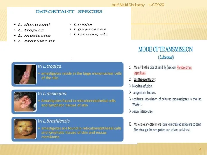
- 4. 4/9/2020 prof. Mahi Ghobashy
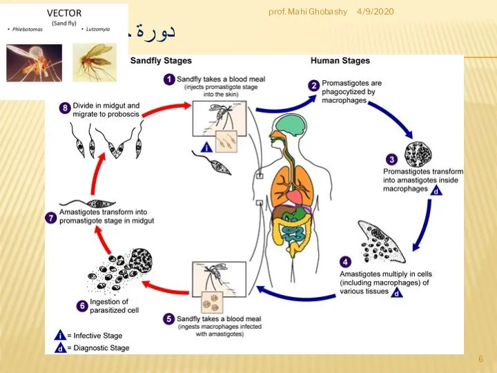
- 6. دورة حياة الليشمانيا 4/9/2020 prof. Mahi Ghobashy
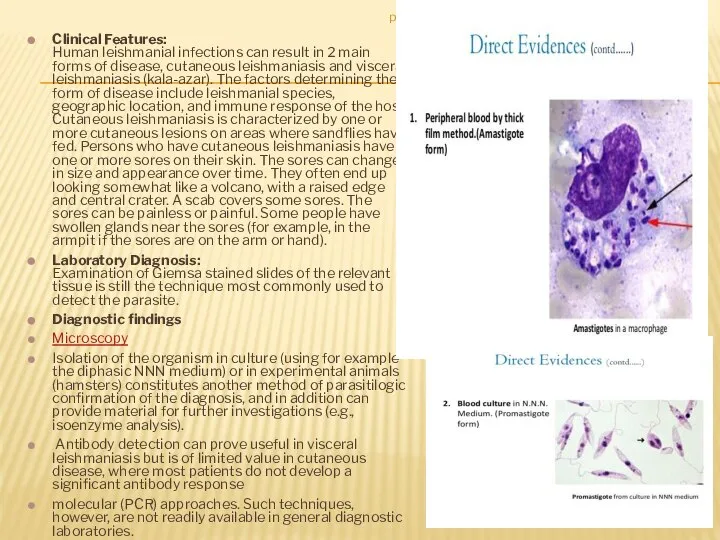
- 8. Clinical Features: Human leishmanial infections can result in 2 main forms of disease, cutaneous leishmaniasis and
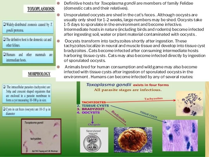
- 9. TOXOPLASMA GONDII Causal Agent: Toxoplasma gondii that infects most species of warm blooded animals, including humans,
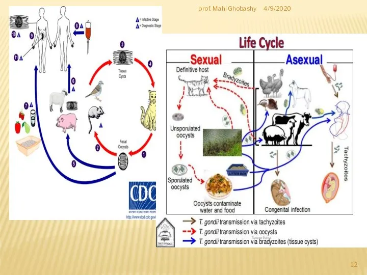
- 10. Definitive hosts for Toxoplasma gondii are members of family Felidae (domestic cats and their relatives). Unsporulated
- 12. 4/9/2020 prof. Mahi Ghobashy
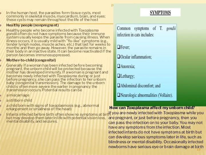
- 13. In the human host, the parasites form tissue cysts, most commonly in skeletal muscle, myocardium, brain,
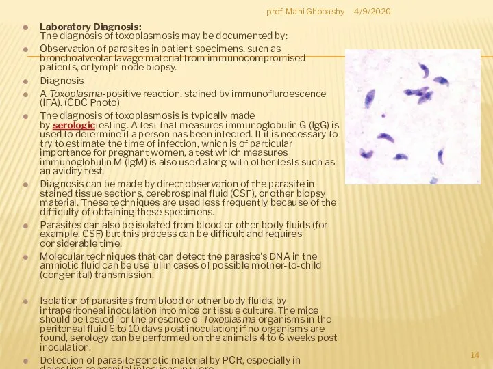
- 14. Laboratory Diagnosis: The diagnosis of toxoplasmosis may be documented by: Observation of parasites in patient specimens,
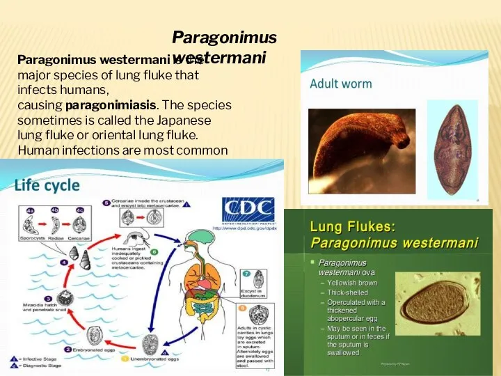
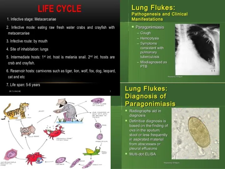
- 15. Paragonimus westermani is the major species of lung fluke that infects humans, causing paragonimiasis. The species
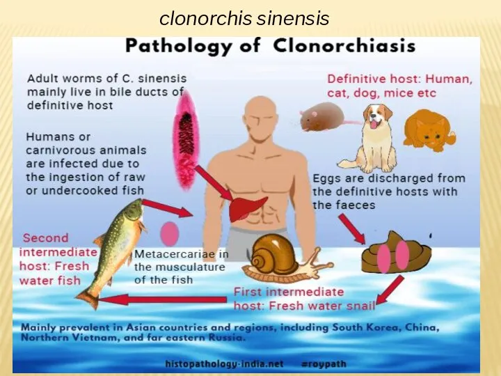
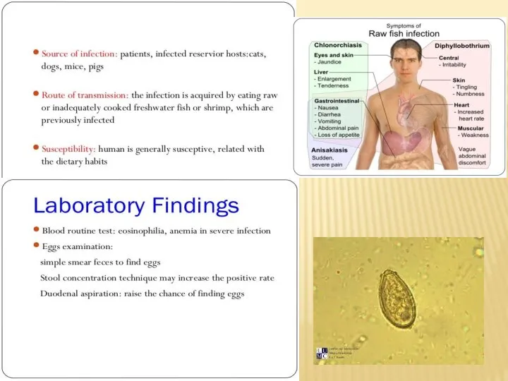
- 17. clonorchis sinensis
- 19. DRACUNCULUS MEDINENSIS 4/16/2020 prof. Mahi Ghobashy
- 23. Скачать презентацию
Слайд 3LEISHMANIA
4/9/2020
prof. Mahi Ghobashy
LEISHMANIA
4/9/2020
prof. Mahi Ghobashy
Слайд 44/9/2020
prof. Mahi Ghobashy
4/9/2020
prof. Mahi Ghobashy
Слайд 6دورة حياة الليشمانيا
4/9/2020
prof. Mahi Ghobashy
دورة حياة الليشمانيا
4/9/2020
prof. Mahi Ghobashy
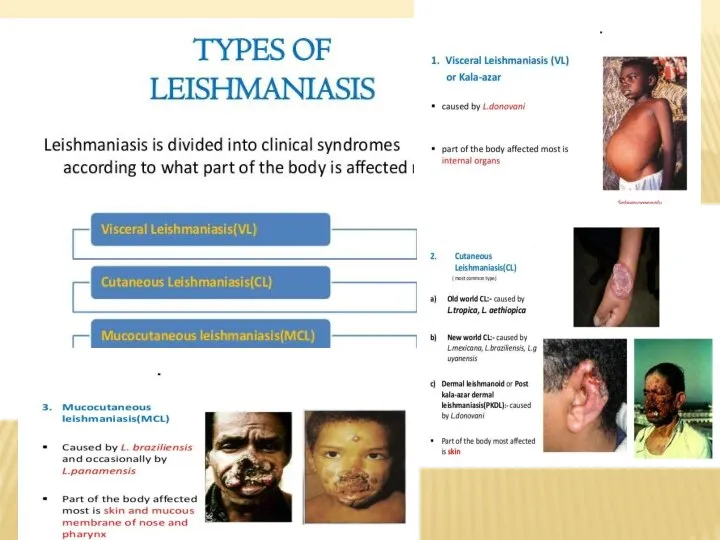
Слайд 8Clinical Features:
Human leishmanial infections can result in 2 main forms of disease,
Clinical Features: Human leishmanial infections can result in 2 main forms of disease,
Слайд 9TOXOPLASMA GONDII
Causal Agent:
Toxoplasma gondii that infects most species of warm blooded animals,
TOXOPLASMA GONDII
Causal Agent: Toxoplasma gondii that infects most species of warm blooded animals,
Слайд 10Definitive hosts for Toxoplasma gondii are members of family Felidae (domestic cats
Definitive hosts for Toxoplasma gondii are members of family Felidae (domestic cats

Слайд 124/9/2020
prof. Mahi Ghobashy
4/9/2020
prof. Mahi Ghobashy
Слайд 13
In the human host, the parasites form tissue cysts, most commonly
In the human host, the parasites form tissue cysts, most commonly
Слайд 14Laboratory Diagnosis:
The diagnosis of toxoplasmosis may be documented by:
Observation of parasites in
Laboratory Diagnosis:
The diagnosis of toxoplasmosis may be documented by:
Observation of parasites in
Слайд 15Paragonimus westermani is the major species of lung fluke that infects humans, causing paragonimiasis.
Paragonimus westermani is the major species of lung fluke that infects humans, causing paragonimiasis.
Слайд 17clonorchis sinensis
clonorchis sinensis
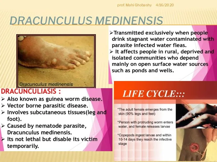
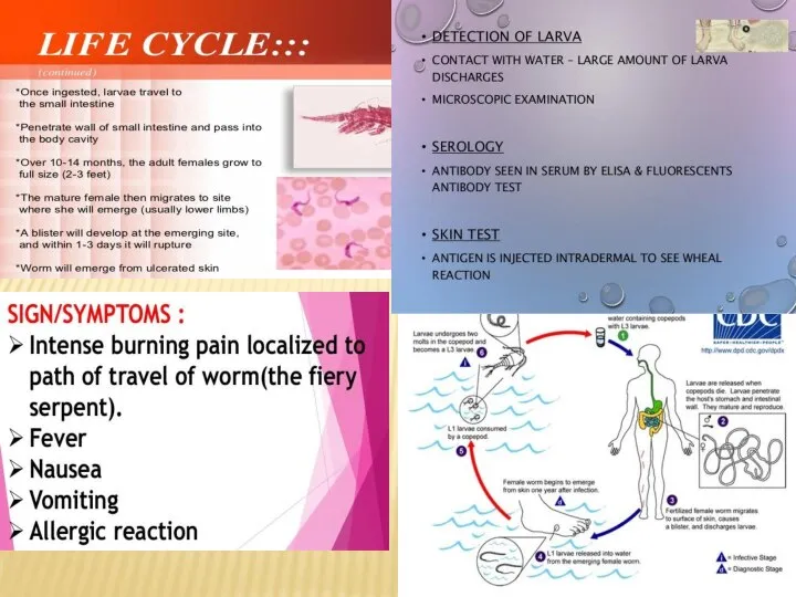
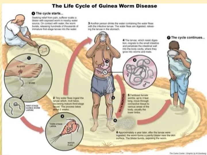
Слайд 19DRACUNCULUS MEDINENSIS
4/16/2020
prof. Mahi Ghobashy
DRACUNCULUS MEDINENSIS
4/16/2020
prof. Mahi Ghobashy
 Help Pippo do the cooking!
 Yesterday at 7 o´clock
 Local authorities in USA
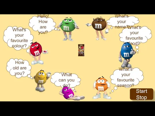 Yuliya Minyuk
 Impressive Phrases
 New York
 Презентация к уроку английского языка "What language do dolphins speak?" -
  Вживання артикля the з географічними назвами
 Superlative
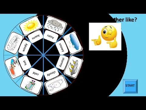 What is the weather like?
 The Ural Mountains
 Презентация к уроку английского языка "Организация самостоятельной работы учащихся на уроках английского языка" -
 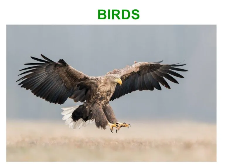 Птицы. Birds
 Legal systems of the world. Common law
 My second project of life
 Квест для марафона
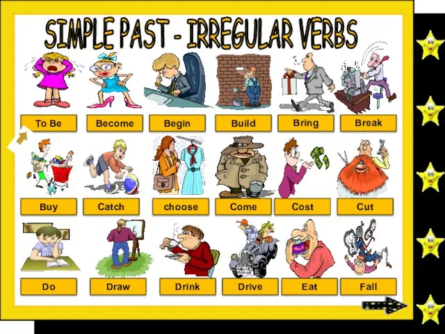 Simple past. Irregular verbs
 The Value of Seismic: From Greenfields to Mine Architecture, Structure, and Direct Targeting
 Speaking Session
 Comparison of heroes
 Reading ESL Classroom
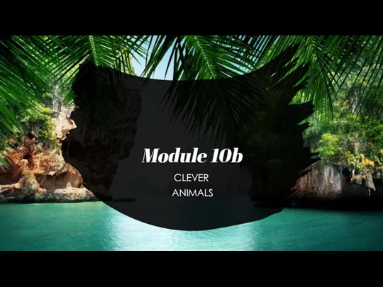 Clever animals
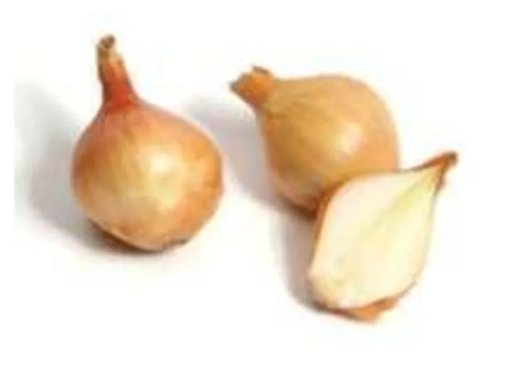 Vegetables. Овощи
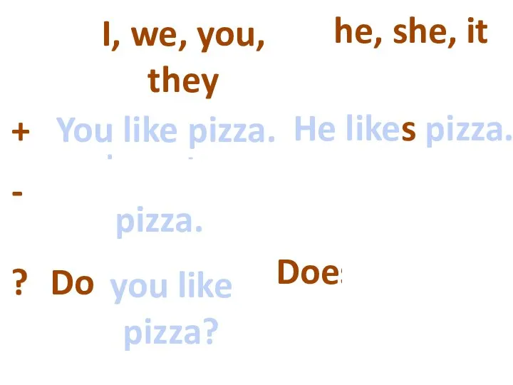 Present simple
 Test What’s time is it?
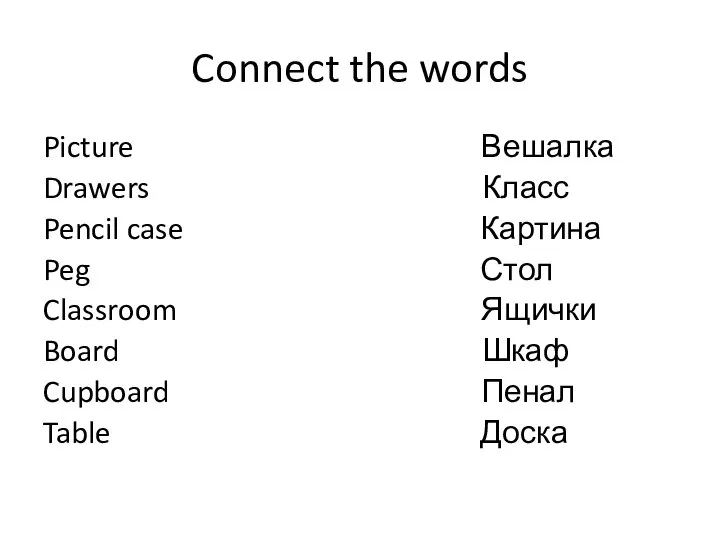 Connect the words
 Easter project by Riko Nomiyama
 FiguraDesign. Summary