- Central Nervous System: CNS

Содержание
- 2. The Spinal Cord Foramen magnum to L1 or L2 Runs through the vertebral canal of the
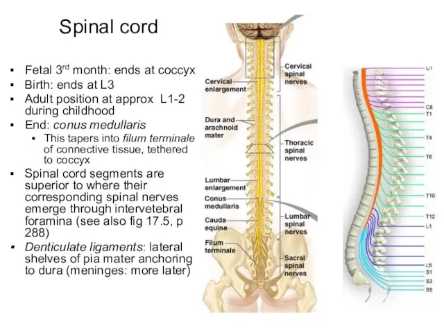
- 3. Fetal 3rd month: ends at coccyx Birth: ends at L3 Adult position at approx L1-2 during
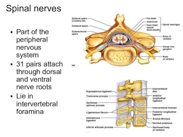
- 4. Spinal nerves Part of the peripheral nervous system 31 pairs attach through dorsal and ventral nerve
- 5. Spinal nerves continued Divided based on vertebral locations 8 cervical 12 thoracic 5 lumbar 5 sacral
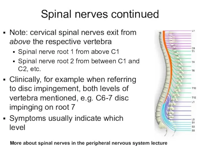
- 6. Spinal nerves continued Note: cervical spinal nerves exit from above the respective vertebra Spinal nerve root
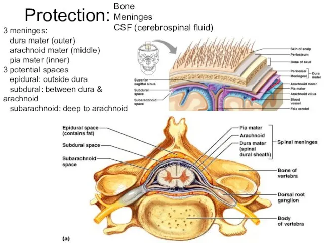
- 7. Protection: Bone Meninges CSF (cerebrospinal fluid) 3 meninges: dura mater (outer) arachnoid mater (middle) pia mater
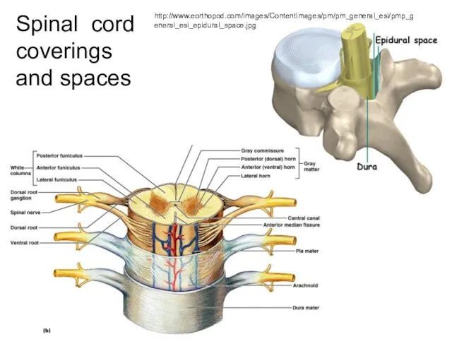
- 8. Dura mater Arachnoid mater Pia mater Spinal cord coverings and spaces http://www.eorthopod.com/images/ContentImages/pm/pm_general_esi/pmp_general_esi_epidural_space.jpg
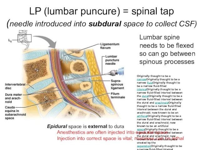
- 9. LP (lumbar puncure) = spinal tap (needle introduced into subdural space to collect CSF) Lumbar spine
- 10. Spinal cord anatomy Posterior median sulcus (“p”) Anterior median fissure (“a”) White matter (yellow here) Gray
- 11. Gray/White in spinal cord Hollow central cavity (“central canal”) Gray matter surrounds cavity White matter surrounds
- 12. Spinal cord anatomy Gray commissure with central canal Columns of gray running the length of the
- 13. White matter of the spinal cord (myelinated and unmyelinated axons) Ascending fibers: sensory information from sensory
- 14. The Brain: embryonic development Develops from neural tube Brain subdivides into Forebrain Midbrain Hindbrain These further
- 15. Brain development Learn forebrain, midbrain and hindbrain in (b) See next color coded pics in reference
- 16. Space restrictions force cerebral hemispheres to grow posteriorly over rest of brain, enveloping it Cerebral hemispheres
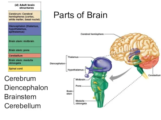
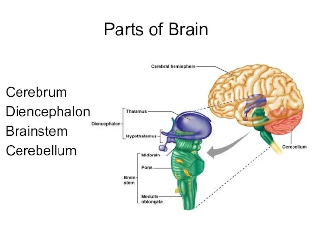
- 17. Anatomical classification Cerebral hemispheres Diencephalon Thalamus Hypothalamus Brain stem Midbrain Pons Medulla Cerebellum Spinal cord
- 18. Parts of Brain Cerebrum Diencephalon Brainstem Cerebellum
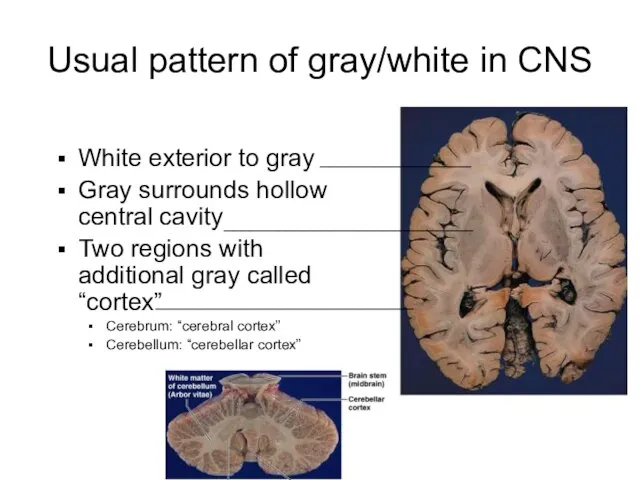
- 19. Usual pattern of gray/white in CNS White exterior to gray Gray surrounds hollow central cavity Two
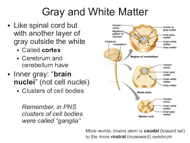
- 20. Gray and White Matter Like spinal cord but with another layer of gray outside the white
- 21. Ventricles Central cavities expanded Filled with CSF (cerebrospinal fluid) Lined by ependymal cells (these cells lining
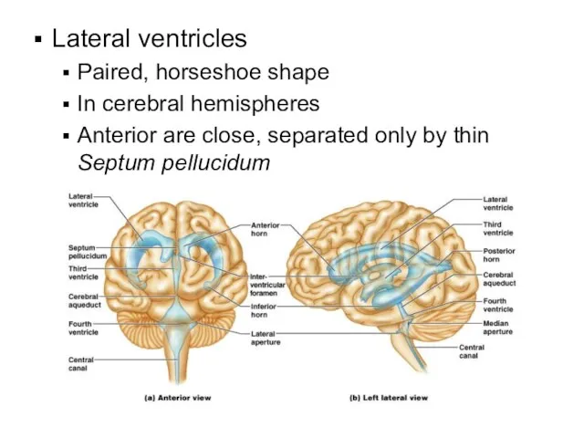
- 22. Lateral ventricles Paired, horseshoe shape In cerebral hemispheres Anterior are close, separated only by thin Septum
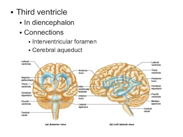
- 23. Third ventricle In diencephalon Connections Interventricular foramen Cerebral aqueduct
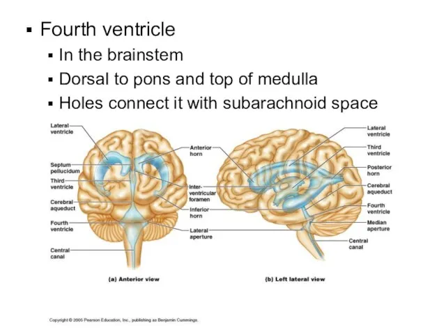
- 24. Fourth ventricle In the brainstem Dorsal to pons and top of medulla Holes connect it with
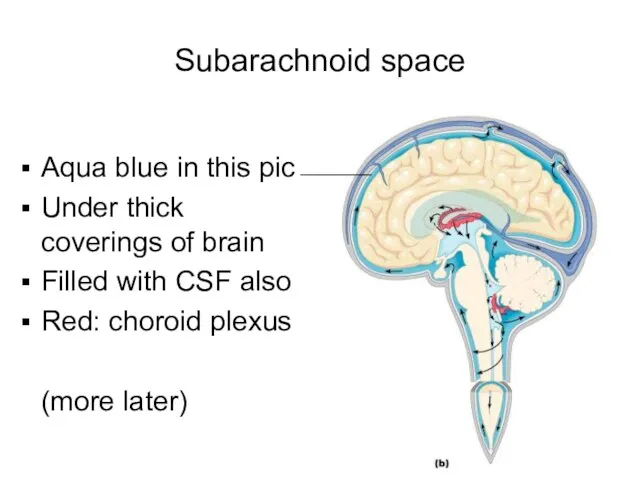
- 25. Subarachnoid space Aqua blue in this pic Under thick coverings of brain Filled with CSF also
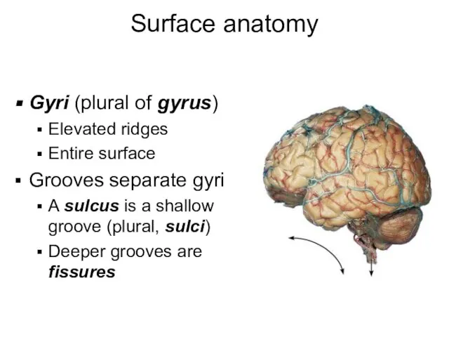
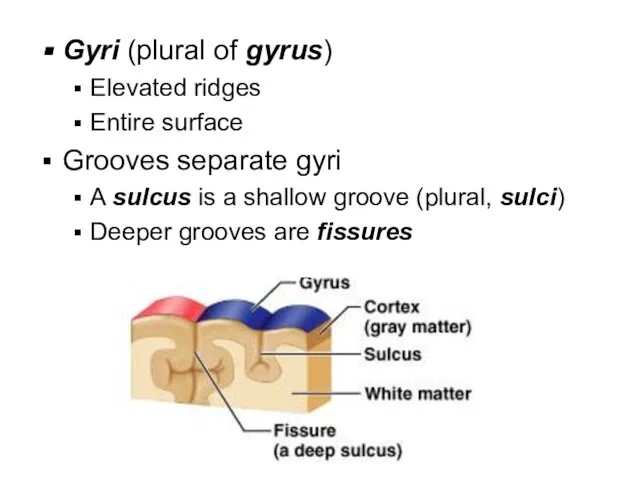
- 26. Surface anatomy Gyri (plural of gyrus) Elevated ridges Entire surface Grooves separate gyri A sulcus is
- 27. Gyri (plural of gyrus) Elevated ridges Entire surface Grooves separate gyri A sulcus is a shallow
- 28. Parts of Brain Cerebrum Diencephalon Brainstem Cerebellum
- 29. simplified… Back of brain: perception Top of brain: movement Front of brain: thinking
- 30. Cerebral hemispheres Lobes: under bones of same name Frontal Parietal Temporal Occipital Plus: Insula (buried deep
- 31. Cerebral hemispheres: note lobes Divided by longitudinal fissure into right & left sides Central sulcus divides
- 32. Lateral sulcus separates temporal lobe from parietal lobe Parieto-occipital sulcus divides occipital and parietal lobes (not
- 33. coronal section Note: longitudinal fissure, lateral sulcus, insula Note: cerebral cortex (external sheet of gray), cerebral
- 34. Cerebral cortex Executive functioning capability Gray matter: of neuron cell bodies, dendrites, short unmyelinated axons 100
- 35. Prenatal life: genes are responsible for creating the architecture of the brain Cortex is the last
- 36. Cerebral cortex All the neurons are interneurons By definition confined to the CNS They have to
- 37. Sensory areas Posterior to central sulcus Primary somatosensory cortex: postcentral gyrus of parietal lobe (allows conscious
- 38. From special sense organs Sight: occipital lobe Primary visual cortex (17) Handles info from contralateral retina
- 39. Refer back to this labeled version as needed
- 40. Smell (olfactory sense): uncus Deep in temporal lobe along medial surface
- 41. fMRI: functional magnetic resonance imaging Cerebral cortex of person speaking & hearing Activity (blood flow) in
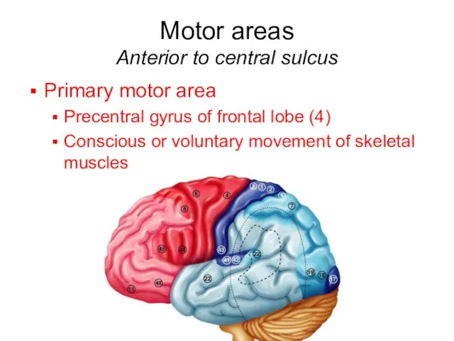
- 42. Motor areas Anterior to central sulcus Primary motor area Precentral gyrus of frontal lobe (4) Conscious
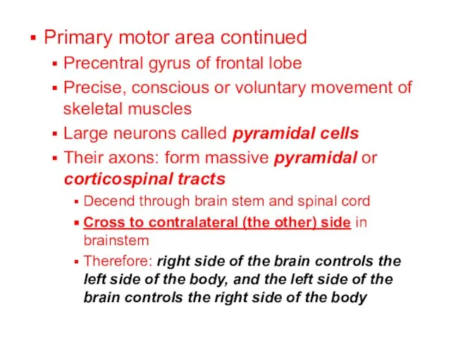
- 43. Primary motor area continued Precentral gyrus of frontal lobe Precise, conscious or voluntary movement of skeletal
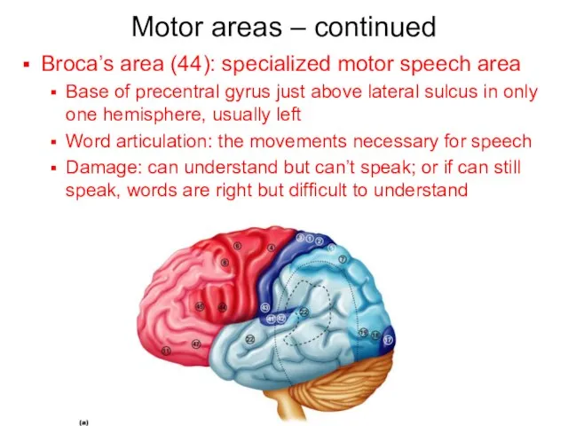
- 44. Motor areas – continued Broca’s area (44): specialized motor speech area Base of precentral gyrus just
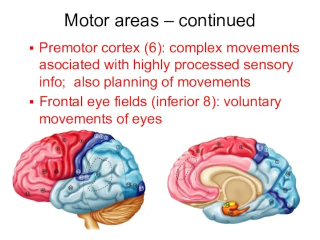
- 45. Motor areas – continued Premotor cortex (6): complex movements asociated with highly processed sensory info; also
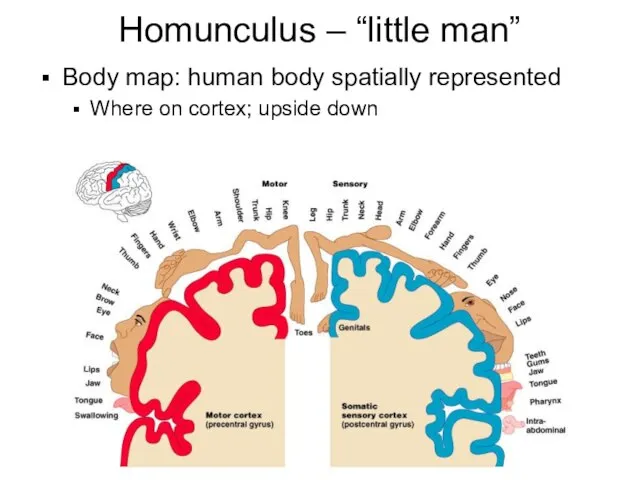
- 46. Homunculus – “little man” Body map: human body spatially represented Where on cortex; upside down
- 47. Association Areas Remember… Three kinds of functional areas (cerebrum) Motor areas: movement Sensory areas: perception Association
- 48. Association Areas Tie together different kinds of sensory input Associate new input with memories Is to
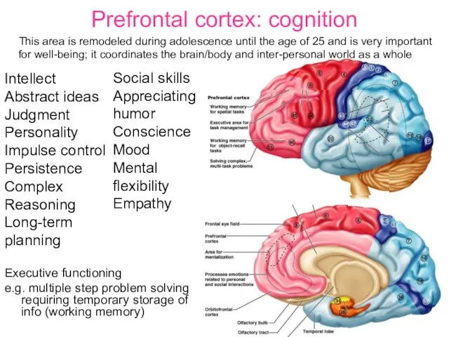
- 49. Prefrontal cortex: cognition Executive functioning e.g. multiple step problem solving requiring temporary storage of info (working
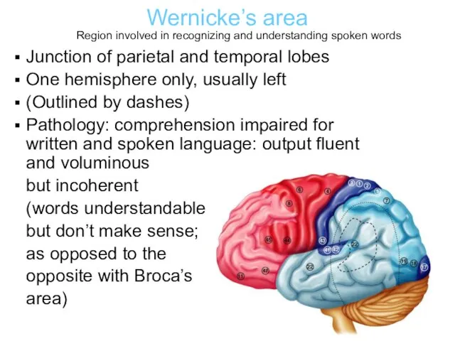
- 50. Wernicke’s area Junction of parietal and temporal lobes One hemisphere only, usually left (Outlined by dashes)
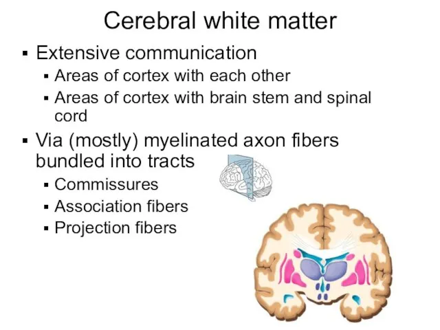
- 51. Cerebral white matter Extensive communication Areas of cortex with each other Areas of cortex with brain
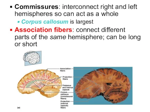
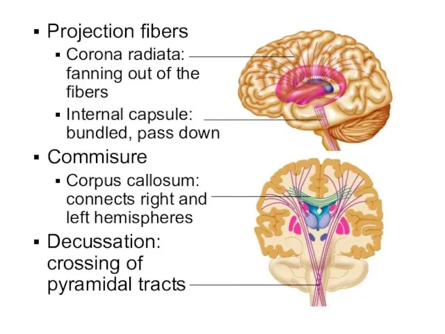
- 52. Commissures: interconnect right and left hemispheres so can act as a whole Corpus callosum is largest
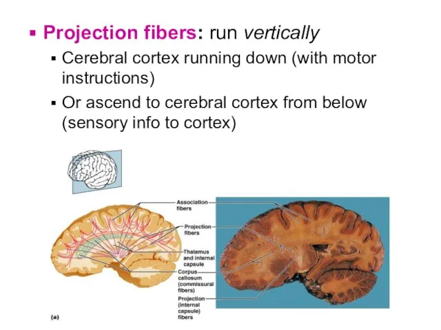
- 53. Projection fibers: run vertically Cerebral cortex running down (with motor instructions) Or ascend to cerebral cortex
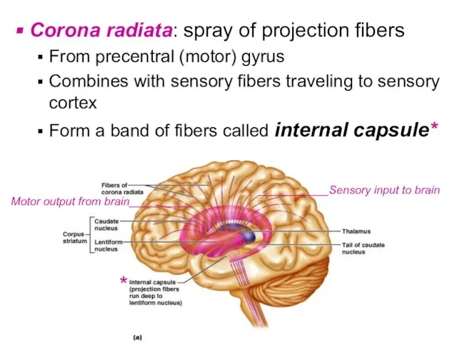
- 54. Corona radiata: spray of projection fibers From precentral (motor) gyrus Combines with sensory fibers traveling to
- 55. Projection fibers Corona radiata: fanning out of the fibers Internal capsule: bundled, pass down Commisure Corpus
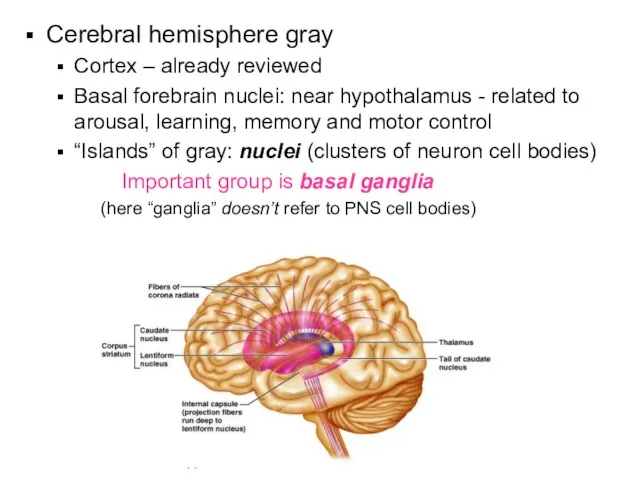
- 56. Cerebral hemisphere gray Cortex – already reviewed Basal forebrain nuclei: near hypothalamus - related to arousal,
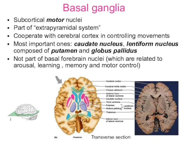
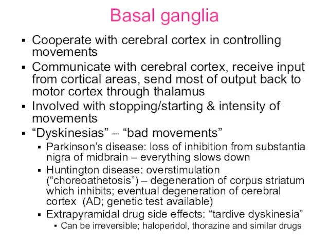
- 57. Basal ganglia Subcortical motor nuclei Part of “extrapyramidal system” Cooperate with cerebral cortex in controlling movements
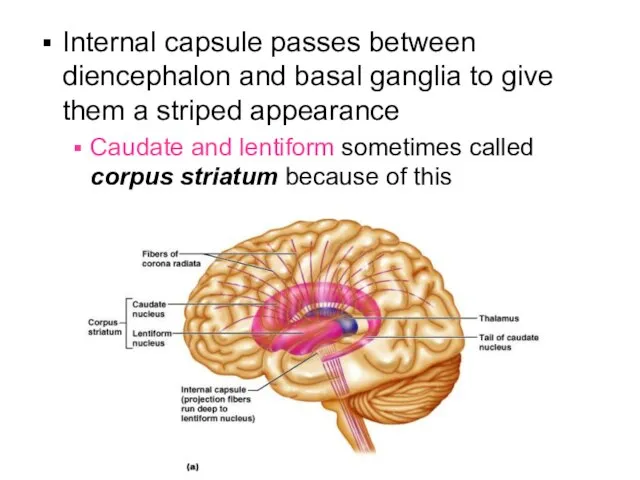
- 58. Internal capsule passes between diencephalon and basal ganglia to give them a striped appearance Caudate and
- 59. Basal ganglia Cooperate with cerebral cortex in controlling movements Communicate with cerebral cortex, receive input from
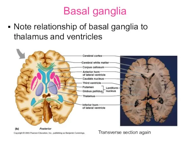
- 60. Basal ganglia Note relationship of basal ganglia to thalamus and ventricles Transverse section again
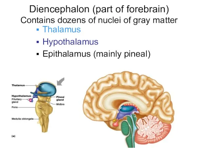
- 61. Diencephalon (part of forebrain) Contains dozens of nuclei of gray matter Thalamus Hypothalamus Epithalamus (mainly pineal)
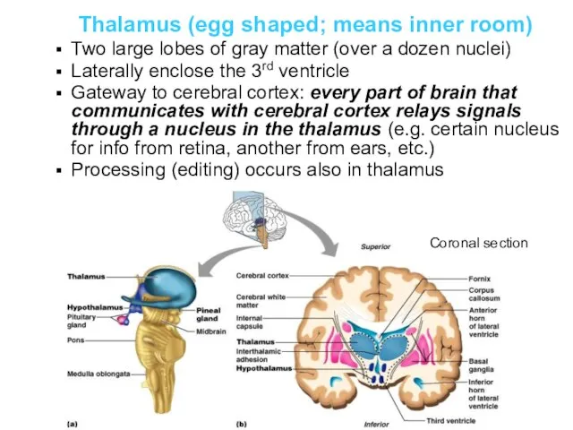
- 62. Thalamus (egg shaped; means inner room) Two large lobes of gray matter (over a dozen nuclei)
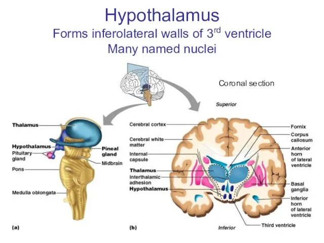
- 63. Hypothalamus Forms inferolateral walls of 3rd ventricle Many named nuclei Coronal section
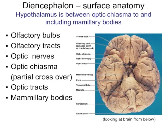
- 64. Diencephalon – surface anatomy Hypothalamus is between optic chiasma to and including mamillary bodies Olfactory bulbs
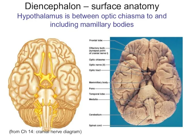
- 65. Diencephalon – surface anatomy Hypothalamus is between optic chiasma to and including mamillary bodies (from Ch
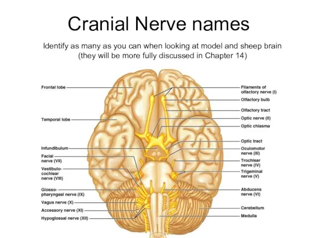
- 66. Cranial Nerve names Identify as many as you can when looking at model and sheep brain
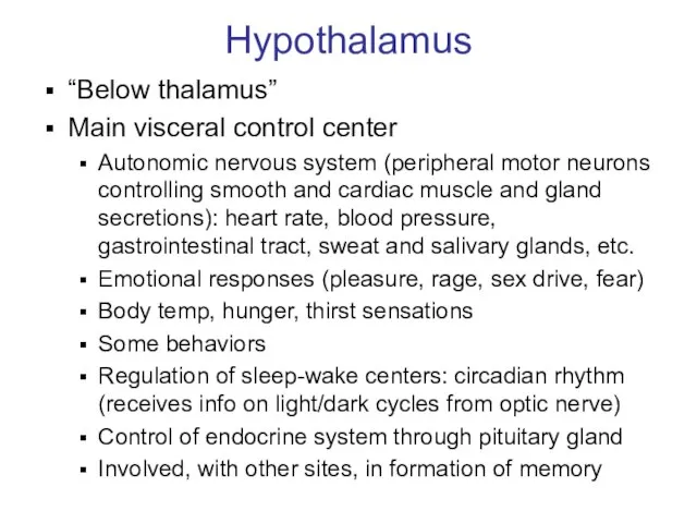
- 67. Hypothalamus “Below thalamus” Main visceral control center Autonomic nervous system (peripheral motor neurons controlling smooth and
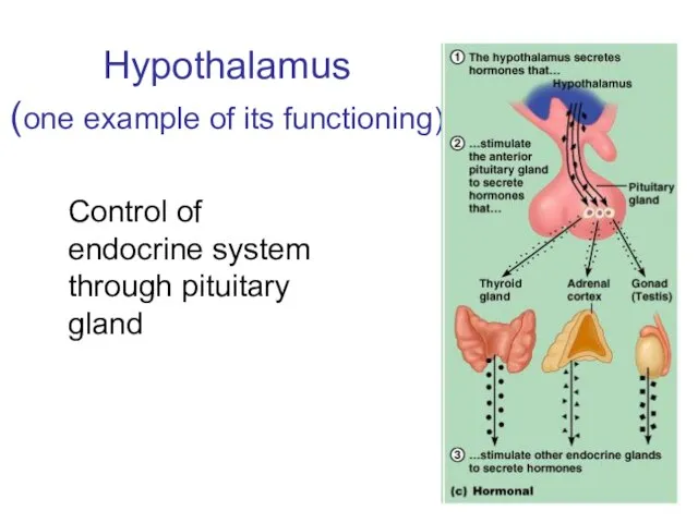
- 68. Hypothalamus (one example of its functioning) Control of endocrine system through pituitary gland
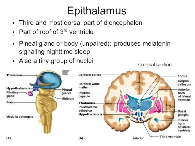
- 69. Epithalamus Third and most dorsal part of diencephalon Part of roof of 3rd ventricle Pineal gland
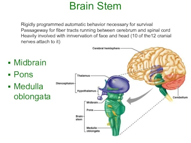
- 70. Brain Stem Midbrain Pons Medulla oblongata Rigidly programmed automatic behavior necessary for survival Passageway for fiber
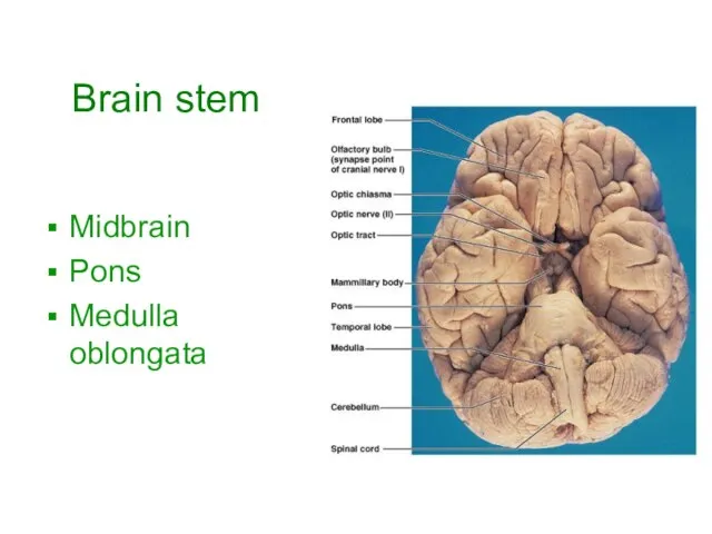
- 71. Brain stem Midbrain Pons Medulla oblongata
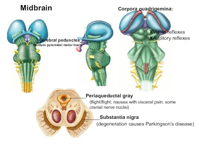
- 72. __Cerebral peduncles____ Contain pyramidal motor tracts Corpora quadrigemina: XVisual reflexes XAuditory reflexes Midbrain ______Substantia nigra (degeneration
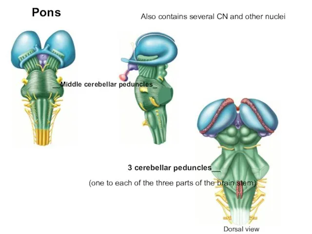
- 73. __Middle cerebellar peduncles_ Pons 3 cerebellar peduncles__ Also contains several CN and other nuclei (one to
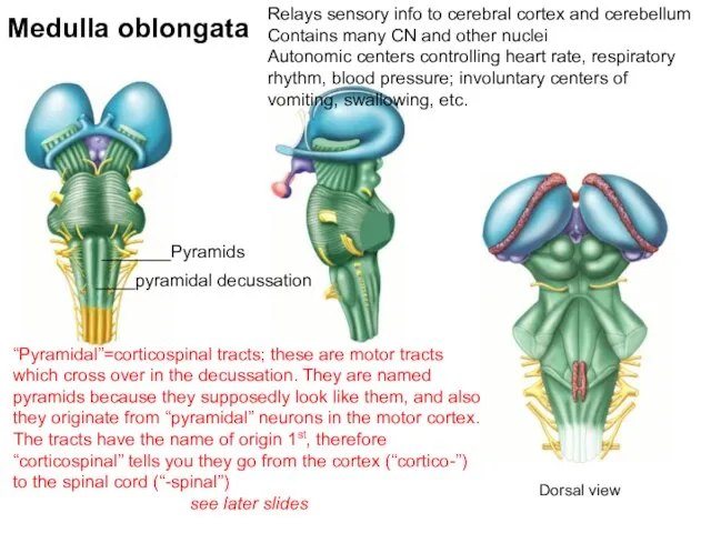
- 74. Medulla oblongata Relays sensory info to cerebral cortex and cerebellum Contains many CN and other nuclei
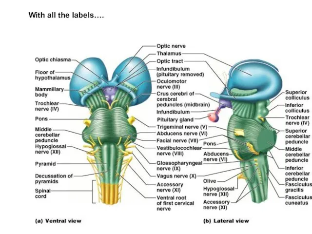
- 75. With all the labels….
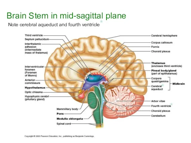
- 76. Brain Stem in mid-sagittal plane Note cerebral aqueduct and fourth ventricle* * *
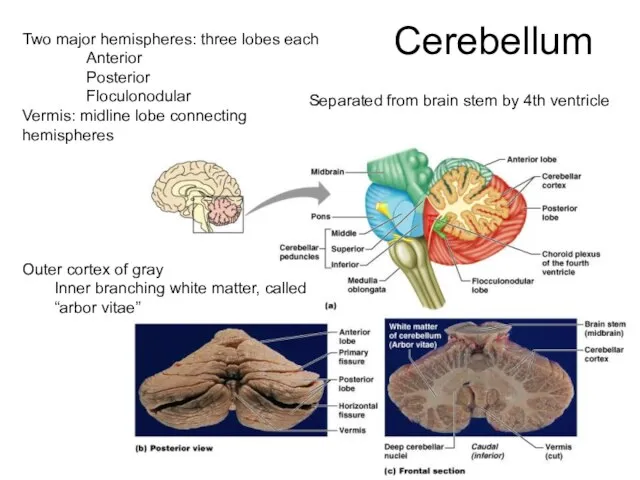
- 77. Cerebellum Two major hemispheres: three lobes each Anterior Posterior Floculonodular Vermis: midline lobe connecting hemispheres Outer
- 78. Functions of cerebellum Smooths, coordinates & fine tunes bodily movements Helps maintain body posture Helps maintain
- 79. Functional brain systems (as opposed to anatomical ones) Networks of distant neurons that function together Limbic
- 80. Limbic system (not a discrete structure - includes many brain areas) Most important parts: Hipocampus Amygdala
- 81. Limbic system continued Called the “emotional” brain Is essential for flexible, stable, adaptive functioning Links different
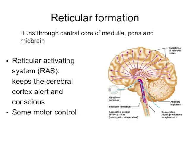
- 82. Reticular formation Runs through central core of medulla, pons and midbrain Reticular activating system (RAS): keeps
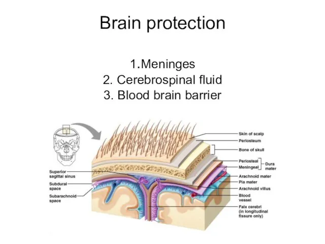
- 83. Brain protection 1.Meninges 2. Cerebrospinal fluid 3. Blood brain barrier
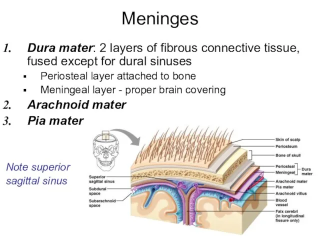
- 84. Meninges Dura mater: 2 layers of fibrous connective tissue, fused except for dural sinuses Periosteal layer
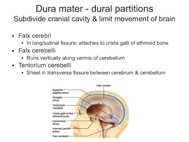
- 85. Dura mater - dural partitions Subdivide cranial cavity & limit movement of brain Falx cerebri In
- 86. Arachnoid mater Between dura and arachnoid: subdural space Dura and arachnoid cover brain loosely Deep to
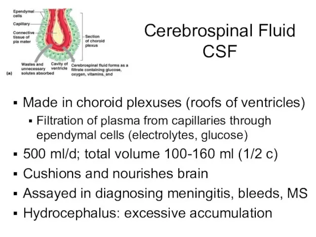
- 87. Cerebrospinal Fluid CSF Made in choroid plexuses (roofs of ventricles) Filtration of plasma from capillaries through
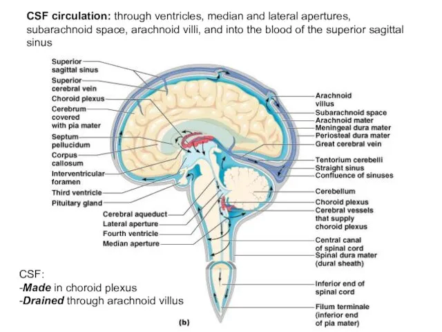
- 88. CSF circulation: through ventricles, median and lateral apertures, subarachnoid space, arachnoid villi, and into the blood
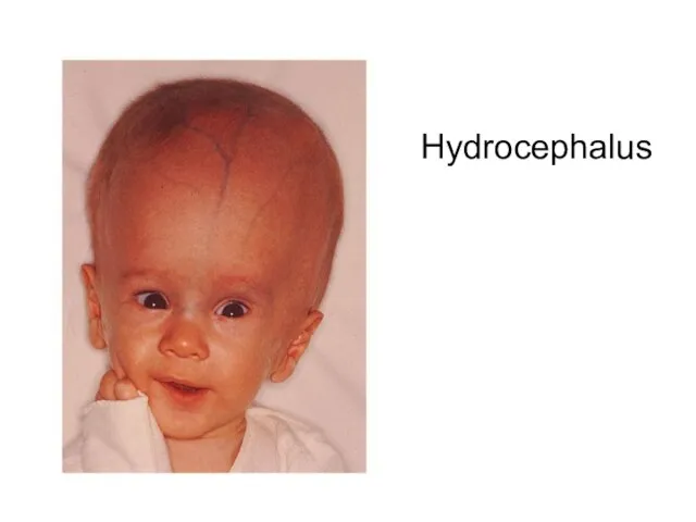
- 89. Hydrocephalus
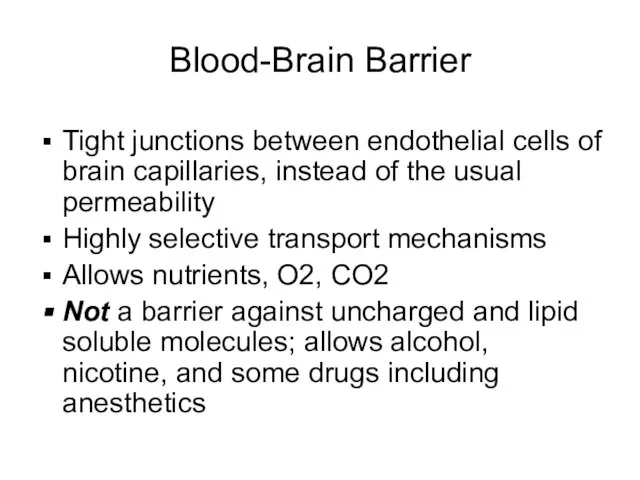
- 90. Blood-Brain Barrier Tight junctions between endothelial cells of brain capillaries, instead of the usual permeability Highly
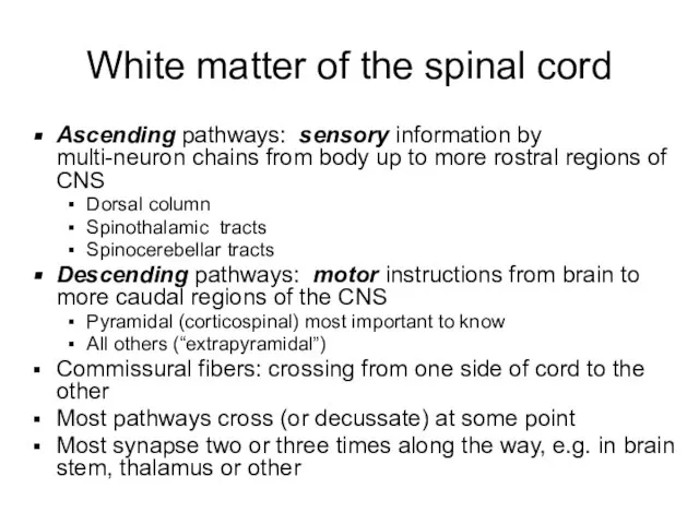
- 91. White matter of the spinal cord Ascending pathways: sensory information by multi-neuron chains from body up
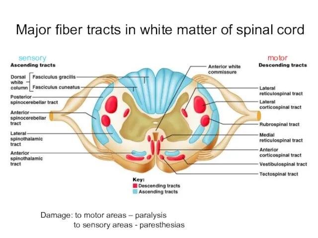
- 92. Major fiber tracts in white matter of spinal cord Damage: to motor areas – paralysis to
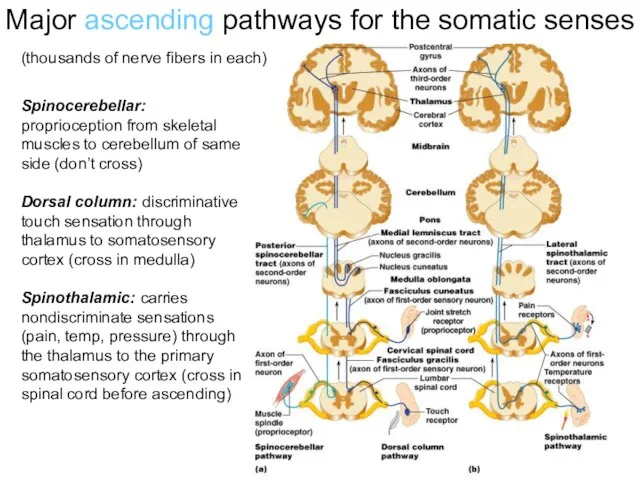
- 93. Major ascending pathways for the somatic senses Spinocerebellar: proprioception from skeletal muscles to cerebellum of same
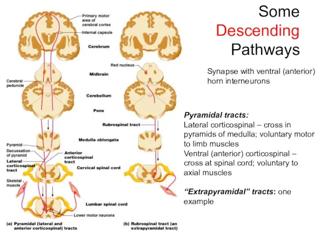
- 94. Some Descending Pathways Pyramidal tracts: Lateral corticospinal – cross in pyramids of medulla; voluntary motor to
- 95. Check out: Medical gross anatomy atlas images (good teaching pics): http://anatomy.med.umich.edu/atlas/atlas_index.html (can access from Paul Wissman’s
- 96. Hints & additional pics Unless your prints of the slides are very large and clear, look
- 97. Know the names of the ventricles and which ones connect to which, in order You don’t
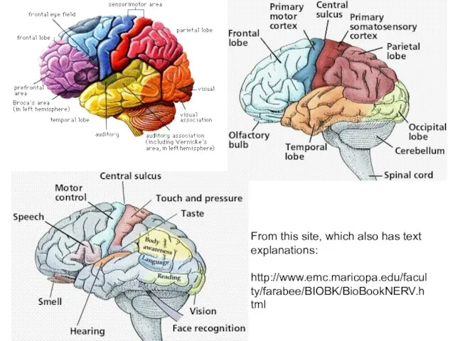
- 98. From this site, which also has text explanations: http://www.emc.maricopa.edu/faculty/farabee/BIOBK/BioBookNERV.html
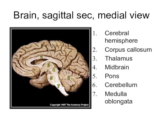
- 99. Brain, sagittal sec, medial view Cerebral hemisphere Corpus callosum Thalamus Midbrain Pons Cerebellum Medulla oblongata
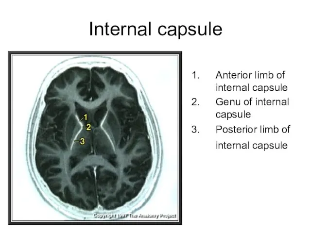
- 100. Internal capsule Anterior limb of internal capsule Genu of internal capsule Posterior limb of internal capsule
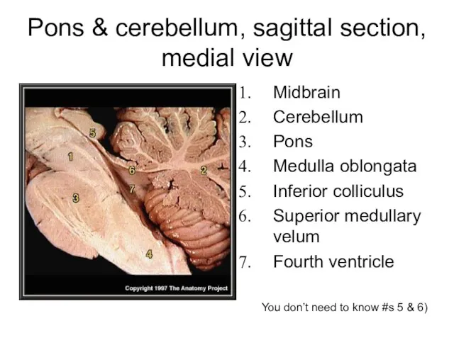
- 101. Pons & cerebellum, sagittal section, medial view Midbrain Cerebellum Pons Medulla oblongata Inferior colliculus Superior medullary
- 103. Скачать презентацию
Слайд 3Fetal 3rd month: ends at coccyx
Birth: ends at L3
Adult position at approx
Fetal 3rd month: ends at coccyx
Birth: ends at L3
Adult position at approx
Слайд 4Spinal nerves
Part of the peripheral nervous system
31 pairs attach through dorsal and
Spinal nerves
Part of the peripheral nervous system
31 pairs attach through dorsal and
Слайд 5Spinal nerves continued
Divided based on vertebral locations
8 cervical
12 thoracic
5 lumbar
5 sacral
1 coccygeal
Cauda
Spinal nerves continued
Divided based on vertebral locations
8 cervical
12 thoracic
5 lumbar
5 sacral
1 coccygeal
Cauda
Слайд 6Spinal nerves continued
Note: cervical spinal nerves exit from above the respective vertebra
Spinal
Spinal nerves continued
Note: cervical spinal nerves exit from above the respective vertebra
Spinal
Слайд 7Protection:
Bone
Meninges
CSF (cerebrospinal fluid)
3 meninges:
dura mater (outer)
arachnoid mater (middle)
pia mater
Protection:
Bone
Meninges
CSF (cerebrospinal fluid)
3 meninges:
dura mater (outer)
arachnoid mater (middle)
pia mater
Слайд 8Dura mater
Arachnoid mater
Pia mater
Spinal cord coverings and spaces
http://www.eorthopod.com/images/ContentImages/pm/pm_general_esi/pmp_general_esi_epidural_space.jpg
Dura mater
Arachnoid mater
Pia mater
Spinal cord coverings and spaces
http://www.eorthopod.com/images/ContentImages/pm/pm_general_esi/pmp_general_esi_epidural_space.jpg
Слайд 9LP (lumbar puncure) = spinal tap
(needle introduced into subdural space to collect
LP (lumbar puncure) = spinal tap (needle introduced into subdural space to collect
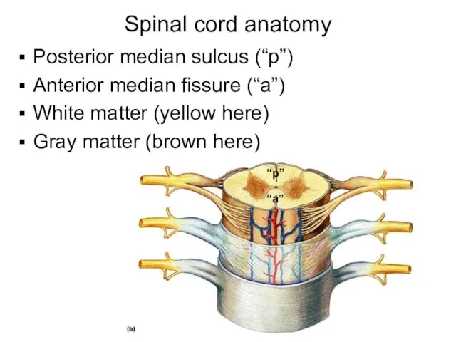
Слайд 10Spinal cord anatomy
Posterior median sulcus (“p”)
Anterior median fissure (“a”)
White matter (yellow here)
Gray
Spinal cord anatomy
Posterior median sulcus (“p”)
Anterior median fissure (“a”)
White matter (yellow here)
Gray
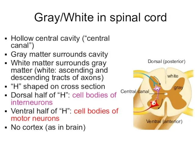
Слайд 11Gray/White in spinal cord
Hollow central cavity (“central canal”)
Gray matter surrounds cavity
White matter
Gray/White in spinal cord
Hollow central cavity (“central canal”)
Gray matter surrounds cavity
White matter
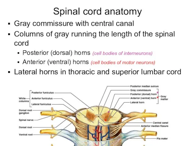
Слайд 12Spinal cord anatomy
Gray commissure with central canal
Columns of gray running the length
Spinal cord anatomy
Gray commissure with central canal
Columns of gray running the length
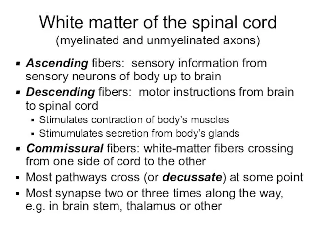
Слайд 13White matter of the spinal cord
(myelinated and unmyelinated axons)
Ascending fibers: sensory information
White matter of the spinal cord
(myelinated and unmyelinated axons)
Ascending fibers: sensory information
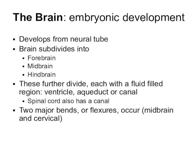
Слайд 14The Brain: embryonic development
Develops from neural tube
Brain subdivides into
Forebrain
Midbrain
Hindbrain
These further divide,
The Brain: embryonic development
Develops from neural tube
Brain subdivides into
Forebrain
Midbrain
Hindbrain
These further divide,
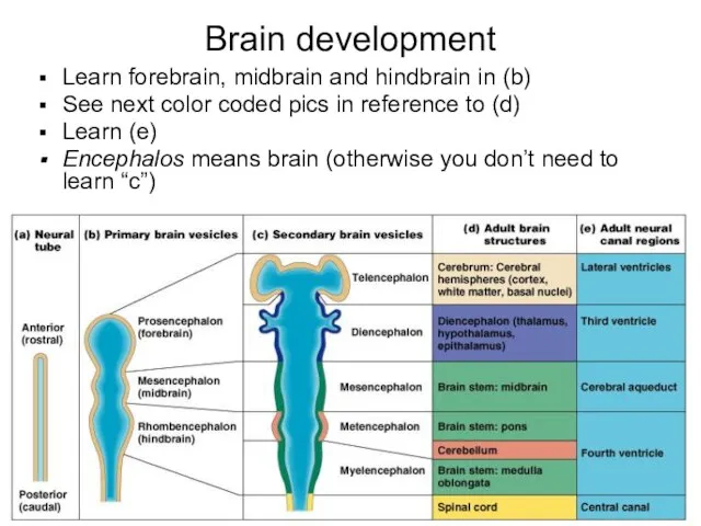
Слайд 15Brain development
Learn forebrain, midbrain and hindbrain in (b)
See next color
Brain development
Learn forebrain, midbrain and hindbrain in (b)
See next color
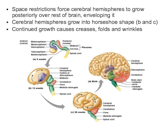
Слайд 16Space restrictions force cerebral hemispheres to grow posteriorly over rest of brain,
Space restrictions force cerebral hemispheres to grow posteriorly over rest of brain,
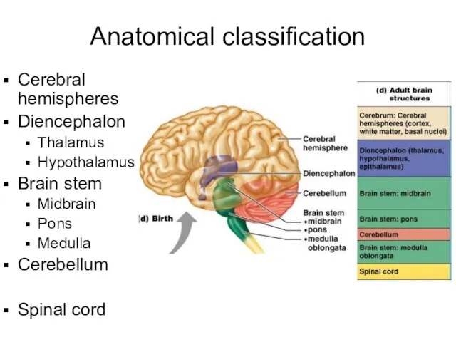
Слайд 17Anatomical classification
Cerebral hemispheres
Diencephalon
Thalamus
Hypothalamus
Brain stem
Midbrain
Pons
Medulla
Cerebellum
Spinal cord
Anatomical classification
Cerebral hemispheres
Diencephalon
Thalamus
Hypothalamus
Brain stem
Midbrain
Pons
Medulla
Cerebellum
Spinal cord
Слайд 18Parts of Brain
Cerebrum
Diencephalon
Brainstem
Cerebellum
Parts of Brain
Cerebrum
Diencephalon
Brainstem
Cerebellum
Слайд 19Usual pattern of gray/white in CNS
White exterior to gray
Gray surrounds hollow central
Usual pattern of gray/white in CNS
White exterior to gray
Gray surrounds hollow central
Слайд 20Gray and White Matter
Like spinal cord but with another layer of gray
Gray and White Matter
Like spinal cord but with another layer of gray
Слайд 21Ventricles
Central cavities expanded
Filled with CSF (cerebrospinal fluid)
Lined by ependymal cells (these cells
Ventricles
Central cavities expanded
Filled with CSF (cerebrospinal fluid)
Lined by ependymal cells (these cells
Слайд 22Lateral ventricles
Paired, horseshoe shape
In cerebral hemispheres
Anterior are close, separated only by thin
Lateral ventricles
Paired, horseshoe shape
In cerebral hemispheres
Anterior are close, separated only by thin
Слайд 23Third ventricle
In diencephalon
Connections
Interventricular foramen
Cerebral aqueduct
Third ventricle
In diencephalon
Connections
Interventricular foramen
Cerebral aqueduct
Слайд 24Fourth ventricle
In the brainstem
Dorsal to pons and top of medulla
Holes connect it
Fourth ventricle
In the brainstem
Dorsal to pons and top of medulla
Holes connect it
Слайд 25Subarachnoid space
Aqua blue in this pic
Under thick coverings of brain
Filled with CSF
Subarachnoid space
Aqua blue in this pic
Under thick coverings of brain
Filled with CSF
Слайд 26Surface anatomy
Gyri (plural of gyrus)
Elevated ridges
Entire surface
Grooves separate gyri
A sulcus is
Surface anatomy
Gyri (plural of gyrus)
Elevated ridges
Entire surface
Grooves separate gyri
A sulcus is
Слайд 27Gyri (plural of gyrus)
Elevated ridges
Entire surface
Grooves separate gyri
A sulcus is a shallow
Gyri (plural of gyrus)
Elevated ridges
Entire surface
Grooves separate gyri
A sulcus is a shallow
Слайд 28Parts of Brain
Cerebrum
Diencephalon
Brainstem
Cerebellum
Parts of Brain
Cerebrum
Diencephalon
Brainstem
Cerebellum
Слайд 29simplified…
Back of brain: perception
Top of brain: movement
Front of brain: thinking
simplified…
Back of brain: perception
Top of brain: movement
Front of brain: thinking
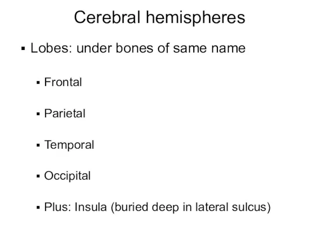
Слайд 30Cerebral hemispheres
Lobes: under bones of same name
Frontal
Parietal
Temporal
Occipital
Plus: Insula (buried deep in lateral
Cerebral hemispheres
Lobes: under bones of same name
Frontal
Parietal
Temporal
Occipital
Plus: Insula (buried deep in lateral
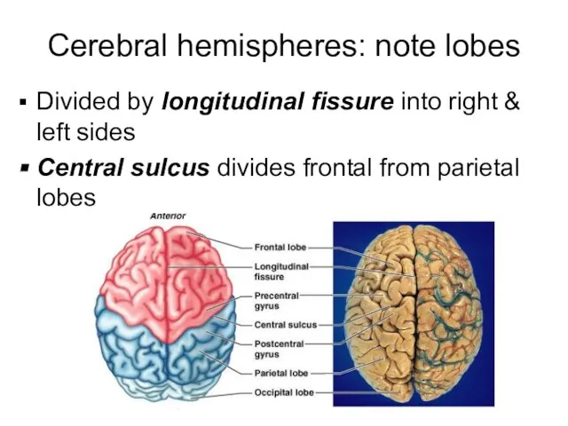
Слайд 31Cerebral hemispheres: note lobes
Divided by longitudinal fissure into right & left sides
Central
Cerebral hemispheres: note lobes
Divided by longitudinal fissure into right & left sides
Central
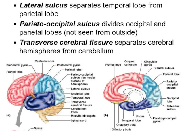
Слайд 32Lateral sulcus separates temporal lobe from parietal lobe
Parieto-occipital sulcus divides occipital and
Lateral sulcus separates temporal lobe from parietal lobe
Parieto-occipital sulcus divides occipital and
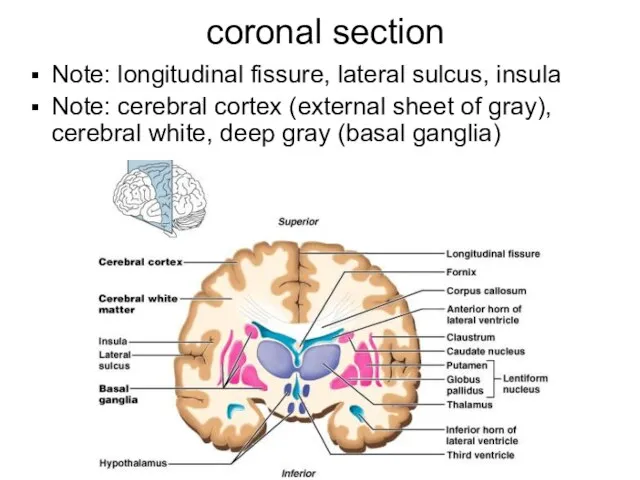
Слайд 33 coronal section
Note: longitudinal fissure, lateral sulcus, insula
Note: cerebral cortex (external sheet
coronal section
Note: longitudinal fissure, lateral sulcus, insula
Note: cerebral cortex (external sheet
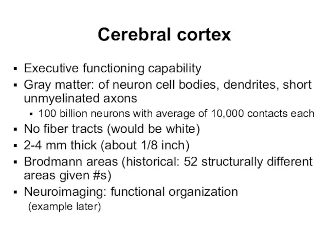
Слайд 34Cerebral cortex
Executive functioning capability
Gray matter: of neuron cell bodies, dendrites, short unmyelinated
Cerebral cortex
Executive functioning capability
Gray matter: of neuron cell bodies, dendrites, short unmyelinated
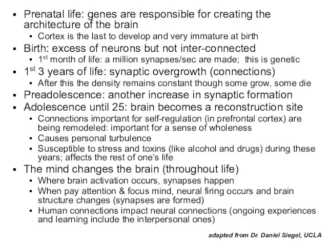
Слайд 35Prenatal life: genes are responsible for creating the architecture of the brain
Cortex
Prenatal life: genes are responsible for creating the architecture of the brain
Cortex
Слайд 36Cerebral cortex
All the neurons are interneurons
By definition confined to the CNS
They have
Cerebral cortex
All the neurons are interneurons
By definition confined to the CNS
They have
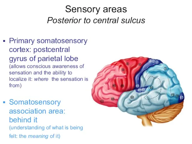
Слайд 37Sensory areas
Posterior to central sulcus
Primary somatosensory cortex: postcentral gyrus of parietal
Sensory areas
Posterior to central sulcus
Primary somatosensory cortex: postcentral gyrus of parietal
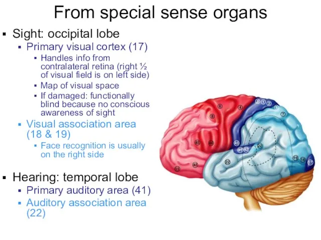
Слайд 38From special sense organs
Sight: occipital lobe
Primary visual cortex (17)
Handles info from contralateral
From special sense organs
Sight: occipital lobe
Primary visual cortex (17)
Handles info from contralateral
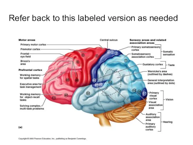
Слайд 39Refer back to this labeled version as needed
Refer back to this labeled version as needed
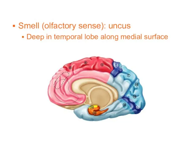
Слайд 40Smell (olfactory sense): uncus
Deep in temporal lobe along medial surface
Smell (olfactory sense): uncus
Deep in temporal lobe along medial surface
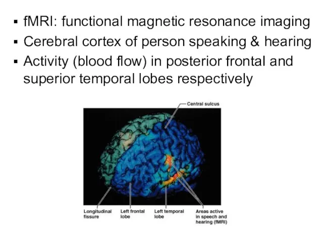
Слайд 41fMRI: functional magnetic resonance imaging
Cerebral cortex of person speaking & hearing
Activity (blood
fMRI: functional magnetic resonance imaging
Cerebral cortex of person speaking & hearing
Activity (blood
Слайд 42Motor areas
Anterior to central sulcus
Primary motor area
Precentral gyrus of frontal lobe
Motor areas
Anterior to central sulcus
Primary motor area
Precentral gyrus of frontal lobe
Слайд 43Primary motor area continued
Precentral gyrus of frontal lobe
Precise, conscious or voluntary movement
Primary motor area continued
Precentral gyrus of frontal lobe
Precise, conscious or voluntary movement
Слайд 44Motor areas – continued
Broca’s area (44): specialized motor speech area
Base of
Motor areas – continued
Broca’s area (44): specialized motor speech area
Base of
Слайд 45Motor areas – continued
Premotor cortex (6): complex movements asociated with highly processed
Motor areas – continued
Premotor cortex (6): complex movements asociated with highly processed
Слайд 46Homunculus – “little man”
Body map: human body spatially represented
Where on cortex; upside
Homunculus – “little man”
Body map: human body spatially represented
Where on cortex; upside
Слайд 47Association Areas
Remember…
Three kinds of functional areas (cerebrum)
Motor areas: movement
Sensory areas: perception
Association areas:
Association Areas
Remember…
Three kinds of functional areas (cerebrum)
Motor areas: movement
Sensory areas: perception
Association areas:
Слайд 48Association Areas
Tie together different kinds of sensory input
Associate new input with memories
Is
Association Areas
Tie together different kinds of sensory input
Associate new input with memories
Is
Слайд 49Prefrontal cortex: cognition
Executive functioning
e.g. multiple step problem solving requiring temporary storage of
Prefrontal cortex: cognition
Executive functioning
e.g. multiple step problem solving requiring temporary storage of
Слайд 50Wernicke’s area
Junction of parietal and temporal lobes
One hemisphere only, usually left
(Outlined by
Wernicke’s area
Junction of parietal and temporal lobes
One hemisphere only, usually left
(Outlined by
Слайд 51Cerebral white matter
Extensive communication
Areas of cortex with each other
Areas of cortex with
Cerebral white matter
Extensive communication
Areas of cortex with each other
Areas of cortex with
Слайд 52Commissures: interconnect right and left hemispheres so can act as a whole
Corpus
Commissures: interconnect right and left hemispheres so can act as a whole
Corpus
Слайд 53Projection fibers: run vertically
Cerebral cortex running down (with motor instructions)
Or ascend to
Projection fibers: run vertically
Cerebral cortex running down (with motor instructions)
Or ascend to
Слайд 54Corona radiata: spray of projection fibers
From precentral (motor) gyrus
Combines with sensory fibers
Corona radiata: spray of projection fibers
From precentral (motor) gyrus
Combines with sensory fibers
Слайд 55Projection fibers
Corona radiata: fanning out of the fibers
Internal capsule: bundled, pass
Projection fibers
Corona radiata: fanning out of the fibers
Internal capsule: bundled, pass
Слайд 56Cerebral hemisphere gray
Cortex – already reviewed
Basal forebrain nuclei: near hypothalamus - related
Cerebral hemisphere gray
Cortex – already reviewed
Basal forebrain nuclei: near hypothalamus - related
Слайд 57Basal ganglia
Subcortical motor nuclei
Part of “extrapyramidal system”
Cooperate with cerebral cortex in controlling
Basal ganglia
Subcortical motor nuclei
Part of “extrapyramidal system”
Cooperate with cerebral cortex in controlling
Слайд 58Internal capsule passes between diencephalon and basal ganglia to give them a
Internal capsule passes between diencephalon and basal ganglia to give them a
Слайд 59Basal ganglia
Cooperate with cerebral cortex in controlling movements
Communicate with cerebral cortex, receive
Basal ganglia
Cooperate with cerebral cortex in controlling movements
Communicate with cerebral cortex, receive
Слайд 60Basal ganglia
Note relationship of basal ganglia to thalamus and ventricles
Transverse section again
Basal ganglia
Note relationship of basal ganglia to thalamus and ventricles
Transverse section again
Слайд 61Diencephalon (part of forebrain)
Contains dozens of nuclei of gray matter
Thalamus
Hypothalamus
Epithalamus (mainly pineal)
Diencephalon (part of forebrain)
Contains dozens of nuclei of gray matter
Thalamus
Hypothalamus
Epithalamus (mainly pineal)
Слайд 62Thalamus (egg shaped; means inner room)
Two large lobes of gray matter (over
Thalamus (egg shaped; means inner room)
Two large lobes of gray matter (over
Слайд 63Hypothalamus
Forms inferolateral walls of 3rd ventricle
Many named nuclei
Coronal section
Hypothalamus
Forms inferolateral walls of 3rd ventricle
Many named nuclei
Coronal section
Слайд 64Diencephalon – surface anatomy
Hypothalamus is between optic chiasma to and including mamillary
Diencephalon – surface anatomy Hypothalamus is between optic chiasma to and including mamillary
Слайд 65Diencephalon – surface anatomy
Hypothalamus is between optic chiasma to and including mamillary
Diencephalon – surface anatomy Hypothalamus is between optic chiasma to and including mamillary
Слайд 66Cranial Nerve names
Identify as many as you can when looking at model
Cranial Nerve names
Identify as many as you can when looking at model
Слайд 67Hypothalamus
“Below thalamus”
Main visceral control center
Autonomic nervous system (peripheral motor neurons controlling smooth
Hypothalamus
“Below thalamus”
Main visceral control center
Autonomic nervous system (peripheral motor neurons controlling smooth
Слайд 68Hypothalamus
(one example of its functioning)
Control of endocrine system through pituitary gland
Hypothalamus
(one example of its functioning)
Control of endocrine system through pituitary gland
Слайд 69Epithalamus
Third and most dorsal part of diencephalon
Part of roof of 3rd ventricle
Pineal
Epithalamus
Third and most dorsal part of diencephalon
Part of roof of 3rd ventricle
Pineal
Слайд 70Brain Stem
Midbrain
Pons
Medulla oblongata
Rigidly programmed automatic behavior necessary for survival
Passageway for fiber tracts
Brain Stem
Midbrain
Pons
Medulla oblongata
Rigidly programmed automatic behavior necessary for survival
Passageway for fiber tracts
Слайд 71Brain stem
Midbrain
Pons
Medulla oblongata
Brain stem
Midbrain
Pons
Medulla oblongata
Слайд 72__Cerebral peduncles____
Contain pyramidal motor tracts
Corpora quadrigemina:
XVisual reflexes
XAuditory reflexes
Midbrain
______Substantia nigra
(degeneration causes Parkingson’s
__Cerebral peduncles____
Contain pyramidal motor tracts
Corpora quadrigemina:
XVisual reflexes
XAuditory reflexes
Midbrain
______Substantia nigra
(degeneration causes Parkingson’s
Слайд 73__Middle cerebellar peduncles_
Pons
3 cerebellar peduncles__
Also contains several CN and other nuclei
(one
__Middle cerebellar peduncles_
Pons
3 cerebellar peduncles__
Also contains several CN and other nuclei
(one
Слайд 74Medulla oblongata
Relays sensory info to cerebral cortex and cerebellum
Contains many CN
Medulla oblongata
Relays sensory info to cerebral cortex and cerebellum
Contains many CN
Слайд 75With all the labels….
With all the labels….
Слайд 76Brain Stem in mid-sagittal plane
Note cerebral aqueduct and fourth ventricle*
*
*
Brain Stem in mid-sagittal plane
Note cerebral aqueduct and fourth ventricle*
*
*
Слайд 77Cerebellum
Two major hemispheres: three lobes each
Anterior
Posterior
Floculonodular
Vermis: midline lobe connecting hemispheres
Outer cortex
Cerebellum
Two major hemispheres: three lobes each
Anterior
Posterior
Floculonodular
Vermis: midline lobe connecting hemispheres
Outer cortex
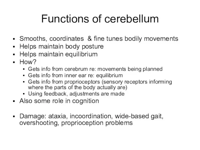
Слайд 78Functions of cerebellum
Smooths, coordinates & fine tunes bodily movements
Helps maintain body posture
Helps
Functions of cerebellum
Smooths, coordinates & fine tunes bodily movements
Helps maintain body posture
Helps
Слайд 79Functional brain systems
(as opposed to anatomical ones)
Networks of distant neurons that function
Functional brain systems
(as opposed to anatomical ones)
Networks of distant neurons that function
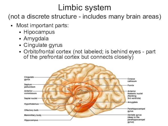
Слайд 80Limbic system
(not a discrete structure - includes many brain areas)
Most important
Limbic system
(not a discrete structure - includes many brain areas)
Most important
Слайд 81Limbic system continued
Called the “emotional” brain
Is essential for flexible, stable, adaptive functioning
Links
Limbic system continued
Called the “emotional” brain
Is essential for flexible, stable, adaptive functioning
Links
Слайд 82Reticular formation
Runs through central core of medulla, pons and midbrain
Reticular activating
system (RAS):
Reticular formation
Runs through central core of medulla, pons and midbrain
Reticular activating
system (RAS):
Слайд 83Brain protection
1.Meninges
2. Cerebrospinal fluid
3. Blood brain barrier
Brain protection
1.Meninges
2. Cerebrospinal fluid
3. Blood brain barrier
Слайд 84Meninges
Dura mater: 2 layers of fibrous connective tissue, fused except for dural
Meninges
Dura mater: 2 layers of fibrous connective tissue, fused except for dural
Слайд 85Dura mater - dural partitions
Subdivide cranial cavity & limit movement of brain
Falx
Dura mater - dural partitions
Subdivide cranial cavity & limit movement of brain
Falx
Слайд 86Arachnoid mater
Between dura and arachnoid: subdural space
Dura and arachnoid cover brain loosely
Deep
Arachnoid mater
Between dura and arachnoid: subdural space
Dura and arachnoid cover brain loosely
Deep

Слайд 87Cerebrospinal Fluid
CSF
Made in choroid plexuses (roofs of ventricles)
Filtration of plasma from capillaries
Cerebrospinal Fluid
CSF
Made in choroid plexuses (roofs of ventricles)
Filtration of plasma from capillaries
Слайд 88CSF circulation: through ventricles, median and lateral apertures, subarachnoid space, arachnoid villi,
CSF circulation: through ventricles, median and lateral apertures, subarachnoid space, arachnoid villi,
Слайд 89 Hydrocephalus
Hydrocephalus
Слайд 90Blood-Brain Barrier
Tight junctions between endothelial cells of brain capillaries, instead of the
Blood-Brain Barrier
Tight junctions between endothelial cells of brain capillaries, instead of the
Слайд 91White matter of the spinal cord
Ascending pathways: sensory information by multi-neuron chains
White matter of the spinal cord
Ascending pathways: sensory information by multi-neuron chains
Слайд 92Major fiber tracts in white matter of spinal cord
Damage: to motor areas
Major fiber tracts in white matter of spinal cord
Damage: to motor areas
Слайд 93Major ascending pathways for the somatic senses
Spinocerebellar: proprioception from skeletal muscles to
Major ascending pathways for the somatic senses
Spinocerebellar: proprioception from skeletal muscles to
Слайд 94Some Descending Pathways
Pyramidal tracts:
Lateral corticospinal – cross in pyramids of medulla;
Some Descending Pathways
Pyramidal tracts:
Lateral corticospinal – cross in pyramids of medulla;
Слайд 95Check out: Medical gross anatomy atlas images (good teaching pics):
http://anatomy.med.umich.edu/atlas/atlas_index.html
(can access from
Check out: Medical gross anatomy atlas images (good teaching pics):
http://anatomy.med.umich.edu/atlas/atlas_index.html
(can access from
Слайд 96Hints & additional pics
Unless your prints of the slides are very large
Hints & additional pics
Unless your prints of the slides are very large
Слайд 97Know the names of the ventricles and which ones connect to which,
Know the names of the ventricles and which ones connect to which,
Слайд 98From this site, which also has text explanations:
http://www.emc.maricopa.edu/faculty/farabee/BIOBK/BioBookNERV.html
From this site, which also has text explanations:
http://www.emc.maricopa.edu/faculty/farabee/BIOBK/BioBookNERV.html
Слайд 99Brain, sagittal sec, medial view
Cerebral hemisphere
Corpus callosum
Thalamus
Midbrain
Pons
Cerebellum
Brain, sagittal sec, medial view
Cerebral hemisphere
Corpus callosum
Thalamus
Midbrain
Pons
Cerebellum
Слайд 100Internal capsule
Anterior limb of internal capsule
Genu of internal capsule
Posterior limb
Internal capsule
Anterior limb of internal capsule
Genu of internal capsule
Posterior limb
Слайд 101Pons & cerebellum, sagittal section, medial view
Midbrain
Cerebellum
Pons
Medulla oblongata
Inferior
Pons & cerebellum, sagittal section, medial view
Midbrain
Cerebellum
Pons
Medulla oblongata
Inferior
 00079766-3ed2eb83 (1)
 Продажа помещения. Фото (11)
 Как подготовиться к сдаче ЕГЭ
 видеоролик
 Понятие культуры труда
 Публичный отчет за 2020-2021 годы
 M
M Проект информатизации Образовательного учреждения
 2019 декабрь ООО Жилкомсервис Кронштадтского района
 Frohe Weihnachten!
 Северная чернь
 Тема урока « Наука и семья»8 КЛАСС( химия и литература)
 Буддизм в России
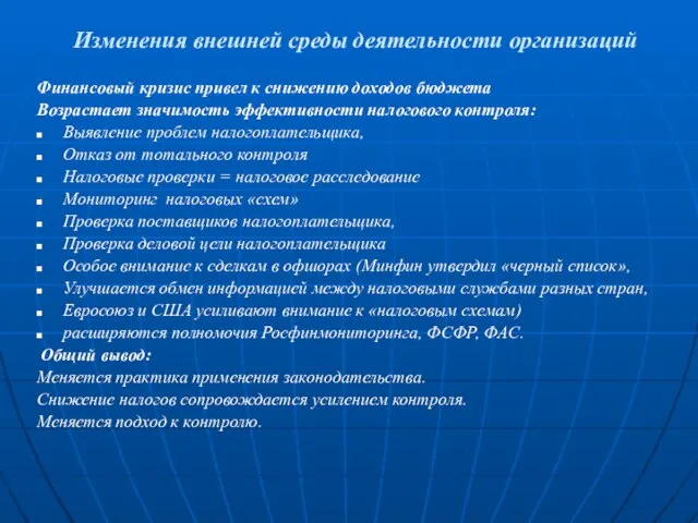 Изменения внешней среды деятельности организаций
 Независимая оценка качества образования. Этапы формирования 1-х классов
 1-high
 Образец оформления конспекта
 Технология индустриального программирования
  Презентация на тему "mala akademiya" - скачать презентации по Педагогике
 Unknown Company The Next Phase
 Фрагмент Лекции СМО
 Акция Service Clinic
 О жизни и деятельности (1885-1969 гг.)
 Оформление документации по итогам ежемесячного пересчета
 Недемократические режимы
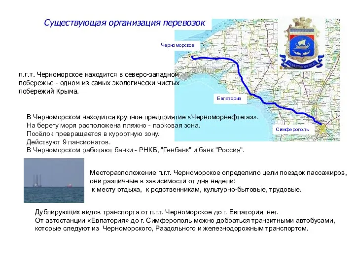 Существующая организация перевозок п.г.т. Черноморское
 Подмосковные промыслы
 Танцы, 4 класс