- Donetsk National Medical University Department of Radiology X-RAY

Содержание
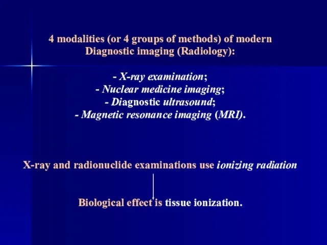
- 2. 4 modalities (or 4 groups of methods) of modern Diagnostic imaging (Radiology): - X-ray examination; -
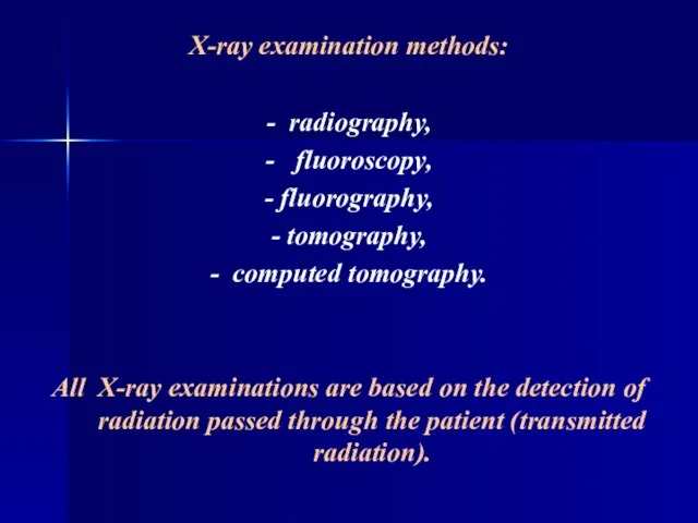
- 3. X-ray examination methods: - radiography, - fluoroscopy, - fluorography, - tomography, - computed tomography. All X-ray
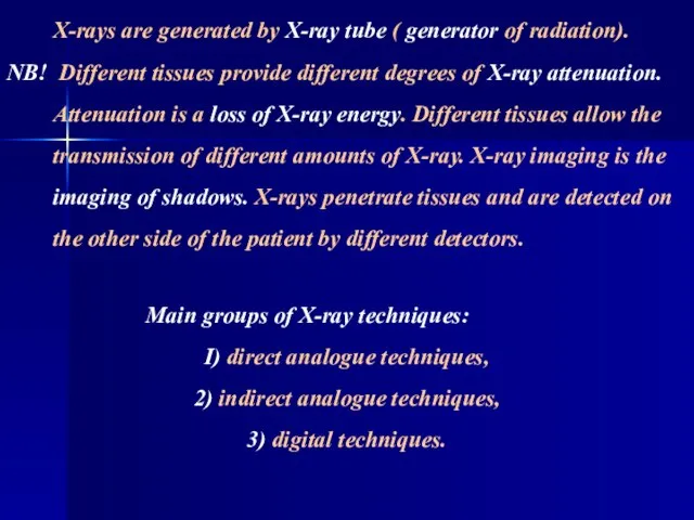
- 4. X-rays are generated by X-ray tube ( generator of radiation). NB! Different tissues provide different degrees
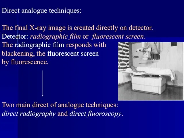
- 5. Direct analogue techniques: The final X-ray image is created directly on detector. Detector: radiographic film or
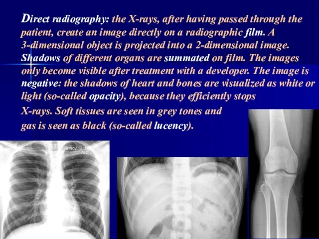
- 6. Direct radiography: the X-rays, after having passed through the patient, create an image directly on a
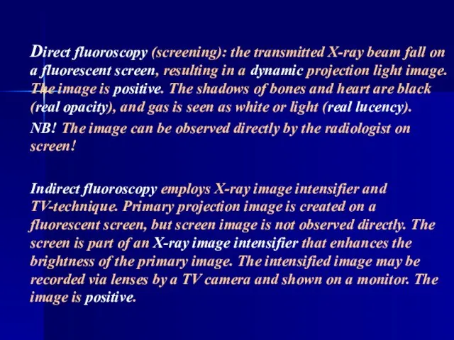
- 7. Direct fluoroscopy (screening): the transmitted X-ray beam fall on a fluorescent screen, resulting in a dynamic
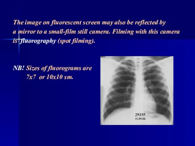
- 8. The image on fluorescent screen may also be reflected by a mirror to a small-film still
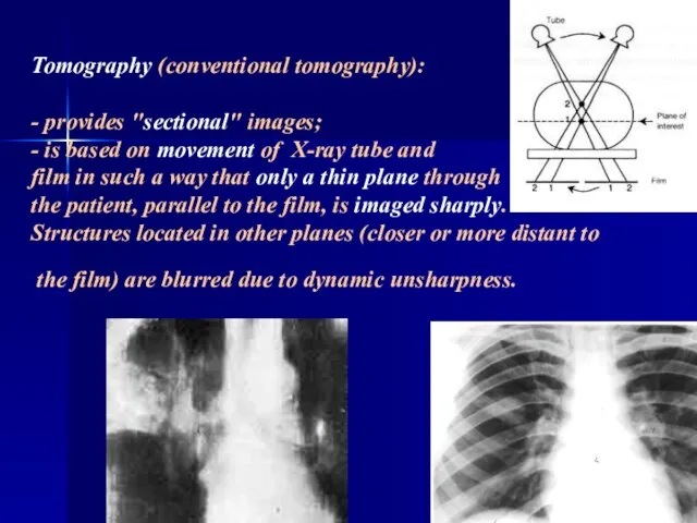
- 9. Tomography (conventional tomography): - provides "sectional" images; - is based on movement of X-ray tube and
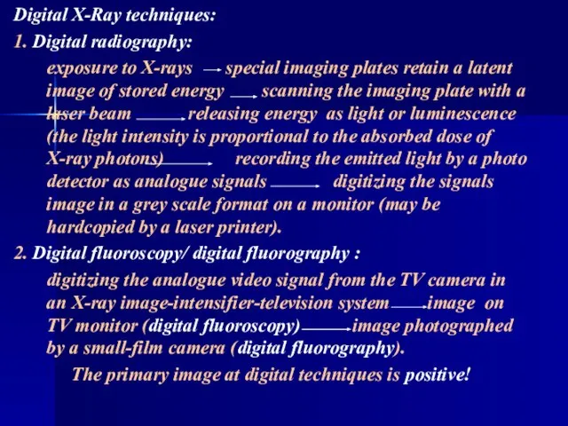
- 10. Digital X-Ray techniques: 1. Digital radiography: exposure to X-rays special imaging plates retain a latent image
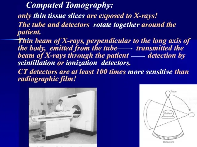
- 11. Computed Tomography: only thin tissue slices are exposed to X-rays! The tube and detectors rotate together
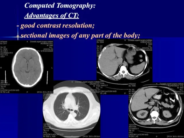
- 12. Computed Tomography: Advantages of CT: - good contrast resolution; - sectional images of any part of
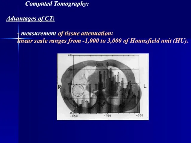
- 13. Computed Tomography: Advantages of CT: - measurement of tissue attenuation: linear scale ranges from -1,000 to
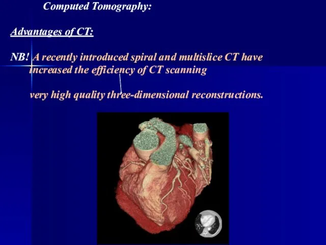
- 14. Computed Tomography: Advantages of CT: NB! A recently introduced spiral and multislice CT have increased the
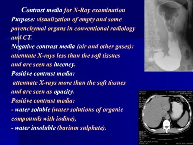
- 15. Contrast media for X-Ray examination Purpose: visualization of empty and some parenchymal organs in conventional radiology
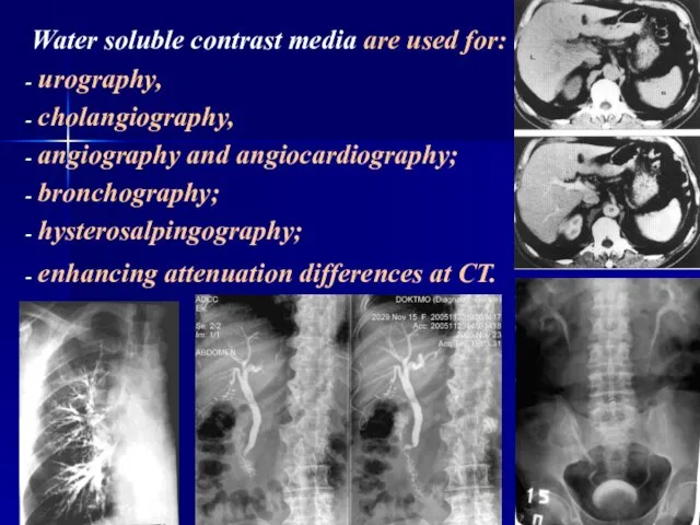
- 16. Water soluble contrast media are used for: urography, cholangiography, angiography and angiocardiography; bronchography; hysterosalpingography; enhancing attenuation
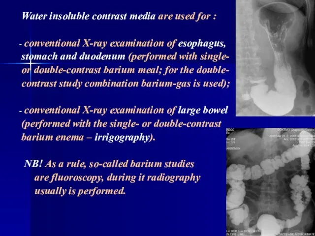
- 17. Water insoluble contrast media are used for : conventional X-ray examination of esophagus, stomach and duodenum
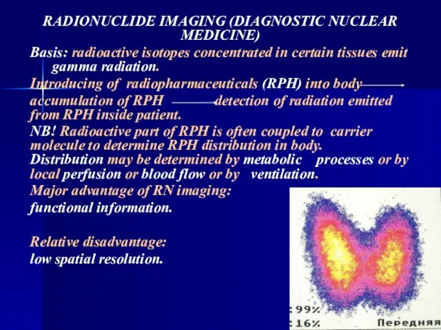
- 18. RADIONUCLIDE IMAGING (DIAGNOSTIC NUCLEAR MEDICINE) Basis: radioactive isotopes concentrated in certain tissues emit gamma radiation. Introducing
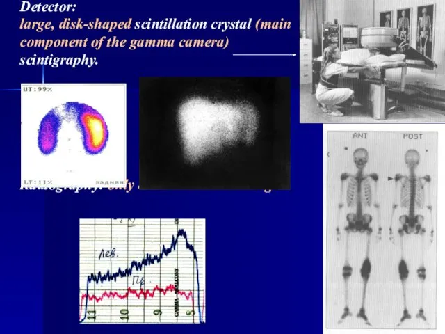
- 19. Detector: large, disk-shaped scintillation crystal (main component of the gamma camera) scintigraphy. Radiography: only curves –
- 21. Скачать презентацию
Слайд 3X-ray examination methods:
- radiography,
- fluoroscopy,
- fluorography,
- tomography,
- computed tomography.
All X-ray examinations
X-ray examination methods:
- radiography,
- fluoroscopy,
- fluorography,
- tomography,
- computed tomography.
All X-ray examinations
Слайд 4
X-rays are generated by X-ray tube ( generator of radiation).
NB!
X-rays are generated by X-ray tube ( generator of radiation).
NB!
Слайд 5
Direct analogue techniques:
The final X-ray image is created directly on detector.
Detector:
Direct analogue techniques: The final X-ray image is created directly on detector. Detector:
Слайд 6 Direct radiography: the X-rays, after having passed through the patient, create an
Direct radiography: the X-rays, after having passed through the patient, create an
Слайд 7
Direct fluoroscopy (screening): the transmitted X-ray beam fall on a fluorescent screen,
Direct fluoroscopy (screening): the transmitted X-ray beam fall on a fluorescent screen,
Слайд 8 The image on fluorescent screen may also be reflected by
a mirror
a mirror
Слайд 9
Tomography (conventional tomography):
- provides "sectional" images;
- is based on movement of X-ray
Tomography (conventional tomography): - provides "sectional" images; - is based on movement of X-ray
Слайд 10Digital X-Ray techniques:
1. Digital radiography:
exposure to X-rays special imaging plates retain
Digital X-Ray techniques:
1. Digital radiography:
exposure to X-rays special imaging plates retain
Слайд 11 Computed Tomography:
only thin tissue slices are exposed to X-rays!
The tube
Computed Tomography:
only thin tissue slices are exposed to X-rays!
The tube
Слайд 12 Computed Tomography:
Advantages of CT:
- good contrast resolution;
- sectional images of any
Computed Tomography:
Advantages of CT:
- good contrast resolution;
- sectional images of any
Слайд 13 Computed Tomography:
Advantages of CT:
- measurement of tissue attenuation:
linear scale ranges
Computed Tomography:
Advantages of CT:
- measurement of tissue attenuation:
linear scale ranges
Слайд 14 Computed Tomography:
Advantages of CT:
NB! A recently introduced spiral and multislice CT
Computed Tomography:
Advantages of CT:
NB! A recently introduced spiral and multislice CT
Слайд 15 Contrast media for X-Ray examination
Purpose: visualization of empty and some
Contrast media for X-Ray examination
Purpose: visualization of empty and some
Слайд 16Water soluble contrast media are used for:
urography,
cholangiography,
Water soluble contrast media are used for:
urography,
cholangiography,
Слайд 17Water insoluble contrast media are used for :
conventional X-ray examination of
Water insoluble contrast media are used for :
conventional X-ray examination of
Слайд 18RADIONUCLIDE IMAGING (DIAGNOSTIC NUCLEAR MEDICINE)
Basis: radioactive isotopes concentrated in certain tissues
RADIONUCLIDE IMAGING (DIAGNOSTIC NUCLEAR MEDICINE)
Basis: radioactive isotopes concentrated in certain tissues
Слайд 19
Detector:
large, disk-shaped scintillation crystal (main
component of the gamma camera)
scintigraphy.
Detector: large, disk-shaped scintillation crystal (main component of the gamma camera) scintigraphy.
 Презентация1 (1)
 70 лет снятия блокады Ленинграда
 Обучение в сотрудничестве
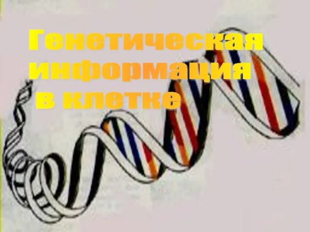 Генетическая информация в клетке
 Осень 2020 г
 Современные технологии мерчендайзинга и программные инструменты для их реализации
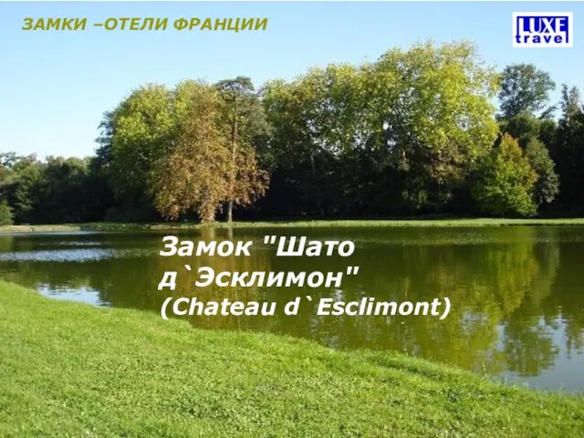 Замок "Шато д`Эсклимон"(Chateau d`Esclimont)
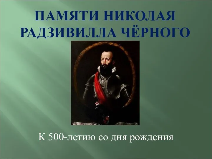 Радзивилл
 Приглашение к участию в тендере ГК «АвтоСпецЦентр»
 Цена и ценность иликак объяснить клиенту за что он платит?
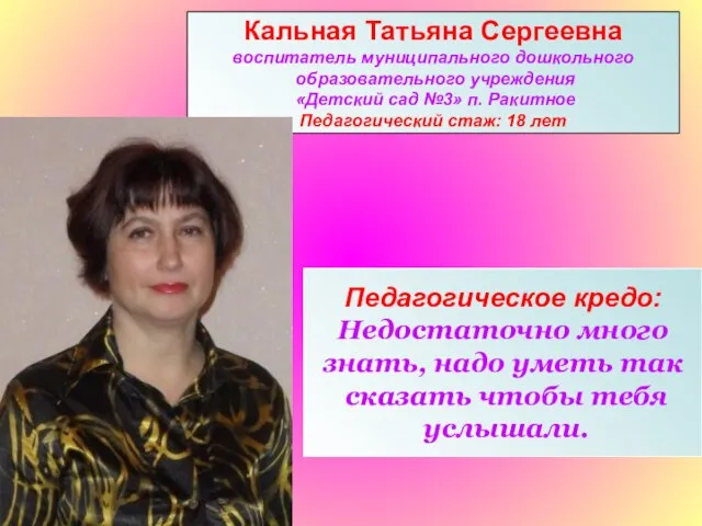 Кальная Татьяна Сергеевна воспитатель муниципального дошкольного образовательного учреждения «Детский сад №3» п. Ракитное Пед
 Многообразие в Болонском процессе
 Закон об административных правонарушениях в Вологодской области
 Воспалительные заболевания кишечника: Энтериты и колиты
 Многогранный талант Н.В. Гоголя
 Dyakovich_Margarita_Indiv_Poekt
 УДОБСТВО ДЛЯ ПОКУПАТЕЛЯ – ЗАЛОГ ВАШЕГО УСПЕХА
 Понятие о местоимении. Личные местоимения.
 Как вести себя во время теракта
 Элективный курс по информатике
 Рынок субфедерального долга: возможности для эмитентовМай 2011 года
 Условия плавания тел
 Путешествие в страну Знаний.
 Проект вариативной части Учебного плана АМОУ СОШ №8 на 2011-2012 учебный год
 Презентация по Географии.
 Декоративный пейзаж
 Теория дифференциальных эмоций Кэррола Э. Изарда
 Итоги реализации лицейского проекта Одаренные дети