- Microbiology

Содержание
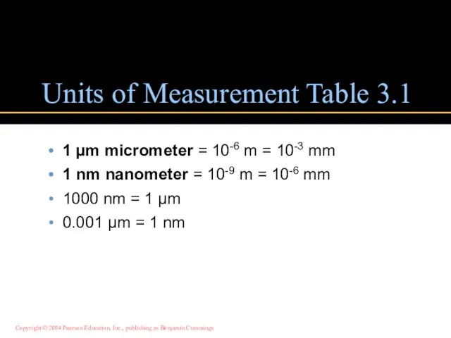
- 2. Units of Measurement Table 3.1 1 µm micrometer = 10-6 m = 10-3 mm 1 nm
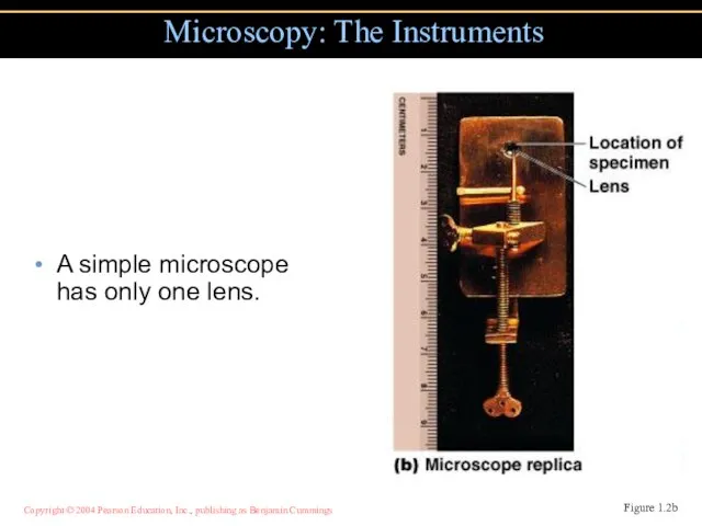
- 3. A simple microscope has only one lens. Microscopy: The Instruments Figure 1.2b
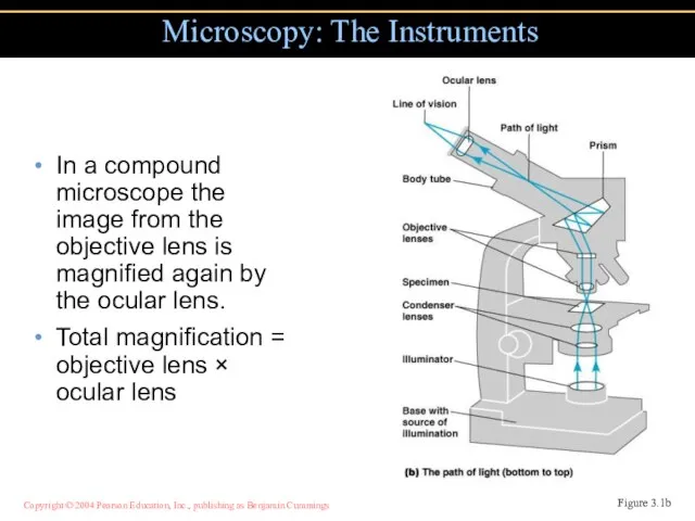
- 4. In a compound microscope the image from the objective lens is magnified again by the ocular
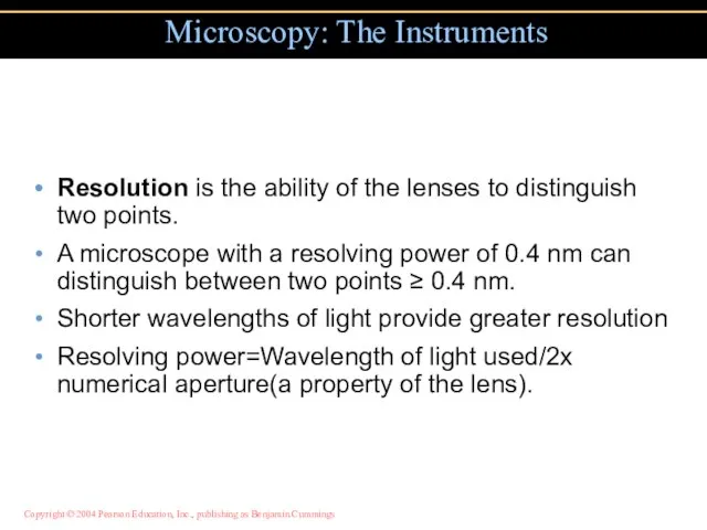
- 5. Resolution is the ability of the lenses to distinguish two points. A microscope with a resolving
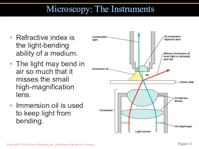
- 6. Refractive index is the light-bending ability of a medium. The light may bend in air so
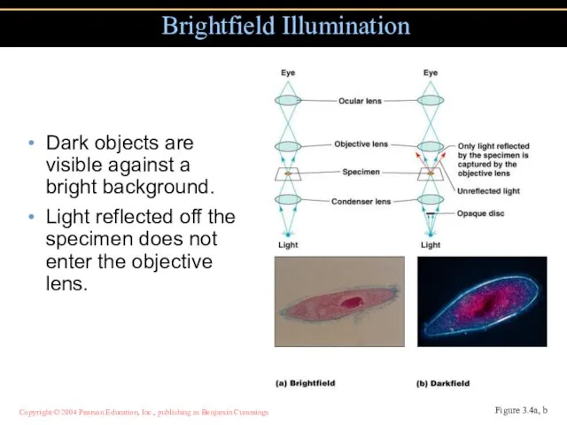
- 7. Dark objects are visible against a bright background. Light reflected off the specimen does not enter
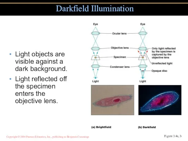
- 8. Light objects are visible against a dark background. Light reflected off the specimen enters the objective
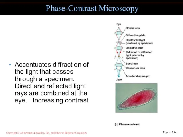
- 9. Accentuates diffraction of the light that passes through a specimen. Direct and reflected light rays are
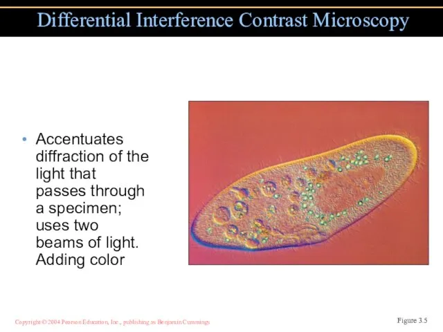
- 10. Accentuates diffraction of the light that passes through a specimen; uses two beams of light. Adding
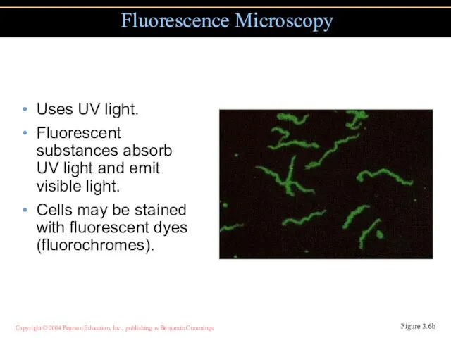
- 11. Uses UV light. Fluorescent substances absorb UV light and emit visible light. Cells may be stained
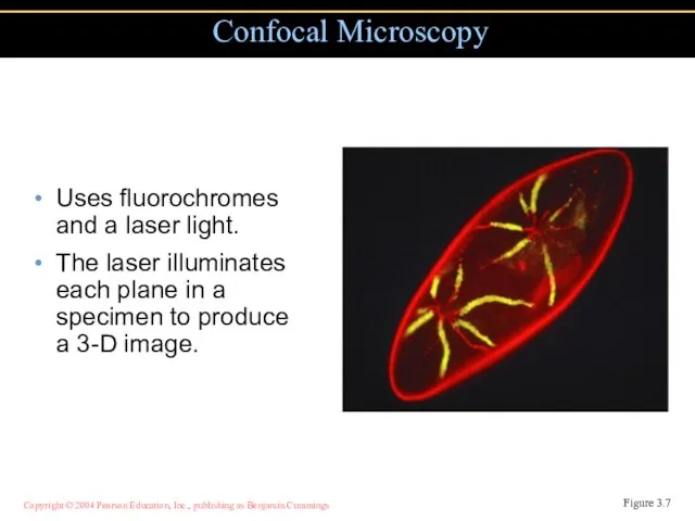
- 12. Uses fluorochromes and a laser light. The laser illuminates each plane in a specimen to produce
- 13. Uses electrons instead of light. The shorter wavelength of electrons gives greater resolution. Why? Electron Microscopy
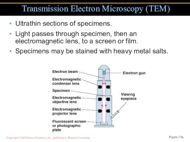
- 14. Ultrathin sections of specimens. Light passes through specimen, then an electromagnetic lens, to a screen or
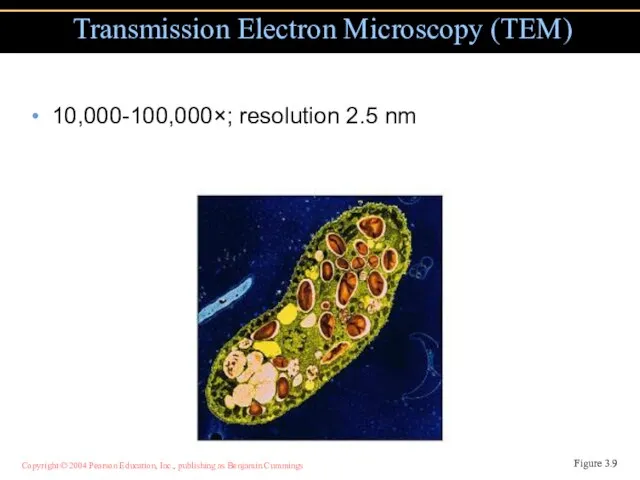
- 15. 10,000-100,000×; resolution 2.5 nm Transmission Electron Microscopy (TEM) Figure 3.9
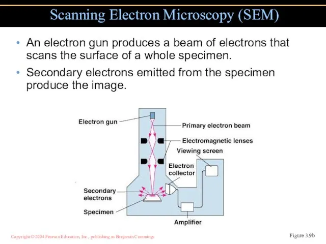
- 16. An electron gun produces a beam of electrons that scans the surface of a whole specimen.
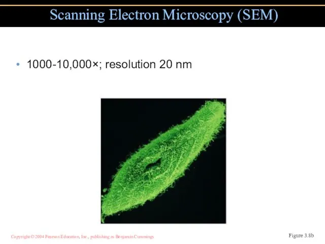
- 17. 1000-10,000×; resolution 20 nm Scanning Electron Microscopy (SEM) Figure 3.8b
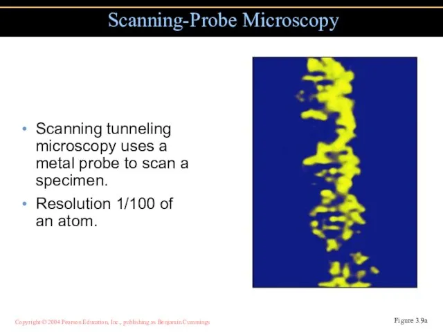
- 18. Scanning tunneling microscopy uses a metal probe to scan a specimen. Resolution 1/100 of an atom.
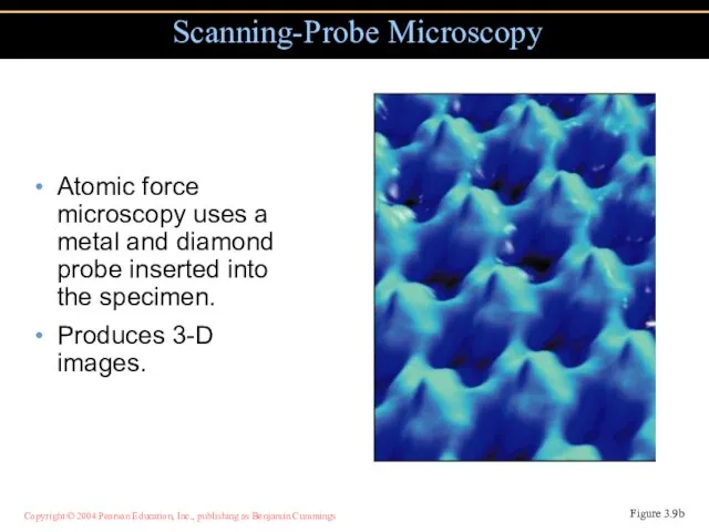
- 19. Atomic force microscopy uses a metal and diamond probe inserted into the specimen. Produces 3-D images.
- 20. Preparation of Specimens for Light Microscopy A thin film of a solution of microbes on a
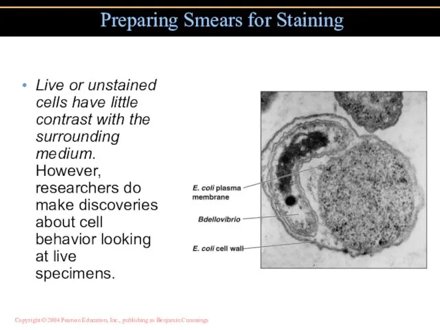
- 21. Live or unstained cells have little contrast with the surrounding medium. However, researchers do make discoveries
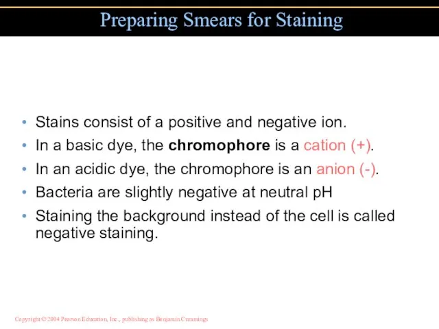
- 22. Stains consist of a positive and negative ion. In a basic dye, the chromophore is a
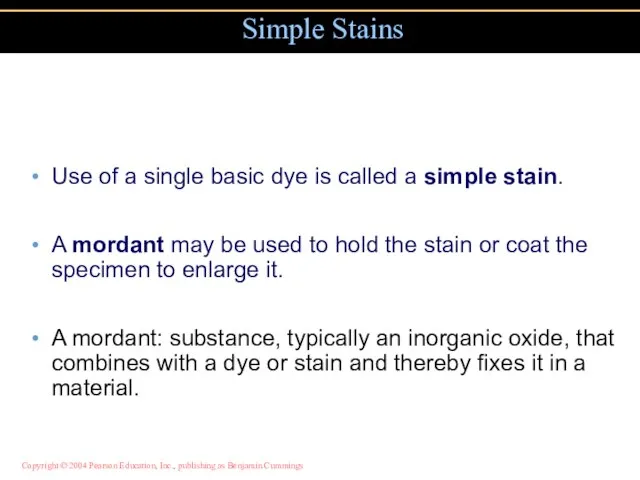
- 23. Use of a single basic dye is called a simple stain. A mordant may be used
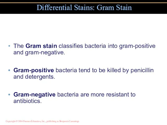
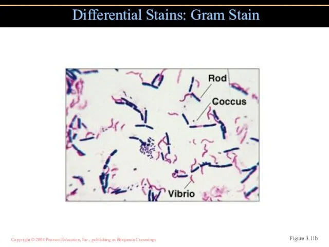
- 24. The Gram stain classifies bacteria into gram-positive and gram-negative. Gram-positive bacteria tend to be killed by
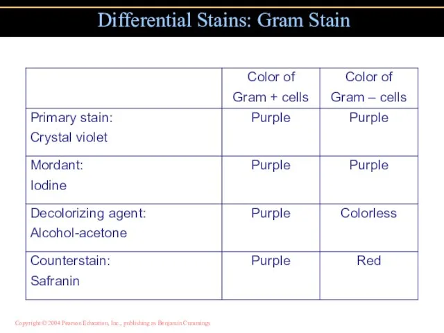
- 25. Differential Stains: Gram Stain
- 26. Differential Stains: Gram Stain Figure 3.11b
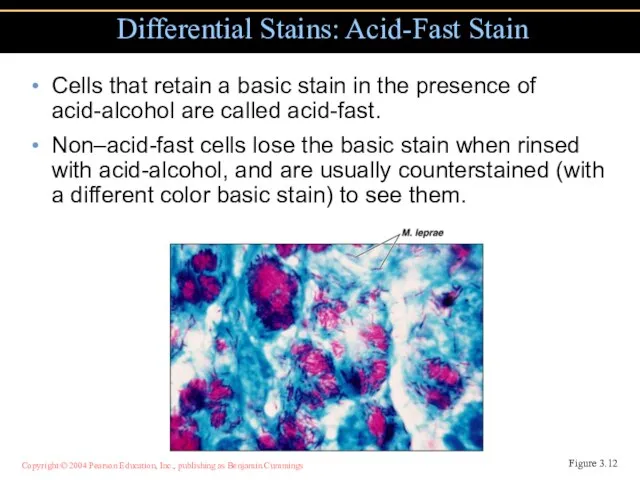
- 27. Cells that retain a basic stain in the presence of acid-alcohol are called acid-fast. Non–acid-fast cells
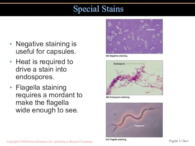
- 28. Negative staining is useful for capsules. Heat is required to drive a stain into endospores. Flagella
- 30. Скачать презентацию
Слайд 3A simple microscope has only one lens.
Microscopy: The Instruments
Figure 1.2b
A simple microscope has only one lens.
Microscopy: The Instruments
Figure 1.2b
Слайд 4In a compound microscope the image from the objective lens is magnified
In a compound microscope the image from the objective lens is magnified
Слайд 5Resolution is the ability of the lenses to distinguish two points.
A microscope
Resolution is the ability of the lenses to distinguish two points.
A microscope
Слайд 6Refractive index is the light-bending ability of a medium.
The light may bend
Refractive index is the light-bending ability of a medium.
The light may bend
Слайд 7Dark objects are visible against a bright background.
Light reflected off the specimen
Dark objects are visible against a bright background.
Light reflected off the specimen
Слайд 8Light objects are visible against a dark background.
Light reflected off the specimen
Light objects are visible against a dark background.
Light reflected off the specimen
Слайд 9Accentuates diffraction of the light that passes through a specimen. Direct and
Accentuates diffraction of the light that passes through a specimen. Direct and
Слайд 10Accentuates diffraction of the light that passes through a specimen; uses two
Accentuates diffraction of the light that passes through a specimen; uses two
Слайд 11Uses UV light.
Fluorescent substances absorb UV light and emit visible light.
Cells
Uses UV light.
Fluorescent substances absorb UV light and emit visible light.
Cells
Слайд 12Uses fluorochromes and a laser light.
The laser illuminates each plane in a
Uses fluorochromes and a laser light.
The laser illuminates each plane in a
Слайд 13Uses electrons instead of light.
The shorter wavelength of electrons gives greater resolution.
Uses electrons instead of light.
The shorter wavelength of electrons gives greater resolution.
Слайд 14Ultrathin sections of specimens.
Light passes through specimen, then an electromagnetic lens, to
Ultrathin sections of specimens.
Light passes through specimen, then an electromagnetic lens, to
Слайд 1510,000-100,000×; resolution 2.5 nm
Transmission Electron Microscopy (TEM)
Figure 3.9
10,000-100,000×; resolution 2.5 nm
Transmission Electron Microscopy (TEM)
Figure 3.9
Слайд 16An electron gun produces a beam of electrons that scans the surface
An electron gun produces a beam of electrons that scans the surface
Слайд 171000-10,000×; resolution 20 nm
Scanning Electron Microscopy (SEM)
Figure 3.8b
1000-10,000×; resolution 20 nm
Scanning Electron Microscopy (SEM)
Figure 3.8b
Слайд 18Scanning tunneling microscopy uses a metal probe to scan a specimen.
Resolution 1/100
Scanning tunneling microscopy uses a metal probe to scan a specimen.
Resolution 1/100
Слайд 19Atomic force microscopy uses a metal and diamond probe inserted into the
Atomic force microscopy uses a metal and diamond probe inserted into the
Слайд 20Preparation of Specimens for
Light Microscopy
A thin film of a solution of
Preparation of Specimens for
Light Microscopy
A thin film of a solution of
Слайд 21Live or unstained cells have little contrast with the surrounding medium. However,
Live or unstained cells have little contrast with the surrounding medium. However,
Слайд 22Stains consist of a positive and negative ion.
In a basic dye, the
Stains consist of a positive and negative ion.
In a basic dye, the
Слайд 23Use of a single basic dye is called a simple stain.
A mordant
Use of a single basic dye is called a simple stain.
A mordant
Слайд 24The Gram stain classifies bacteria into gram-positive and gram-negative.
Gram-positive bacteria tend to
The Gram stain classifies bacteria into gram-positive and gram-negative.
Gram-positive bacteria tend to
Слайд 25Differential Stains: Gram Stain
Differential Stains: Gram Stain
Слайд 26Differential Stains: Gram Stain
Figure 3.11b
Differential Stains: Gram Stain
Figure 3.11b
Слайд 27Cells that retain a basic stain in the presence of acid-alcohol are
Cells that retain a basic stain in the presence of acid-alcohol are
Слайд 28Negative staining is useful for capsules.
Heat is required to drive a stain
Negative staining is useful for capsules.
Heat is required to drive a stain
 ПАМЯТЬ КОМПЬЮТЕРА
 Методы и алгоритмы интеллектуальной поддержки принятия решений в задачах диагностики технических систем в машиностроении
 Робер Кампен (1378 – 1444)
 История холодной войны
 Информация Информационные процессы
  Формирование муниципального задания и финансового норматива на услуги дошкольного образования, обеспечивающего развитие вариа
 上网课的利与弊
 Презентация на тему Древнейшие люди
 А Вы уже пробовали? П р о д у к ц и ю к о м п а н и и : ?
 "Особенности разработки электронного учебно - методического комплекса для расширения методического и творческого взаимодейст
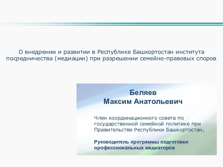 О внедрении и развитии в Республике Башкортостан института посредничества (медиации) при разрешении семейно-правовых споров
 Вильям Шекспир
 Web-сайт Гиперструктура данных
 Фонды. Благотворительній фонд имени Елены Ивановны Рерих
 Казанский собор
 Урал
Урал Магнит на холодильник из пластиковой бутылки
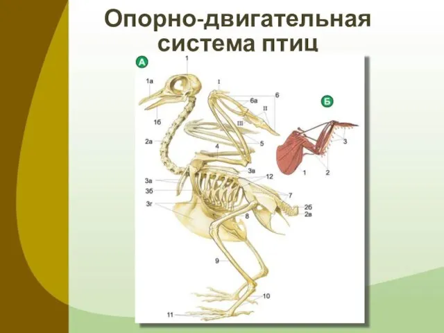 Опорно-двигательная система птиц
 Заказчик: Министерство Экономического Развития и Торговли РФ Разработка: ООО «Дигитал Зон» Формирование единой информационной си
 2010
2010 Ядерная пора Энтропийное исключение.
 Стабильность и новации в условиях кризиса системы государственного и муниципального заказов Калужской области
 Презентация на тему «Любовная лирика Ф. Тютчева. Подготовила учитель русского языка и литературы МОУ СОШ №2 Лыженкова О. Д.
 Лишняя жилая площадь и её правовой режим
 Различие частиц не и ни
 Я ученик школы №4
 Материальная часть стрелкового оружия
 Организация обучения УКНиУ в дистанционном формате