- Radiomics: Extracting more Features using Endoscopic Imaging

Содержание
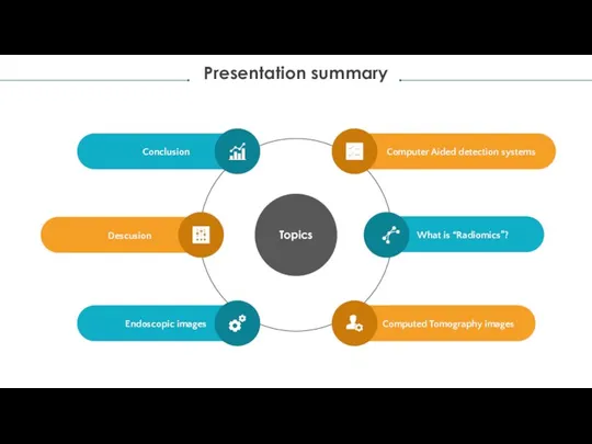
- 2. Presentation summary Topics Computer Aided detection systems What is “Radiomics”? Computed Tomography images Conclusion Descusion Endoscopic
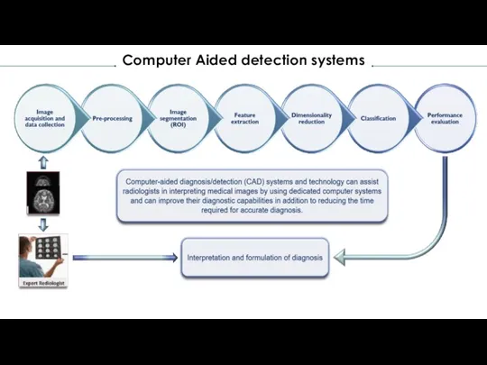
- 3. Computer Aided detection systems
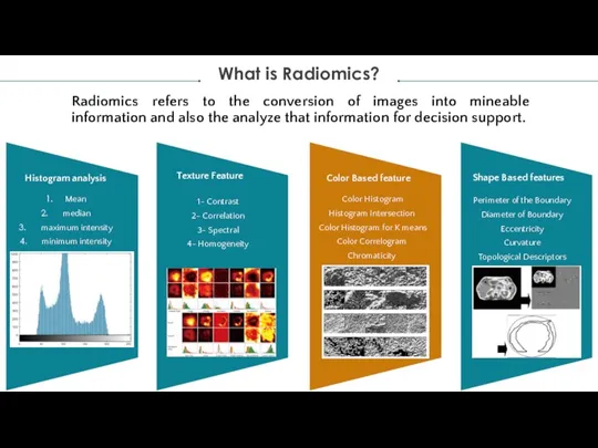
- 4. Radiomics refers to the conversion of images into mineable information and also the analyze that information
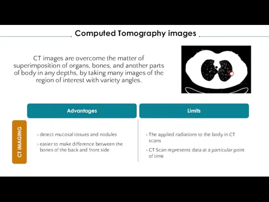
- 5. CT images are overcome the matter of superimposition of organs, bones, and another parts of body
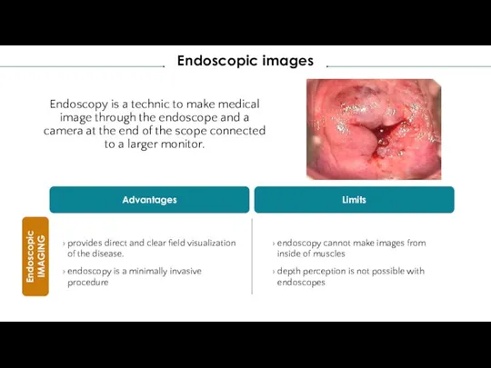
- 6. Endoscopy is a technic to make medical image through the endoscope and a camera at the
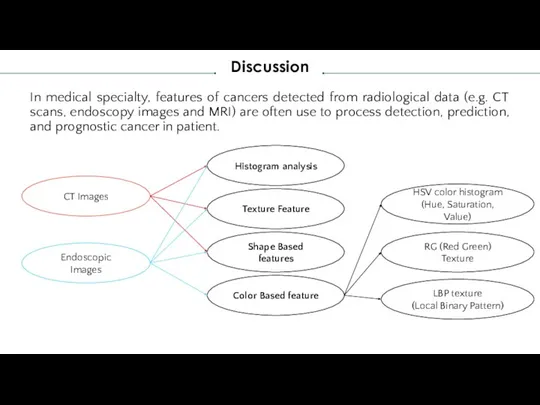
- 7. In medical specialty, features of cancers detected from radiological data (e.g. CT scans, endoscopy images and
- 8. Conclusion According to the features extracted from the medical images reviewed in this Presentation, endoscopic images
- 10. Скачать презентацию
Слайд 2Presentation summary
Topics
Computer Aided detection systems
What is “Radiomics”?
Computed Tomography images
Conclusion
Descusion
Presentation summary
Topics
Computer Aided detection systems
What is “Radiomics”?
Computed Tomography images
Conclusion
Descusion
Слайд 3Computer Aided detection systems
Computer Aided detection systems
Слайд 4Radiomics refers to the conversion of images into mineable information and also
Radiomics refers to the conversion of images into mineable information and also
Слайд 5CT images are overcome the matter of superimposition of organs, bones, and
CT images are overcome the matter of superimposition of organs, bones, and
Слайд 6Endoscopy is a technic to make medical image through the endoscope and
Endoscopy is a technic to make medical image through the endoscope and
Слайд 7In medical specialty, features of cancers detected from radiological data (e.g. CT
In medical specialty, features of cancers detected from radiological data (e.g. CT
Слайд 8Conclusion
According to the features extracted from the medical images reviewed in this
Conclusion
According to the features extracted from the medical images reviewed in this
 Коньки
 Conference on Applied Physics, Information Technologies and Engineering
 Вэб-инфекция
 Тема проекта: «Мораль сей басни такова…» Иван Андреевич Крылов - русский баснописец. предмет: литература
  Опыт построения решений регионального электронного правительства (на примере Калининградской области) Алексей
  Заболевания, передаваемые половым путем (ЗППП), и планирование семьи
 Нагревательные печи и колодцы
 Обзор энергетики Германии
 ДЗЫГА лучшее
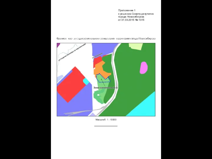 Приложения к решению совета депутатов города Новосибирска. Карты
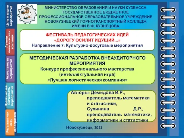 Методическая разработка внеаудиторного мероприятия
 Start up Юрист в Кармане
 ¿Qué hora es?
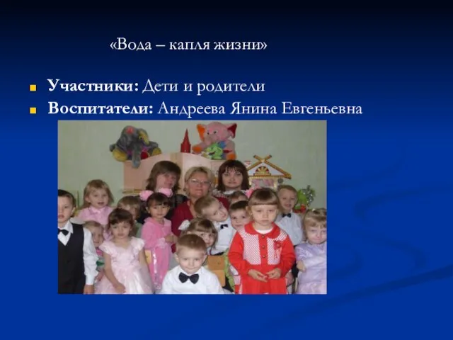 Вода - капля жизни
 Семейные традиции
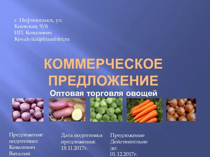 Коммерческое предложение. Оптовая торговля овощей
 Математическое описание случайных явлений
 КЛАССНЫЙ ЧАС: «Россия - всё, чем я живу»
 Здравствуйте!
 Мониторинг уровня воспитанности обучающихсяМБОУ СОШ № 15
 Мониторинг заполнения дневников и журналов на сайте Эпос.Школа
 С 33 сабак 29
 Ртуть
Ртуть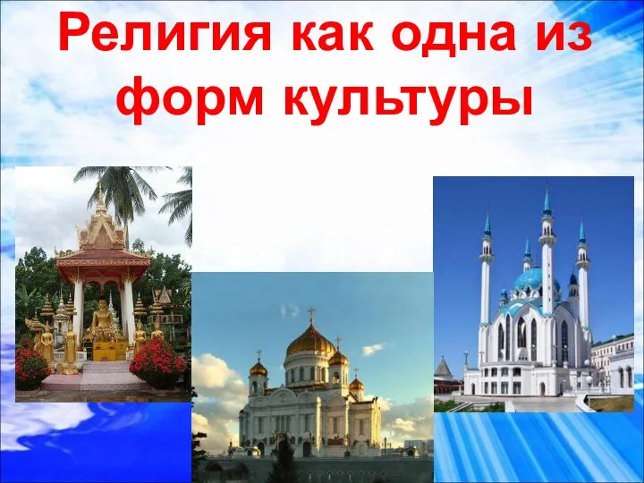 Религия как одна из форм культуры
 Отчет по производственной практике. Дизайн-проект мебели (оборудования) для жилых (общественных) интерьеров
 Furniture in the flat
 ТЕКСТЫ В ПАМЯТИ КОМПЬЮТЕРА
 «Человек есть тайна. Её надо разгадать, и ежели будешь её разгадывать всю жизнь, то не говори, что потерял время: я занимаюсь этой т