- Regional anatomy of neck

Содержание
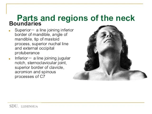
- 2. Parts and regions of the neck Boundaries Superior- a line joining inferior border of mandible, angle
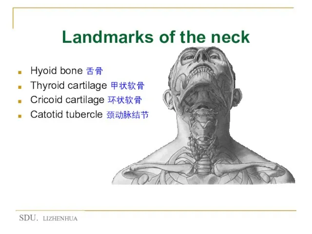
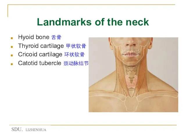
- 3. Landmarks of the neck Hyoid bone 舌骨 Thyroid cartilage 甲状软骨 Cricoid cartilage 环状软骨 Catotid tubercle 颈动脉结节
- 4. Landmarks of the neck Hyoid bone 舌骨 Thyroid cartilage 甲状软骨 Cricoid cartilage 环状软骨 Catotid tubercle 颈动脉结节
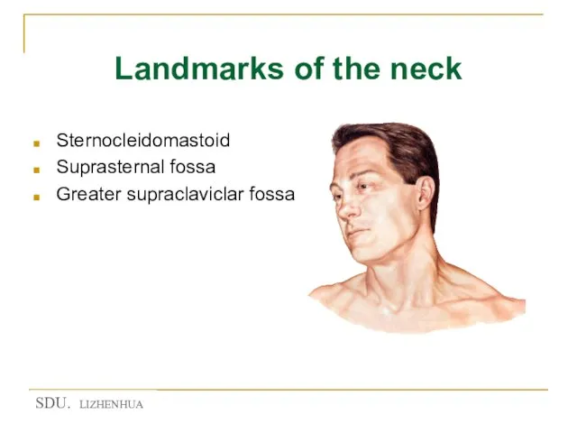
- 5. Landmarks of the neck Sternocleidomastoid Suprasternal fossa Greater supraclaviclar fossa
- 6. Regions of neck Neck 颈 Anterior region of neck 颈前区 Sternocleidomastoid region 胸锁乳突肌区 Lateral region of
- 7. Triangles of anterior region of neck Suprahyoid region 舌骨上区 Submental triangle 颏下三角 Submandibular triangle下颌下三角 Infrahyoid region
- 8. Triangles of lateral region of neck Occipital triangle 枕三角 supraclavicular triangle 锁骨上三角 (greater supraclavicular fossa) 锁骨上大窝
- 9. Skin of the neck The natural line of cleavage of the skin are constant and run
- 10. Superficial fascia Contents Platysma 颈阔肌 Superficial veins Anterior jugular v. 颈前静脉 External jugular v. 颈外静脉 Cutaneous
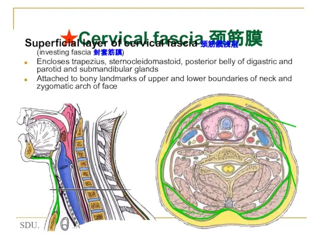
- 11. ★Cervical fascia 颈筋膜 Superficial layer of cervical fascia 颈筋膜浅层 (investing fascia 封套筋膜) Encloses trapezius, sternocleidomastoid, posterior
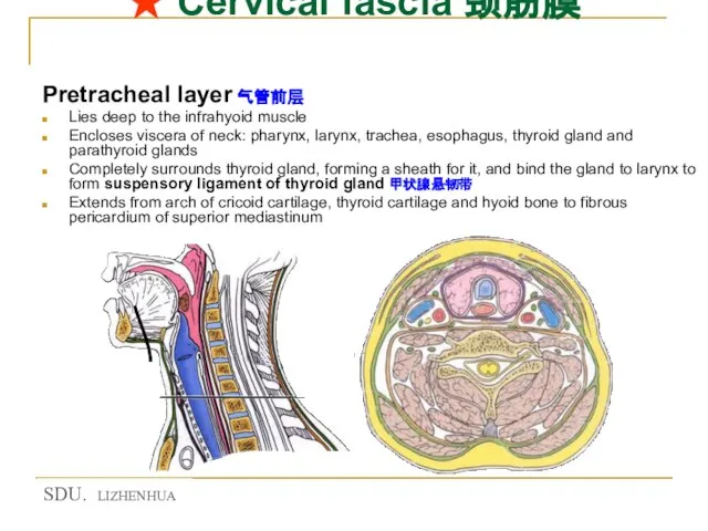
- 12. ★ Cervical fascia 颈筋膜 Pretracheal layer 气管前层 Lies deep to the infrahyoid muscle Encloses viscera of
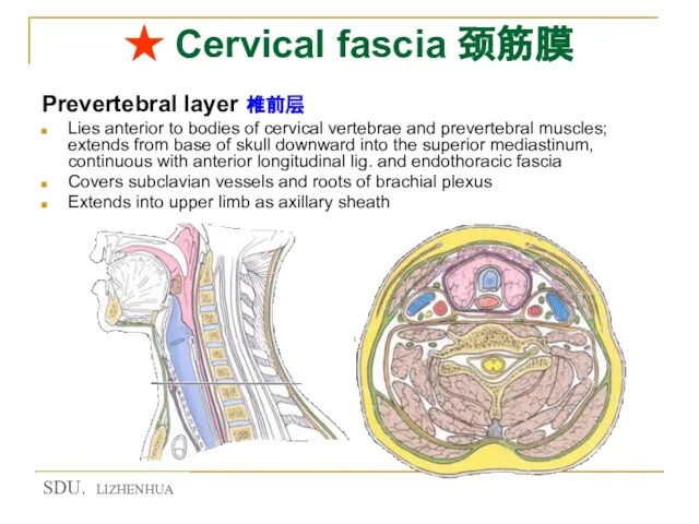
- 13. ★ Cervical fascia 颈筋膜 Prevertebral layer 椎前层 Lies anterior to bodies of cervical vertebrae and prevertebral
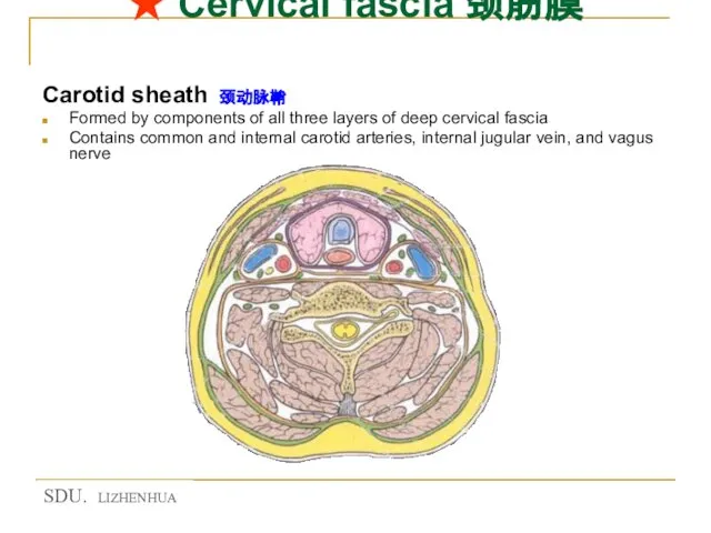
- 14. ★ Cervical fascia 颈筋膜 Carotid sheath 颈动脉鞘 Formed by components of all three layers of deep
- 15. Fascia spaces筋膜间隙 Suprasternal space 胸骨上间隙 3-4cm above manubrium of sterni the investing fascia splits into two
- 16. fascia spaces筋膜间隙 Pretracheal space 气管前间隙 Lies between pretracheal layer and cervical part of trachea Contains arteria
- 17. Fascia spaces筋膜间隙 Retropharyngeal space 咽后间隙 Lies between prevertebral layer and buccopharyngeal fascia Prevertebral space 椎前间隙 Lies
- 18. Anterior region of neck 颈前区
- 19. Suprahyoid region Submental triangle 颏下三角 Lies below the chin Boundaries Laterally by anterior bellies of digastric
- 20. Suprahyoid region Submandibular triangle 下颌下三角 Boundaries Anterior and posterior bellies of digastric Lower border of the
- 21. Infrahyoid region ★ Carotid triangle 颈动脉三角 Boundaries Anterior border of sternocleidomastoid Superior belly of omohyoid Posterior
- 22. Infrahyoid region ★ Carotid triangle 颈动脉三角 Contents Common carotid a. and its branches Internal jugular v.
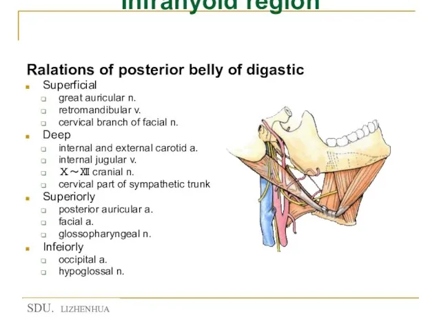
- 23. Infrahyoid region Ralations of posterior belly of digastic Superficial great auricular n. retromandibular v. cervical branch
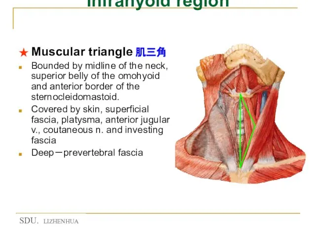
- 24. Infrahyoid region ★ Muscular triangle 肌三角 Bounded by midline of the neck, superior belly of the
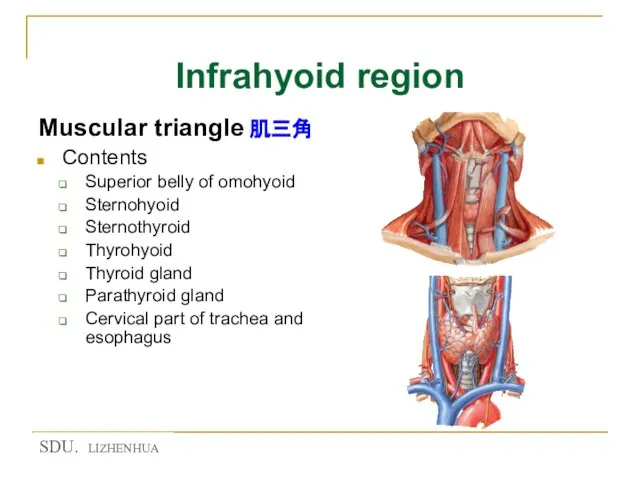
- 25. Infrahyoid region Muscular triangle 肌三角 Contents Superior belly of omohyoid Sternohyoid Sternothyroid Thyrohyoid Thyroid gland Parathyroid
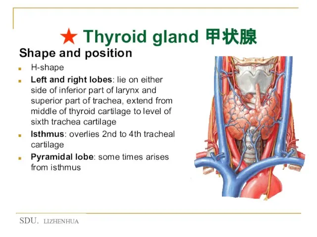
- 26. ★ Thyroid gland 甲状腺 Shape and position H-shape Left and right lobes: lie on either side
- 27. ★ Thyroid gland 甲状腺 Coverings of the thyroid gland False capsule: a sheath of pretracheal fascia
- 28. ★ Thyroid gland 甲状腺 Relations of the thyroid gland Anteriorly: Skin superficial fascia investing fascia Infrahyoid
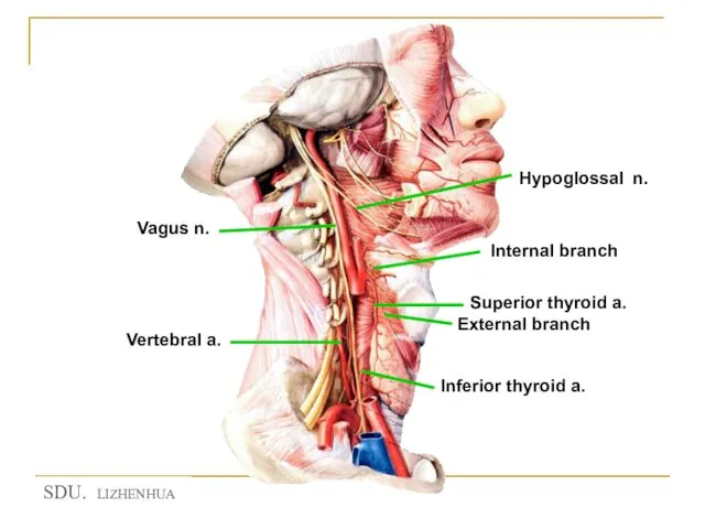
- 29. ★ Arteries of the thyroid gland Superior thyroid a. 甲状腺上动脉 Branch of external carotid a. Runs
- 30. ★ Arteries of the thyroid gland Inferior thyroid artery 甲状腺下动脉 Branch of thyrocervical trunk of subclavian
- 31. ★ Arteries of the thyroid gland Arteria thyroidea ima 甲状腺最下动脉 May arise (4%) from the brachiocephalic
- 32. ★ Nerves of the larynx Superior laryngeal n. 喉上神经 Internal branch 内支:which pierces thyrohyoid membrane to
- 33. ★ Nerves of the larynx Recurrent laryngeal nerves 喉返神经 Ascend in tracheo-esophageal groove Pass deep to
- 34. Venous drainage of the thyroid gland Superior thyroid veins drain into internal jugular vein Middle thyroid
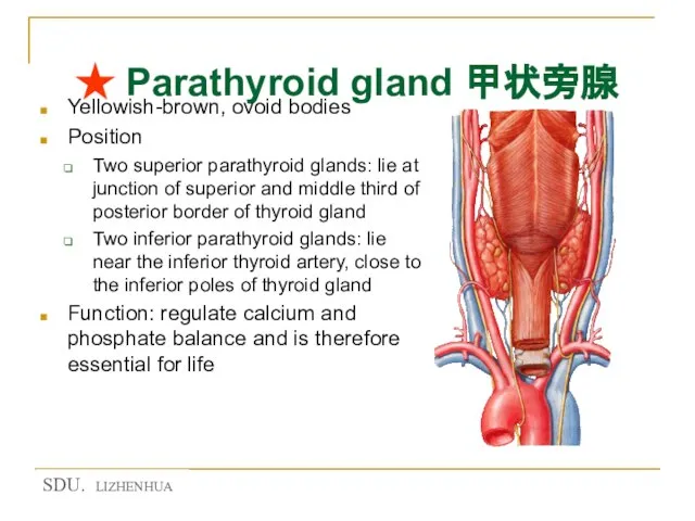
- 35. ★ Parathyroid gland 甲状旁腺 Yellowish-brown, ovoid bodies Position Two superior parathyroid glands: lie at junction of
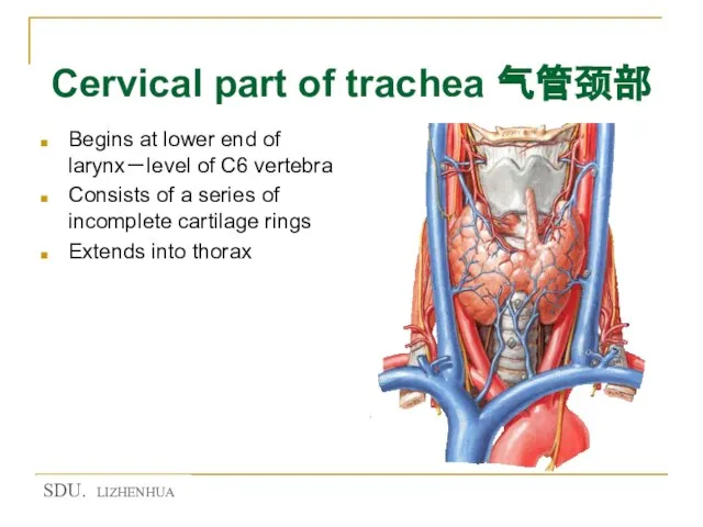
- 36. Cervical part of trachea 气管颈部 Begins at lower end of larynx-level of C6 vertebra Consists of
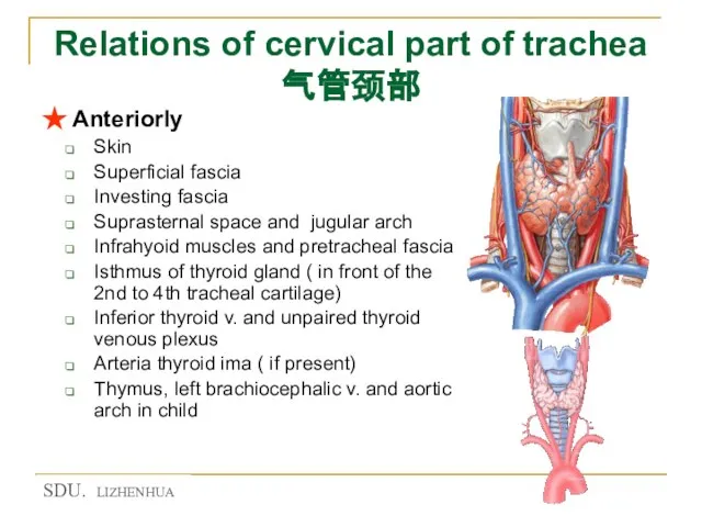
- 37. Relations of cervical part of trachea 气管颈部 ★ Anteriorly Skin Superficial fascia Investing fascia Suprasternal space
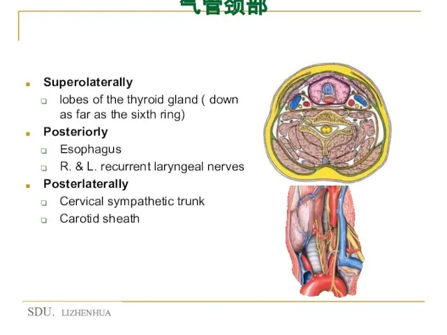
- 38. Relations of cervical part of trachea 气管颈部 Superolaterally lobes of the thyroid gland ( down as
- 39. Cervical part of esophagus 食管颈部 Extending from pharynx at level of C6 vertebra Descends through the
- 40. Sternocleidomastoid region 胸锁乳突肌区 Covered by sternocleidomastoid Contents Ansa cervicalis Carotid sheath Cervical plexus Cervical part of
- 41. Root of neck 颈根部 At thoracic inlet Formed by Anteriorly-manubrium sterni Posteriorly-body of first thoracic vertebra
- 42. Root of neck 颈根部 Contents Cupula of pleura-extends up into the neck, over the apex of
- 43. Triangle of the vertebral a. 椎动脉三角 Boundaries Medially-longus colli Laterally-scalenus anterior Inferiorly-first part of subclavian a.
- 44. Lateral region of neck 颈外侧区 Bounded by posterior border of sternocleidomastoid, anterior border of trapezius and
- 45. Occipital triangle 枕三角 Bounded by posterior border of sternocleidomastoid, anterior border of trapezius and superior border
- 46. Supraclavicular triangle 锁骨上三角 Bounded by posterior border of sternocleidomastoid, inferior belly of omohyoid and middle third
- 47. Skin incisions Make the skin incisions shown in figure Reflect the skin posteriorly to well behind
- 48. Dissection of Superficial Structures Note the underlying platysma muscle, a muscle of facial expression, which has
- 49. Dissection of Superficial Structures Using your scissors incise and spread the tough fascial covering of the
- 50. Cutaneous nerves and superficial veins
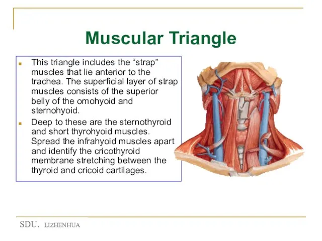
- 51. Muscular Triangle This triangle includes the “strap” muscles that lie anterior to the trachea. The superficial
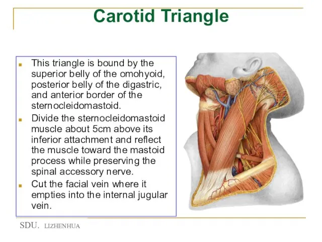
- 52. Carotid Triangle This triangle is bound by the superior belly of the omohyoid, posterior belly of
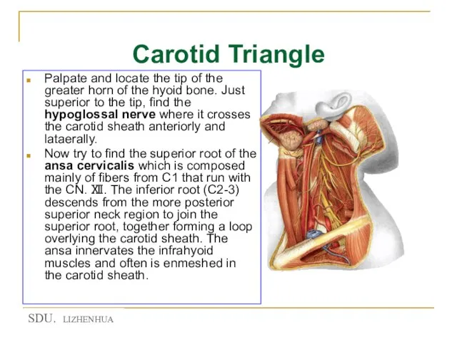
- 53. Carotid Triangle Palpate and locate the tip of the greater horn of the hyoid bone. Just
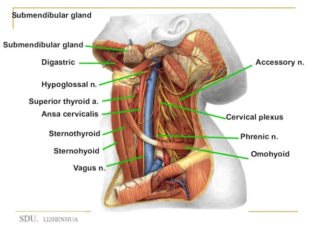
- 54. Submendibular gland
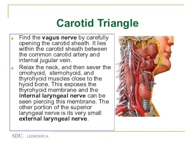
- 55. Carotid Triangle Find the vagus nerve by carefully opening the carotid sheath. It lies within the
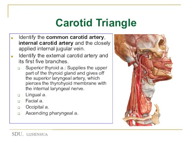
- 56. Carotid Triangle Identify the common carotid artery, internal carotid artery and the closely applied internal jugular
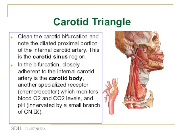
- 58. Carotid Triangle Clean the carotid bifurcation and note the dilated proximal portion of the internal carotid
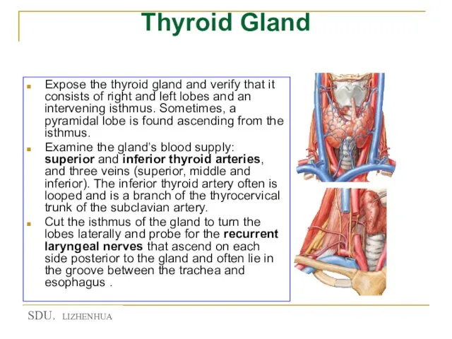
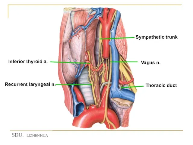
- 59. Thyroid Gland Expose the thyroid gland and verify that it consists of right and left lobes
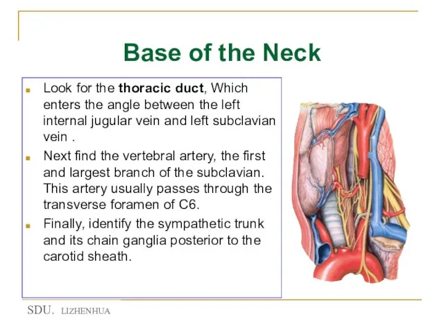
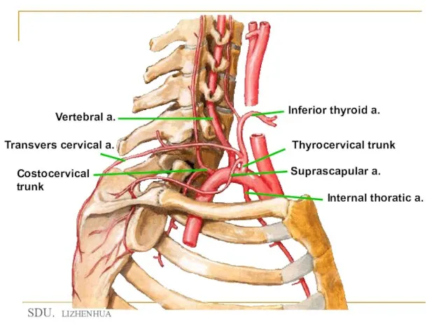
- 60. Base of the Neck Look for the thoracic duct, Which enters the angle between the left
- 61. Sympathetic trunk
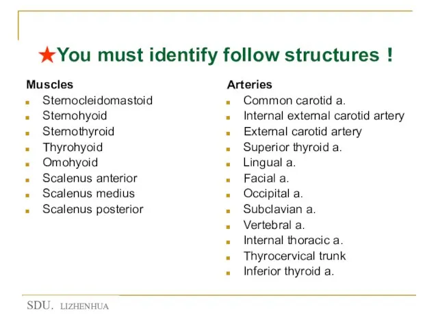
- 63. ★You must identify follow structures! Muscles Sternocleidomastoid Sternohyoid Sternothyroid Thyrohyoid Omohyoid Scalenus anterior Scalenus medius Scalenus
- 65. Скачать презентацию
Слайд 3Landmarks of the neck
Hyoid bone 舌骨
Thyroid cartilage 甲状软骨
Cricoid cartilage 环状软骨
Catotid tubercle
Landmarks of the neck
Hyoid bone 舌骨
Thyroid cartilage 甲状软骨
Cricoid cartilage 环状软骨
Catotid tubercle
Слайд 4Landmarks of the neck
Hyoid bone 舌骨
Thyroid cartilage 甲状软骨
Cricoid cartilage 环状软骨
Catotid tubercle 颈动脉结节
Landmarks of the neck
Hyoid bone 舌骨
Thyroid cartilage 甲状软骨
Cricoid cartilage 环状软骨
Catotid tubercle 颈动脉结节
Слайд 5Landmarks of the neck
Sternocleidomastoid
Suprasternal fossa
Greater supraclaviclar fossa
Landmarks of the neck
Sternocleidomastoid
Suprasternal fossa
Greater supraclaviclar fossa
Слайд 6Regions of neck
Neck 颈
Anterior region of neck 颈前区
Sternocleidomastoid region 胸锁乳突肌区
Lateral region of
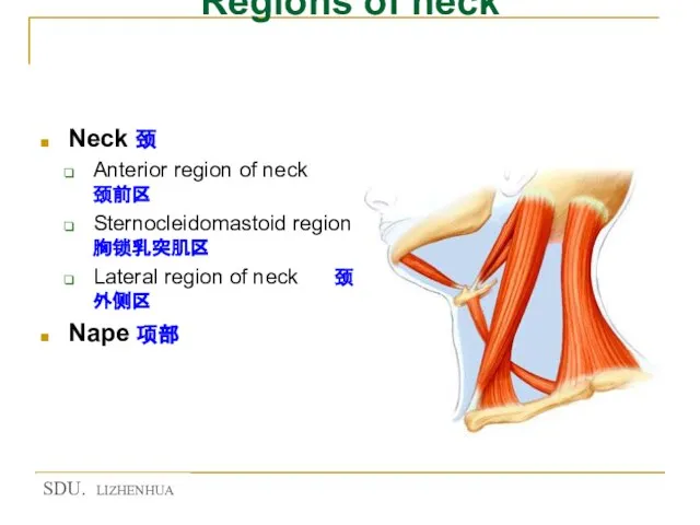
Regions of neck
Neck 颈
Anterior region of neck 颈前区
Sternocleidomastoid region 胸锁乳突肌区
Lateral region of
Слайд 7Triangles of anterior region of neck
Suprahyoid region 舌骨上区
Submental triangle 颏下三角
Submandibular triangle下颌下三角
Infrahyoid region
Triangles of anterior region of neck
Suprahyoid region 舌骨上区
Submental triangle 颏下三角
Submandibular triangle下颌下三角
Infrahyoid region

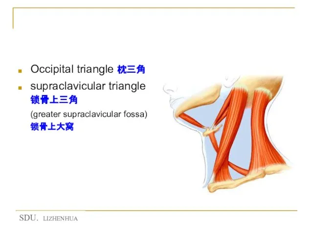
Слайд 8Triangles of lateral region of neck
Occipital triangle 枕三角
supraclavicular triangle 锁骨上三角 (greater
Triangles of lateral region of neck
Occipital triangle 枕三角
supraclavicular triangle 锁骨上三角 (greater
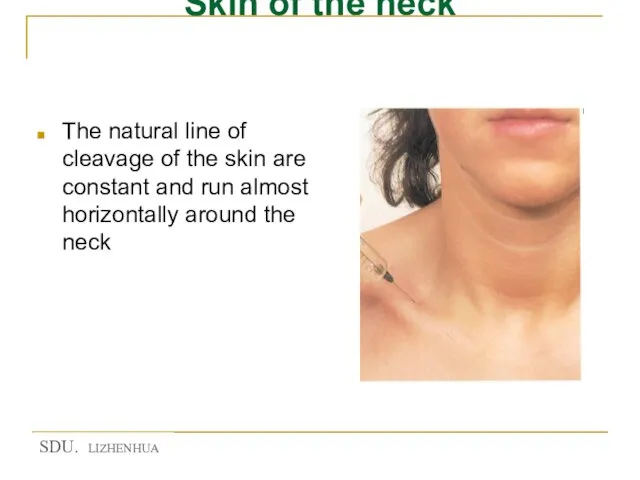
Слайд 9Skin of the neck
The natural line of cleavage of the skin are
Skin of the neck
The natural line of cleavage of the skin are
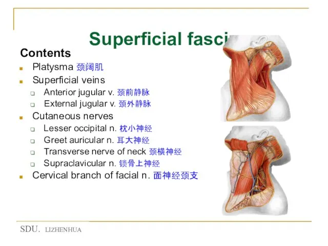
Слайд 10Superficial fascia
Contents
Platysma 颈阔肌
Superficial veins
Anterior jugular v. 颈前静脉
External jugular v. 颈外静脉
Cutaneous
Superficial fascia
Contents
Platysma 颈阔肌
Superficial veins
Anterior jugular v. 颈前静脉
External jugular v. 颈外静脉
Cutaneous
Слайд 11★Cervical fascia 颈筋膜
Superficial layer of cervical fascia 颈筋膜浅层 (investing fascia 封套筋膜)
Encloses trapezius,
★Cervical fascia 颈筋膜
Superficial layer of cervical fascia 颈筋膜浅层 (investing fascia 封套筋膜)
Encloses trapezius,
Слайд 12★ Cervical fascia 颈筋膜
Pretracheal layer 气管前层
Lies deep to the infrahyoid muscle
Encloses
★ Cervical fascia 颈筋膜
Pretracheal layer 气管前层
Lies deep to the infrahyoid muscle
Encloses
Слайд 13★ Cervical fascia 颈筋膜
Prevertebral layer 椎前层
Lies anterior to bodies of cervical vertebrae
★ Cervical fascia 颈筋膜
Prevertebral layer 椎前层
Lies anterior to bodies of cervical vertebrae
Слайд 14★ Cervical fascia 颈筋膜
Carotid sheath 颈动脉鞘
Formed by components of all three layers
★ Cervical fascia 颈筋膜
Carotid sheath 颈动脉鞘
Formed by components of all three layers
Слайд 15Fascia spaces筋膜间隙
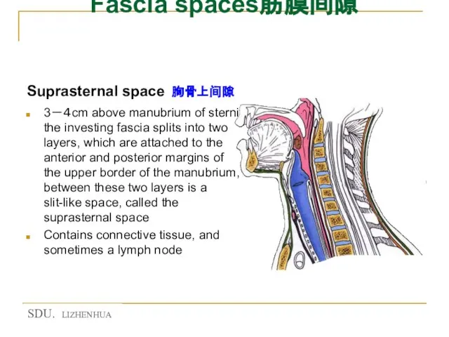
Suprasternal space 胸骨上间隙
3-4cm above manubrium of sterni the investing fascia splits
Fascia spaces筋膜间隙
Suprasternal space 胸骨上间隙
3-4cm above manubrium of sterni the investing fascia splits
Слайд 16fascia spaces筋膜间隙
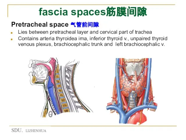
Pretracheal space 气管前间隙
Lies between pretracheal layer and cervical part of trachea
Contains
fascia spaces筋膜间隙
Pretracheal space 气管前间隙
Lies between pretracheal layer and cervical part of trachea
Contains
Слайд 17Fascia spaces筋膜间隙
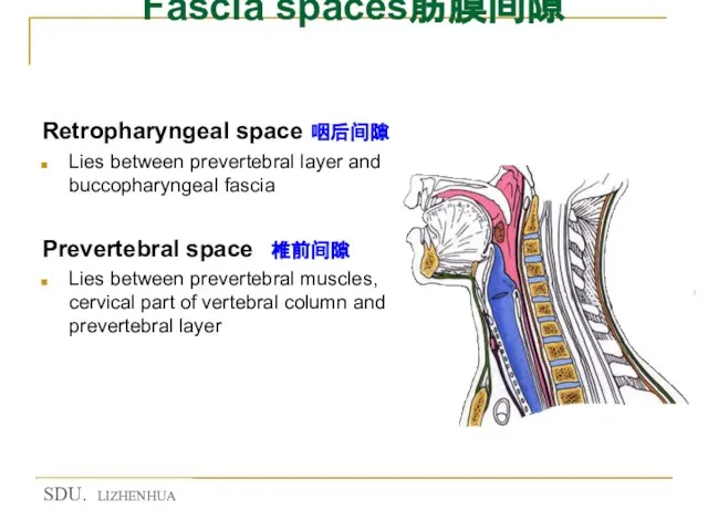
Retropharyngeal space 咽后间隙
Lies between prevertebral layer and buccopharyngeal fascia
Prevertebral space
Fascia spaces筋膜间隙
Retropharyngeal space 咽后间隙
Lies between prevertebral layer and buccopharyngeal fascia
Prevertebral space
Слайд 18Anterior region of neck 颈前区
Anterior region of neck 颈前区
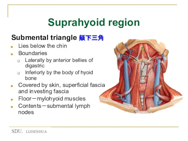
Слайд 19Suprahyoid region
Submental triangle 颏下三角
Lies below the chin
Boundaries
Laterally by anterior bellies
Suprahyoid region
Submental triangle 颏下三角
Lies below the chin
Boundaries
Laterally by anterior bellies
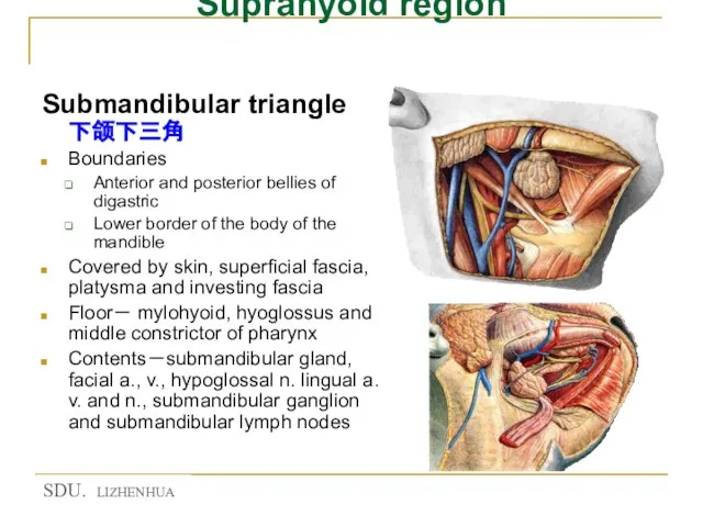
Слайд 20Suprahyoid region
Submandibular triangle 下颌下三角
Boundaries
Anterior and posterior bellies of digastric
Lower border of
Suprahyoid region
Submandibular triangle 下颌下三角
Boundaries
Anterior and posterior bellies of digastric
Lower border of
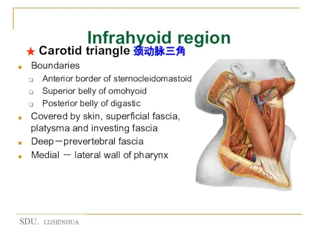
Слайд 21Infrahyoid region
★ Carotid triangle 颈动脉三角
Boundaries
Anterior border of sternocleidomastoid
Superior belly of omohyoid
Infrahyoid region
★ Carotid triangle 颈动脉三角
Boundaries
Anterior border of sternocleidomastoid
Superior belly of omohyoid
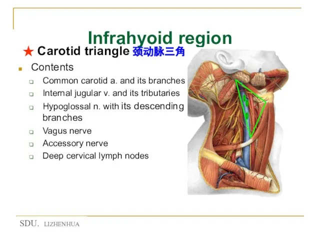
Слайд 22Infrahyoid region
★ Carotid triangle 颈动脉三角
Contents
Common carotid a. and its branches
Internal jugular
Infrahyoid region
★ Carotid triangle 颈动脉三角
Contents
Common carotid a. and its branches
Internal jugular
Слайд 23Infrahyoid region
Ralations of posterior belly of digastic
Superficial
great auricular n.
retromandibular v.
cervical branch
Infrahyoid region
Ralations of posterior belly of digastic
Superficial
great auricular n.
retromandibular v.
cervical branch
Слайд 24Infrahyoid region
★ Muscular triangle 肌三角
Bounded by midline of the neck, superior belly
Infrahyoid region
★ Muscular triangle 肌三角
Bounded by midline of the neck, superior belly
Слайд 25Infrahyoid region
Muscular triangle 肌三角
Contents
Superior belly of omohyoid
Sternohyoid
Sternothyroid
Thyrohyoid
Thyroid gland
Parathyroid gland
Cervical part
Infrahyoid region
Muscular triangle 肌三角
Contents
Superior belly of omohyoid
Sternohyoid
Sternothyroid
Thyrohyoid
Thyroid gland
Parathyroid gland
Cervical part
Слайд 26★ Thyroid gland 甲状腺
Shape and position
H-shape
Left and right lobes: lie on either
★ Thyroid gland 甲状腺
Shape and position
H-shape
Left and right lobes: lie on either
Слайд 27★ Thyroid gland 甲状腺
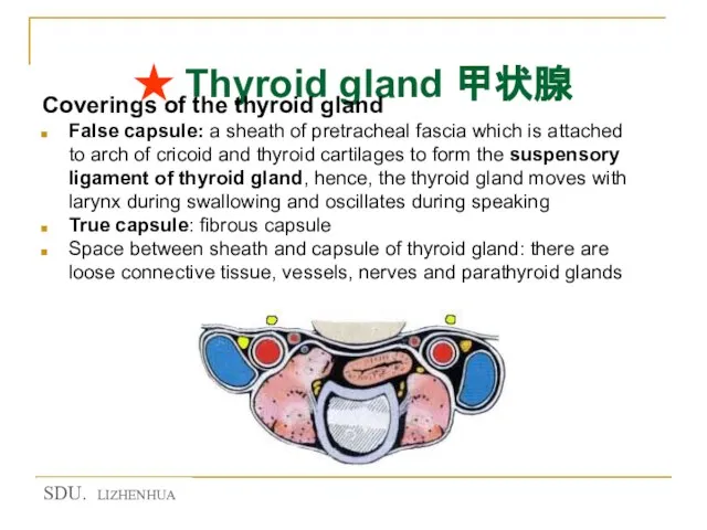
Coverings of the thyroid gland
False capsule: a sheath of
★ Thyroid gland 甲状腺
Coverings of the thyroid gland
False capsule: a sheath of
Слайд 28★ Thyroid gland 甲状腺
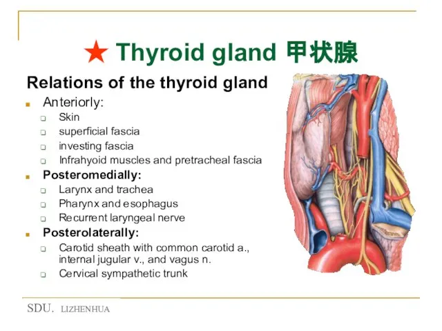
Relations of the thyroid gland
Anteriorly:
Skin
superficial fascia
investing
★ Thyroid gland 甲状腺
Relations of the thyroid gland
Anteriorly:
Skin
superficial fascia
investing
Слайд 29★ Arteries of the thyroid gland
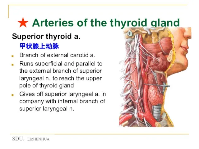
Superior thyroid a.
甲状腺上动脉
Branch of external
★ Arteries of the thyroid gland
Superior thyroid a.
甲状腺上动脉
Branch of external
Слайд 30★ Arteries of the thyroid gland
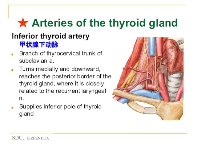
Inferior thyroid artery 甲状腺下动脉
Branch of thyrocervical trunk
★ Arteries of the thyroid gland
Inferior thyroid artery 甲状腺下动脉
Branch of thyrocervical trunk
Слайд 31★ Arteries of the thyroid gland
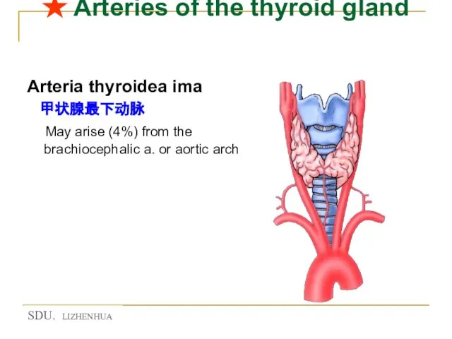
Arteria thyroidea ima
甲状腺最下动脉
May arise
★ Arteries of the thyroid gland
Arteria thyroidea ima
甲状腺最下动脉
May arise
Слайд 32★ Nerves of the larynx
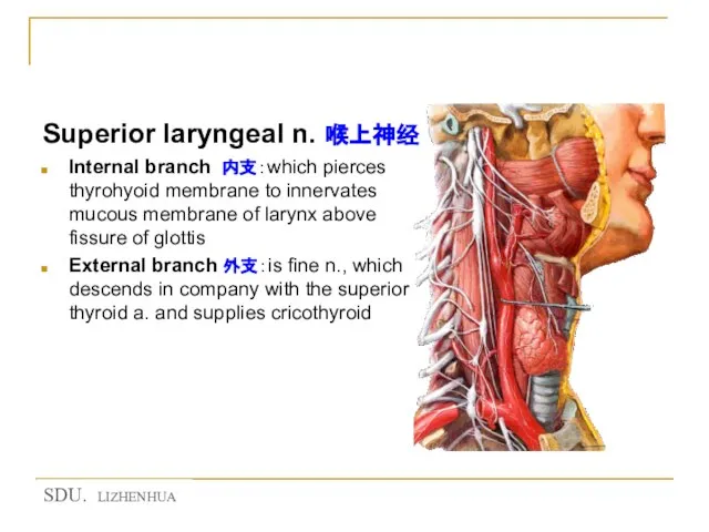
Superior laryngeal n. 喉上神经
Internal branch 内支:which pierces
★ Nerves of the larynx
Superior laryngeal n. 喉上神经
Internal branch 内支:which pierces
Слайд 33★ Nerves of the larynx
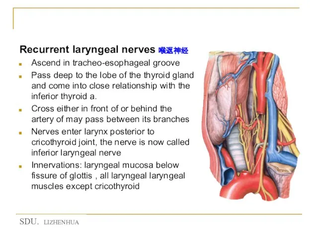
Recurrent laryngeal nerves 喉返神经
Ascend in tracheo-esophageal groove
Pass
★ Nerves of the larynx
Recurrent laryngeal nerves 喉返神经
Ascend in tracheo-esophageal groove
Pass
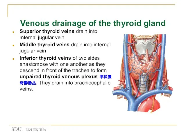
Слайд 34Venous drainage of the thyroid gland
Superior thyroid veins drain into internal jugular
Venous drainage of the thyroid gland
Superior thyroid veins drain into internal jugular
Слайд 35★ Parathyroid gland 甲状旁腺
Yellowish-brown, ovoid bodies
Position
Two superior parathyroid glands: lie at junction
★ Parathyroid gland 甲状旁腺
Yellowish-brown, ovoid bodies
Position
Two superior parathyroid glands: lie at junction
Слайд 36Cervical part of trachea 气管颈部
Begins at lower end of larynx-level of C6
Cervical part of trachea 气管颈部
Begins at lower end of larynx-level of C6
Слайд 37Relations of cervical part of trachea 气管颈部
★ Anteriorly
Skin
Superficial fascia
Investing fascia
Suprasternal space and
Relations of cervical part of trachea 气管颈部
★ Anteriorly
Skin
Superficial fascia
Investing fascia
Suprasternal space and
Слайд 38Relations of cervical part of trachea 气管颈部
Superolaterally
lobes of the thyroid gland
Relations of cervical part of trachea 气管颈部
Superolaterally
lobes of the thyroid gland
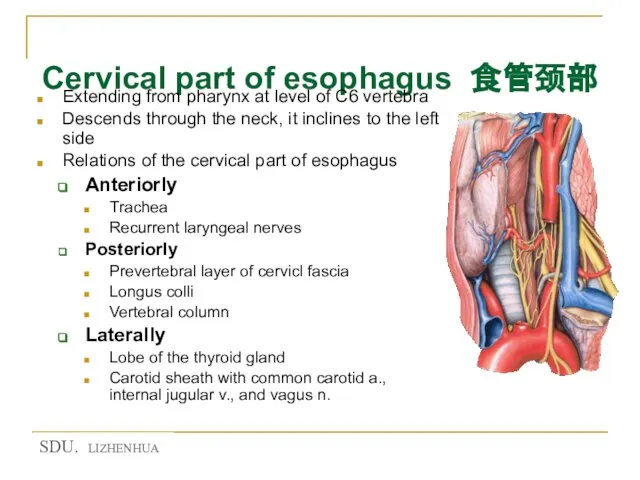
Слайд 39Cervical part of esophagus 食管颈部
Extending from pharynx at level of C6 vertebra
Descends
Cervical part of esophagus 食管颈部
Extending from pharynx at level of C6 vertebra
Descends
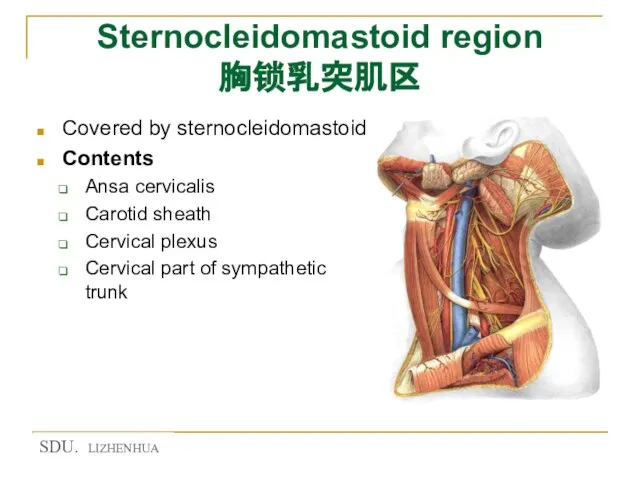
Слайд 40Sternocleidomastoid region 胸锁乳突肌区
Covered by sternocleidomastoid
Contents
Ansa cervicalis
Carotid sheath
Cervical plexus
Cervical
Sternocleidomastoid region 胸锁乳突肌区
Covered by sternocleidomastoid
Contents
Ansa cervicalis
Carotid sheath
Cervical plexus
Cervical
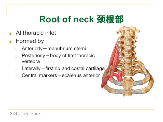
Слайд 41Root of neck 颈根部
At thoracic inlet
Formed by
Anteriorly-manubrium sterni
Posteriorly-body of first thoracic
Root of neck 颈根部
At thoracic inlet
Formed by
Anteriorly-manubrium sterni
Posteriorly-body of first thoracic
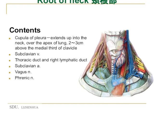
Слайд 42Root of neck 颈根部
Contents
Cupula of pleura-extends up into the neck, over
Root of neck 颈根部
Contents
Cupula of pleura-extends up into the neck, over
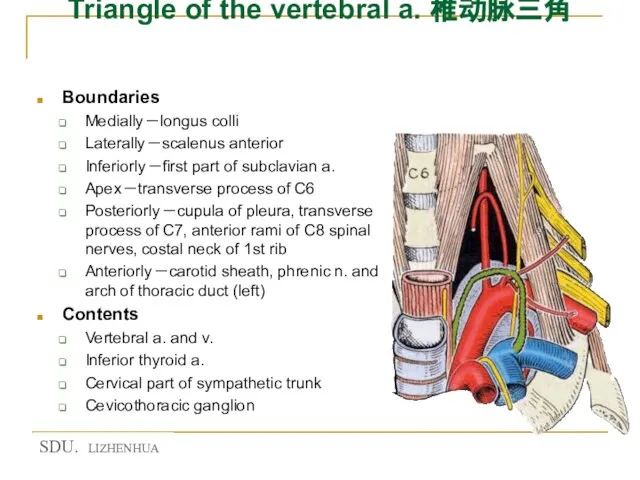
Слайд 43Triangle of the vertebral a. 椎动脉三角
Boundaries
Medially-longus colli
Laterally-scalenus anterior
Inferiorly-first part of
Triangle of the vertebral a. 椎动脉三角
Boundaries
Medially-longus colli
Laterally-scalenus anterior
Inferiorly-first part of
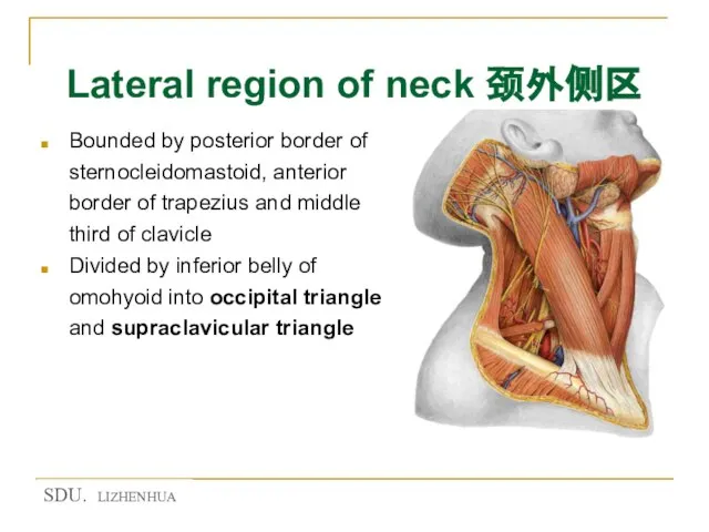
Слайд 44Lateral region of neck 颈外侧区
Bounded by posterior border of sternocleidomastoid, anterior border
Lateral region of neck 颈外侧区
Bounded by posterior border of sternocleidomastoid, anterior border
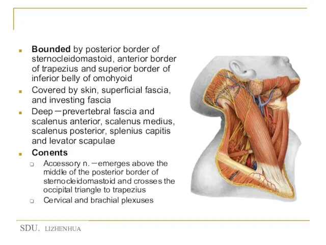
Слайд 45Occipital triangle 枕三角
Bounded by posterior border of sternocleidomastoid, anterior border of trapezius
Occipital triangle 枕三角
Bounded by posterior border of sternocleidomastoid, anterior border of trapezius
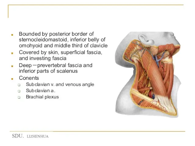
Слайд 46Supraclavicular triangle 锁骨上三角
Bounded by posterior border of sternocleidomastoid, inferior belly of omohyoid
Supraclavicular triangle 锁骨上三角
Bounded by posterior border of sternocleidomastoid, inferior belly of omohyoid
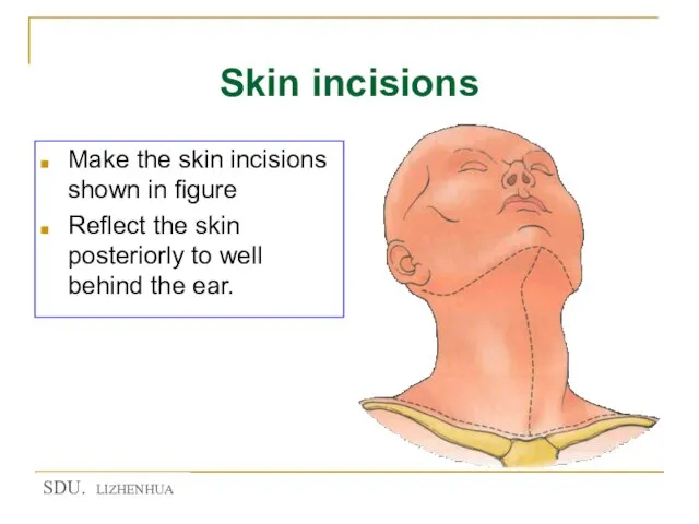
Слайд 47Skin incisions
Make the skin incisions shown in figure
Reflect the skin posteriorly
Skin incisions
Make the skin incisions shown in figure
Reflect the skin posteriorly
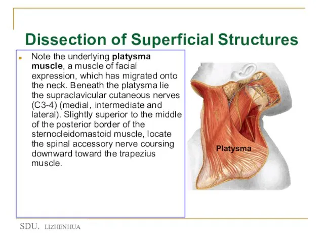
Слайд 48Dissection of Superficial Structures
Note the underlying platysma muscle, a muscle of
Dissection of Superficial Structures
Note the underlying platysma muscle, a muscle of
Слайд 49Dissection of Superficial Structures
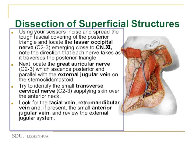
Using your scissors incise and spread the tough fascial
Dissection of Superficial Structures
Using your scissors incise and spread the tough fascial
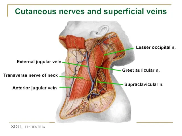
Слайд 50Cutaneous nerves and superficial veins
Cutaneous nerves and superficial veins
Слайд 51Muscular Triangle
This triangle includes the “strap” muscles that lie anterior to the
Muscular Triangle
This triangle includes the “strap” muscles that lie anterior to the
Слайд 52Carotid Triangle
This triangle is bound by the superior belly of the omohyoid,
Carotid Triangle
This triangle is bound by the superior belly of the omohyoid,
Слайд 53Carotid Triangle
Palpate and locate the tip of the greater horn of the
Carotid Triangle
Palpate and locate the tip of the greater horn of the
Слайд 54Submendibular gland
Submendibular gland
Слайд 55Carotid Triangle
Find the vagus nerve by carefully opening the carotid sheath. It
Carotid Triangle
Find the vagus nerve by carefully opening the carotid sheath. It
Слайд 56Carotid Triangle
Identify the common carotid artery, internal carotid artery and the closely
Carotid Triangle
Identify the common carotid artery, internal carotid artery and the closely
Слайд 58Carotid Triangle
Clean the carotid bifurcation and note the dilated proximal portion of
Carotid Triangle
Clean the carotid bifurcation and note the dilated proximal portion of
Слайд 59Thyroid Gland
Expose the thyroid gland and verify that it consists of right
Thyroid Gland
Expose the thyroid gland and verify that it consists of right
Слайд 60Base of the Neck
Look for the thoracic duct, Which enters the angle
Base of the Neck
Look for the thoracic duct, Which enters the angle
Слайд 61 Sympathetic trunk
Sympathetic trunk
Слайд 63★You must identify follow structures!
Muscles
Sternocleidomastoid
Sternohyoid
Sternothyroid
Thyrohyoid
Omohyoid
Scalenus anterior
Scalenus medius
Scalenus posterior
Arteries
Common carotid a.
Internal external
★You must identify follow structures!
Muscles
Sternocleidomastoid
Sternohyoid
Sternothyroid
Thyrohyoid
Omohyoid
Scalenus anterior
Scalenus medius
Scalenus posterior
Arteries
Common carotid a.
Internal external
 Модульная структурасистемы ITAS
 Инвестор, новый уровень
 Разработка и изготовление ансамбля коллекции женской одежды Butterfly
 Илья Муромец и Соловей-разбойник
 Времена года. Лабораторная работа №5
 «СОРОК МГНОВЕНИЙ НАЕДИНЕ С КОРНЕМ СТЕПЕНИ n»
 «Регион 74 в составе Российской Федерации».
 Правовая информация для несовершеннолетних и их родителей
 Дальневосточная пожарно-спасательная академия
 Воспитательный потенциал современного образования:вызов родительской общественности
 СТРЕССОВЫЕ СИТУАЦИИ НА РАБОЧЕМ МЕСТЕ
 Винсент Ван Гог. Подсолнухи
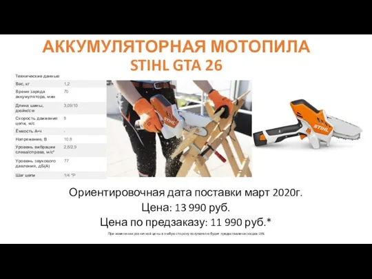 Аккумуляторная мотопила stihl gta 26
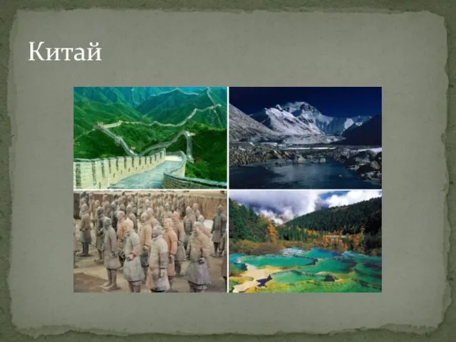 Китай
Китай Об использовании новых организационных форм медицинского обеспечения и оценки результатов новой системы оплаты труда
 Татарское декоративно-прикладное искусство
 Буквы Ч,ч, обозначающие звук [ч’]
 Разрезы в аксонометрических проекциях
 Чему учил китайский мудрец Конфуций
 Порядок обращения за страховой пенсией по случаю потери кормильца
 Презентация на тему Шолохов «Донские рассказы»
 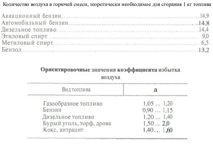 Сгорание топлива. Октановое число
 Налоговый потенциал
 Влияние плавания на здоровье человека
 Топочные устройства ПК
 Иллюстрационный материал к реферату на тему: Эмпирическая школа управления
 Can you swim?
 МОДЕЛИ УПРАВЛЕНИЯ ЗАПАСАМИ МОДЕЛИ УПРАВЛЕНИЯ ЗАПАСАМИ