- Sense organs

Содержание
- 2. Receptors provide information about both external and internal environments. The receptors of the human are located
- 3. It provides more than 80% of the information received about the external environment. THE EYE
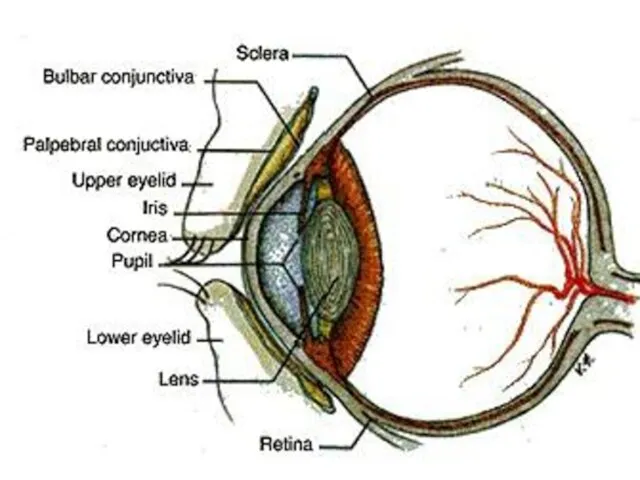
- 4. STRUCTURE OF EYE Eyes contain 3 main parts; Schlerenchyma Choroid Retina
- 5. Schlerenchyma It is supportive structure of eye that protects inner structures of the eye. In the
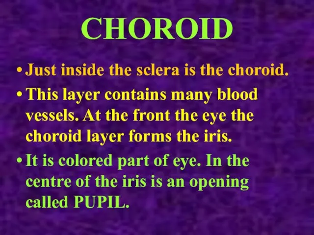
- 6. CHOROID Just inside the sclera is the choroid. This layer contains many blood vessels. At the
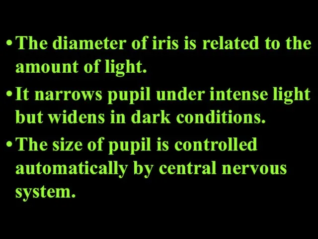
- 7. The diameter of iris is related to the amount of light. It narrows pupil under intense
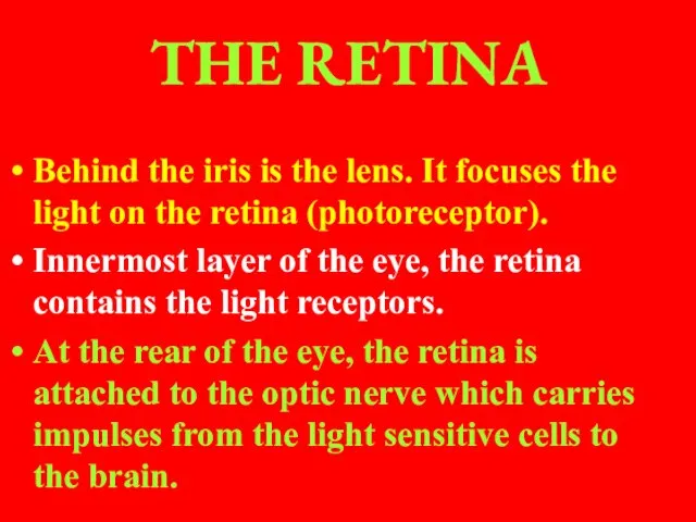
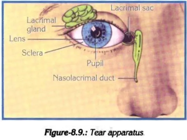
- 8. THE RETINA Behind the iris is the lens. It focuses the light on the retina (photoreceptor).
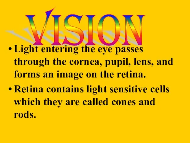
- 11. Light entering the eye passes through the cornea, pupil, lens, and forms an image on the
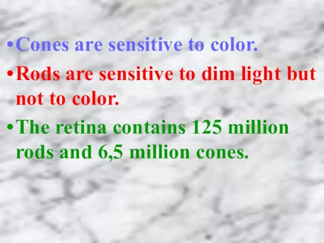
- 12. Cones are sensitive to color. Rods are sensitive to dim light but not to color. The
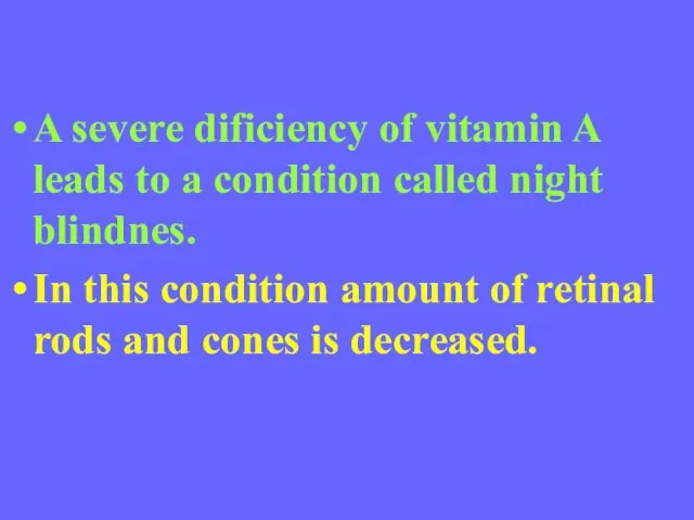
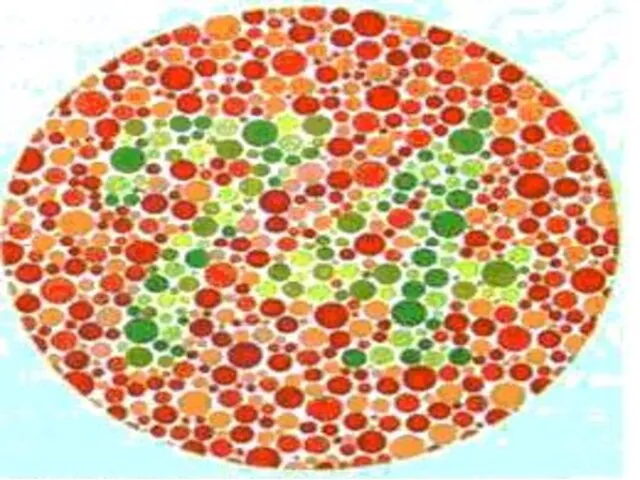
- 13. A severe dificiency of vitamin A leads to a condition called night blindnes. In this condition
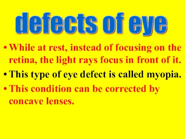
- 15. While at rest, instead of focusing on the retina, the light rays focus in front of
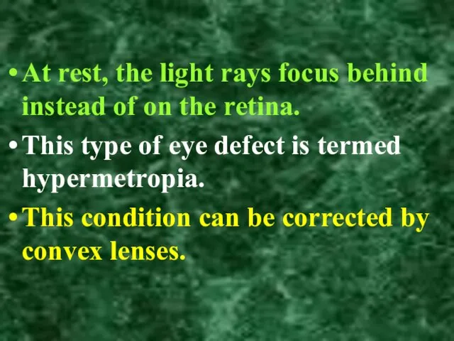
- 16. At rest, the light rays focus behind instead of on the retina. This type of eye
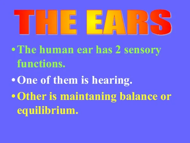
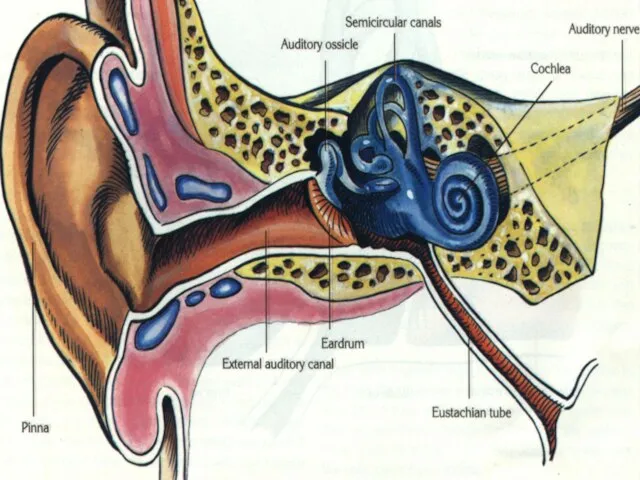
- 17. The human ear has 2 sensory functions. One of them is hearing. Other is maintaning balance
- 18. Structure of ears Ears contains 3 main parts; Outer ear, The middle ear Inner ear
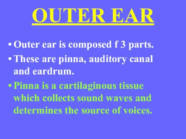
- 20. OUTER EAR Outer ear is composed f 3 parts. These are pinna, auditory canal and eardrum.
- 21. Auditory canal is a canal which is found between pinna and eardrum. It has hairs and
- 22. MIDDLE EAR It contains three small bones which are called the hammer, anvil and stirrup. These
- 24. The hommer attached to the eardrum, the anvil connects the hommer to the stirrup. Stirrup is
- 26. EUSTACHIAN TUBE It is located between pharynx and the middle ear. It equalizes in the middle
- 28. THE INNER EAR It consists of the cochlea and semicircular canals. Cochlea is organ of hearing
- 29. They are separated from another by membranes. Lining of the membranes are specialized hair cells that
- 30. Semicircular canals enable the body to maintain balance. These canals contain fluid and hairlike projenctions that
- 31. Sound waves collected by outer ear pass down the auditory canal to the eardrum. They cause
- 32. Vibration of stirrup cause vibrations in the oval window which in turn cause the fluid within
- 33. Structure of the ear Three regions: Outer ear Middle ear Inner ear
- 34. Process of hearing Sound waves are collected by the ear pinna
- 35. Process of hearing Sound waves pass along the external auditory canal to the ear drum
- 36. Process of hearing Sound waves make the ear drum to vibrate Ear drum converts sound waves
- 37. Process of hearing Ear drum transmits vibration to the ear bones Ear bones transmit and amplify
- 38. Process of hearing Ear bones transmit vibration to the oval windows
- 39. Process of hearing Oval window causes the perilymph in the upper canal of the cochlea to
- 40. Process of hearing Perilymph transmits vibrations to the endolymph in the middle canal
- 41. Process of hearing The sensory hair cells on the bottom membrane of the middle canal are
- 42. Process of hearing The auditory nerve transmits the impulses to the auditory centre of the cerebral
- 43. Process of hearing The vibrations of perilymph are transmitted to the round window Round window bulges
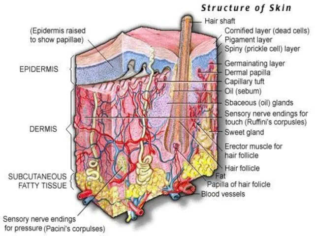
- 44. All multicellular organisms have a skin composed of one or more layers. THE SKIN
- 45. Functions of Skin It protects the inner layers of the body from physical and chemical effects.
- 46. EPIDERMIS is outermost layer of skin. This layer composed of keratinised epithelial cells. Epidermis contains no
- 47. DERMIS is rich in blood vessels and nerve ending. The receptors located in the skin are
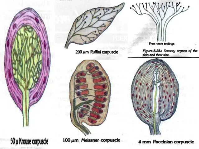
- 49. RECEPTORS Meisner corpuscles: They are involved in reception of touch of the palm sole and lips.
- 50. Ruffini corpuscles: They are involved in recption of heat, touch and pressure. Sweat glands: They are
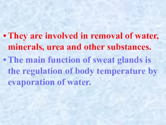
- 52. They are involved in removal of water, minerals, urea and other substances. The main function of
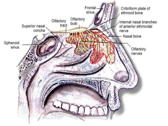
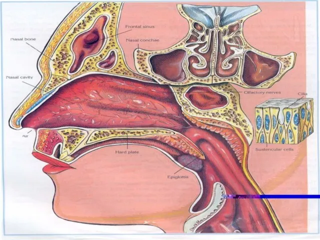
- 53. Nose is the organ of the body involved in botdh respiration and smell. The reception of
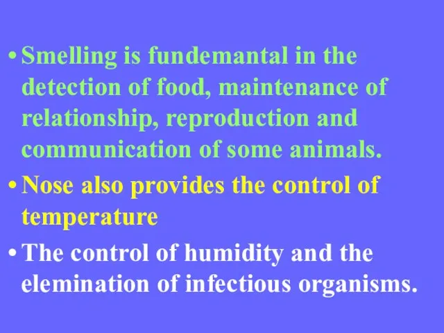
- 55. Smelling is fundemantal in the detection of food, maintenance of relationship, reproduction and communication of some
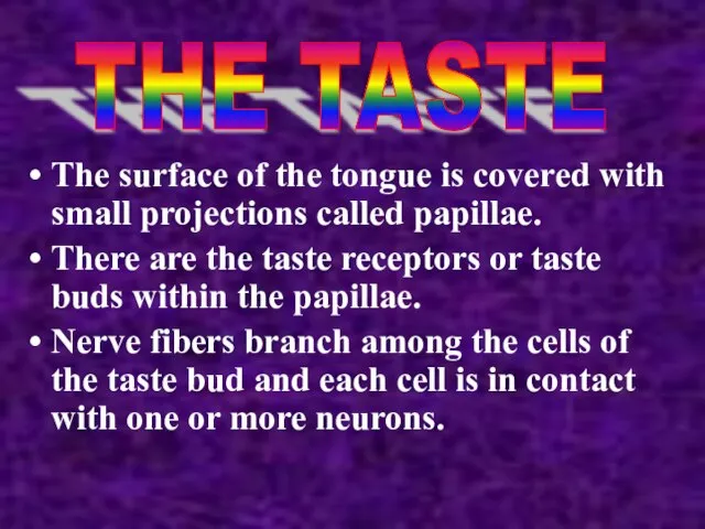
- 57. The surface of the tongue is covered with small projections called papillae. There are the taste
- 58. Only substances that are in solution can stimulate the taste buds. The taste buds are sensitive
- 59. Tend to be localized on specific areas of the tongue, taste buds for sourness are found
- 60. Taste buds for sweetness and saltiness on the tip of the tongue. When taste buds are
- 61. Tongue - the taste organ Detected by taste buds on the upper surface of the tongue
- 63. Скачать презентацию
Слайд 2Receptors provide information about both external and internal environments.
The receptors of the
Receptors provide information about both external and internal environments.
The receptors of the
Слайд 3It provides more than 80% of the information received about the external
It provides more than 80% of the information received about the external
Слайд 4
STRUCTURE OF EYE
Eyes contain 3 main parts;
Schlerenchyma
Choroid
Retina
STRUCTURE OF EYE
Eyes contain 3 main parts;
Schlerenchyma
Choroid
Retina
Слайд 5
Schlerenchyma
It is supportive structure of eye that protects inner structures of the
Schlerenchyma
It is supportive structure of eye that protects inner structures of the
Слайд 6CHOROID
Just inside the sclera is the choroid.
This layer contains many blood vessels.
CHOROID
Just inside the sclera is the choroid.
This layer contains many blood vessels.
Слайд 7The diameter of iris is related to the amount of light.
It narrows
The diameter of iris is related to the amount of light.
It narrows
Слайд 8THE RETINA
Behind the iris is the lens. It focuses the light on
THE RETINA
Behind the iris is the lens. It focuses the light on
Слайд 11Light entering the eye passes through the cornea, pupil, lens, and forms
Light entering the eye passes through the cornea, pupil, lens, and forms
Слайд 12Cones are sensitive to color.
Rods are sensitive to dim light but
Cones are sensitive to color.
Rods are sensitive to dim light but
Слайд 13A severe dificiency of vitamin A leads to a condition called night
A severe dificiency of vitamin A leads to a condition called night
Слайд 15While at rest, instead of focusing on the retina, the light rays
While at rest, instead of focusing on the retina, the light rays
Слайд 16At rest, the light rays focus behind instead of on the retina.
At rest, the light rays focus behind instead of on the retina.
Слайд 17The human ear has 2 sensory functions.
One of them is hearing.
Other
The human ear has 2 sensory functions.
One of them is hearing.
Other
Слайд 18Structure of ears
Ears contains 3 main parts;
Outer ear,
The middle ear
Inner ear
Structure of ears
Ears contains 3 main parts;
Outer ear,
The middle ear
Inner ear
Слайд 20OUTER EAR
Outer ear is composed f 3 parts.
These are pinna, auditory canal
OUTER EAR
Outer ear is composed f 3 parts.
These are pinna, auditory canal
Слайд 21Auditory canal is a canal which is found between pinna and eardrum.
It
Auditory canal is a canal which is found between pinna and eardrum.
It
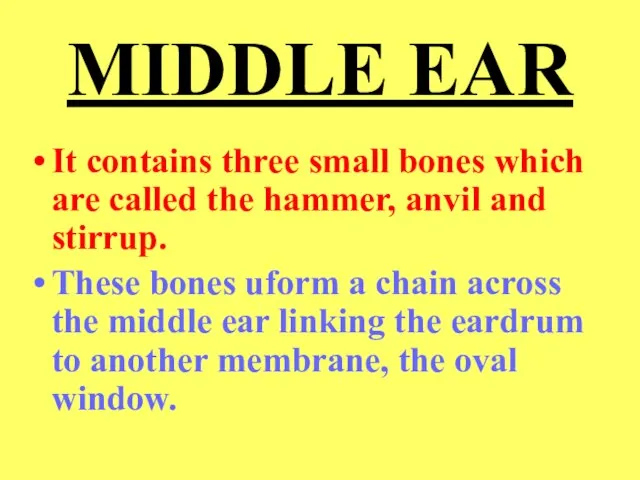
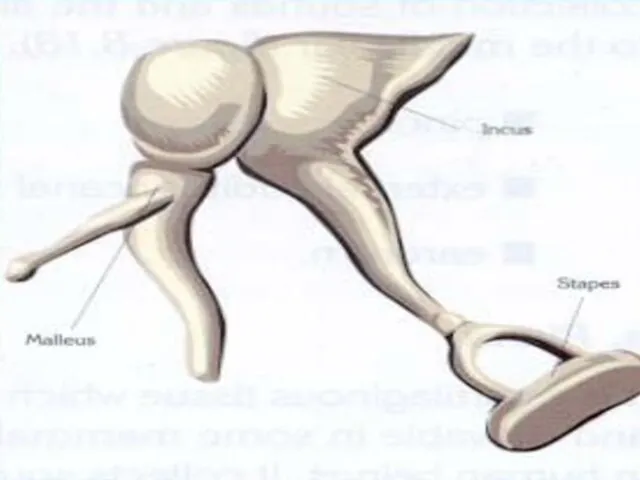
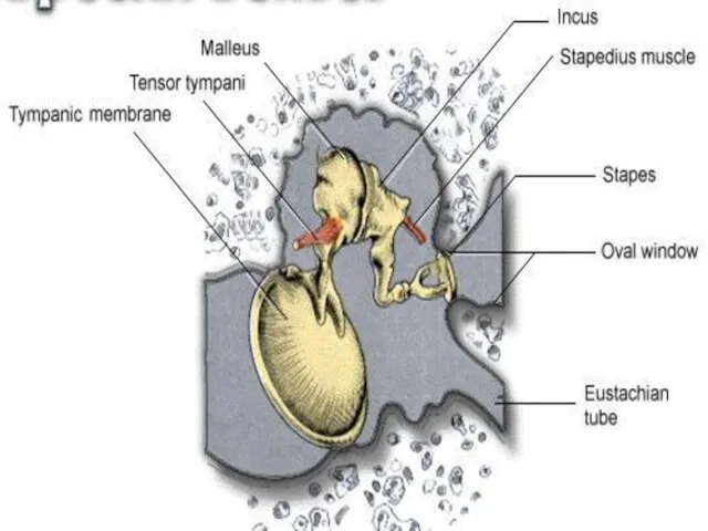
Слайд 22MIDDLE EAR
It contains three small bones which are called the hammer, anvil
MIDDLE EAR
It contains three small bones which are called the hammer, anvil
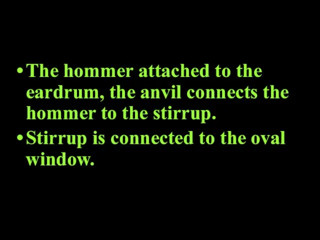
Слайд 24The hommer attached to the eardrum, the anvil connects the hommer to
The hommer attached to the eardrum, the anvil connects the hommer to
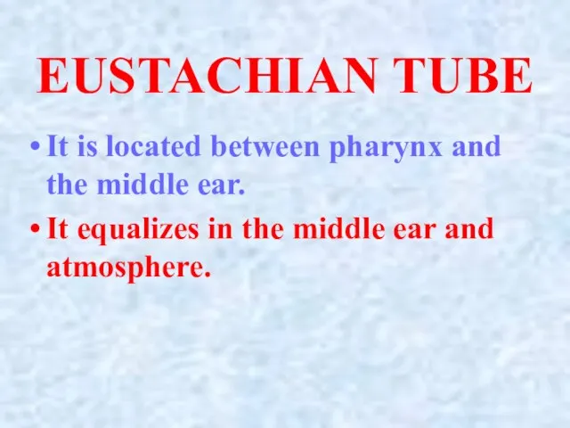
Слайд 26EUSTACHIAN TUBE
It is located between pharynx and the middle ear.
It equalizes in
EUSTACHIAN TUBE
It is located between pharynx and the middle ear.
It equalizes in
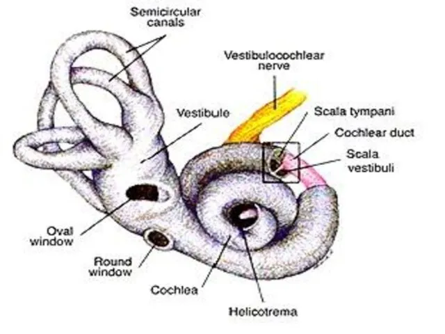
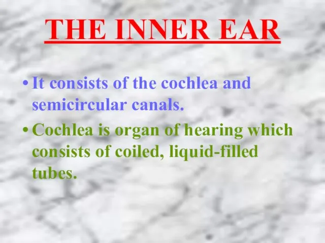
Слайд 28THE INNER EAR
It consists of the cochlea and semicircular canals.
Cochlea is organ
THE INNER EAR
It consists of the cochlea and semicircular canals.
Cochlea is organ
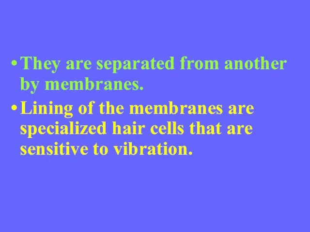
Слайд 29They are separated from another by membranes.
Lining of the membranes are specialized
They are separated from another by membranes.
Lining of the membranes are specialized
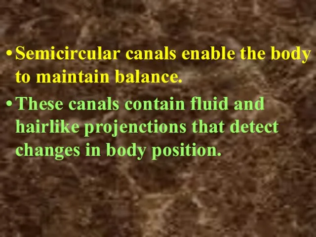
Слайд 30Semicircular canals enable the body to maintain balance.
These canals contain fluid and
Semicircular canals enable the body to maintain balance.
These canals contain fluid and
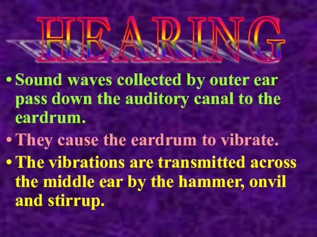
Слайд 31Sound waves collected by outer ear pass down the auditory canal to
Sound waves collected by outer ear pass down the auditory canal to
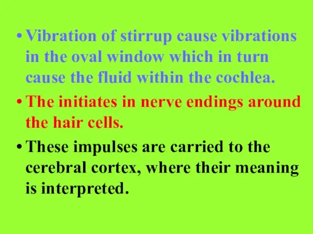
Слайд 32Vibration of stirrup cause vibrations in the oval window which in turn
Vibration of stirrup cause vibrations in the oval window which in turn
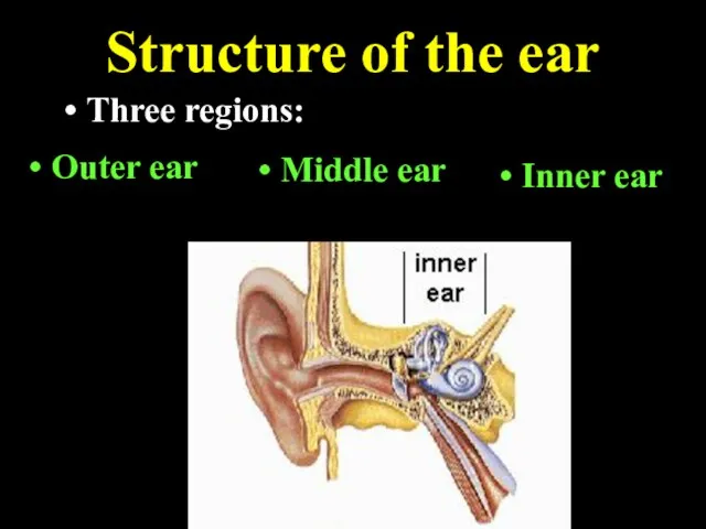
Слайд 33Structure of the ear
Three regions:
Outer ear
Middle ear
Inner ear
Structure of the ear
Three regions:
Outer ear
Middle ear
Inner ear
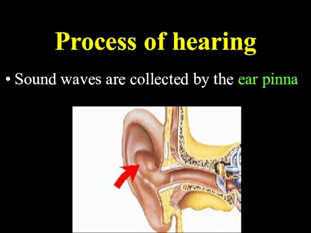
Слайд 34Process of hearing
Sound waves are collected by the ear pinna
Process of hearing
Sound waves are collected by the ear pinna
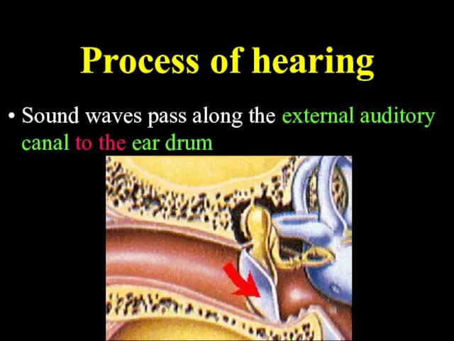
Слайд 35Process of hearing
Sound waves pass along the external auditory canal to the
Process of hearing
Sound waves pass along the external auditory canal to the
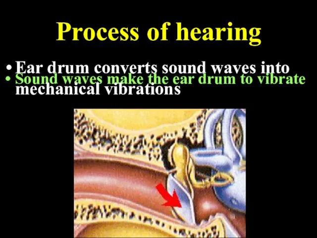
Слайд 36Process of hearing
Sound waves make the ear drum to vibrate
Ear drum converts
Process of hearing
Sound waves make the ear drum to vibrate
Ear drum converts
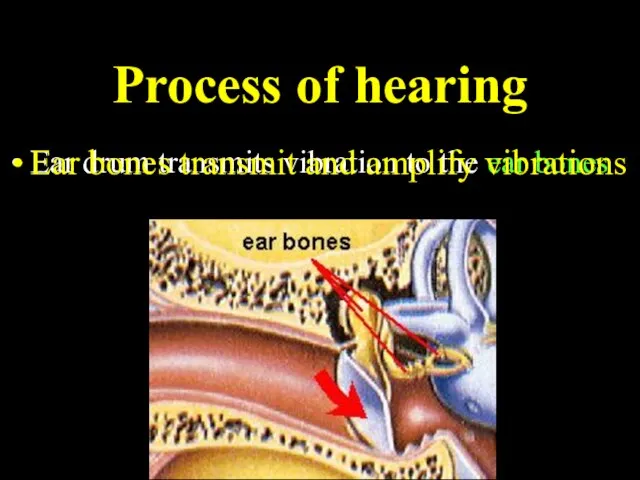
Слайд 37Process of hearing
Ear drum transmits vibration to the ear bones
Ear bones transmit
Process of hearing
Ear drum transmits vibration to the ear bones
Ear bones transmit
Слайд 38Process of hearing
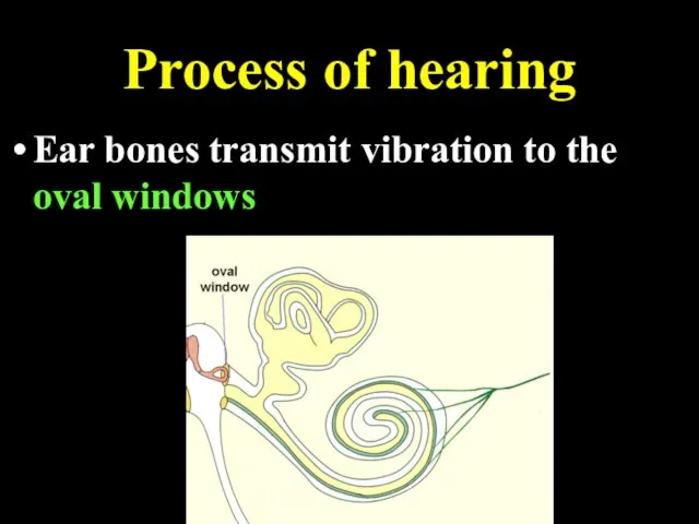
Ear bones transmit vibration to the oval windows
Process of hearing
Ear bones transmit vibration to the oval windows
Слайд 39Process of hearing
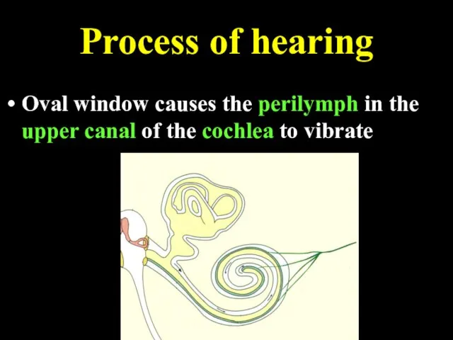
Oval window causes the perilymph in the upper canal of
Process of hearing
Oval window causes the perilymph in the upper canal of
Слайд 40Process of hearing
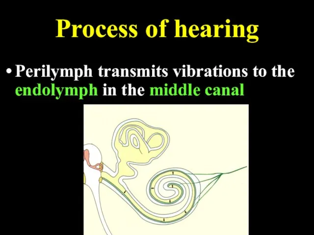
Perilymph transmits vibrations to the endolymph in the middle canal
Process of hearing
Perilymph transmits vibrations to the endolymph in the middle canal
Слайд 41Process of hearing
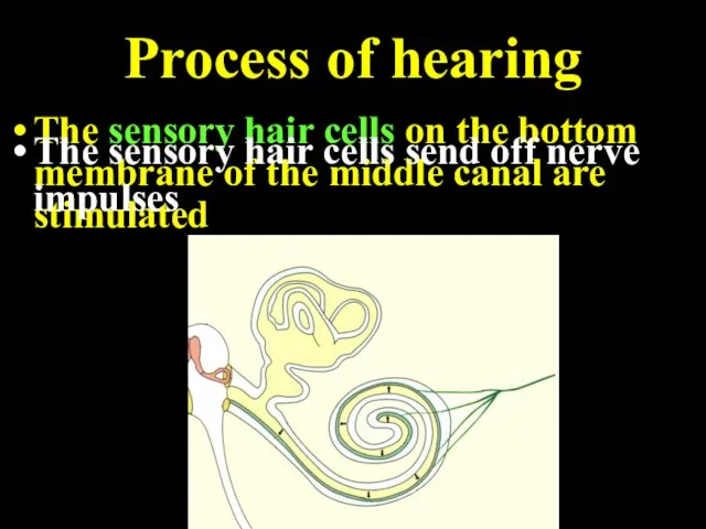
The sensory hair cells on the bottom membrane of the
Process of hearing
The sensory hair cells on the bottom membrane of the
Слайд 42Process of hearing
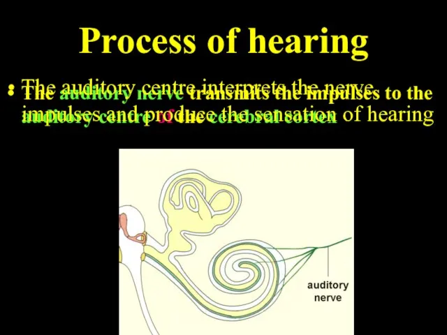
The auditory nerve transmits the impulses to the auditory centre
Process of hearing
The auditory nerve transmits the impulses to the auditory centre
Слайд 43Process of hearing
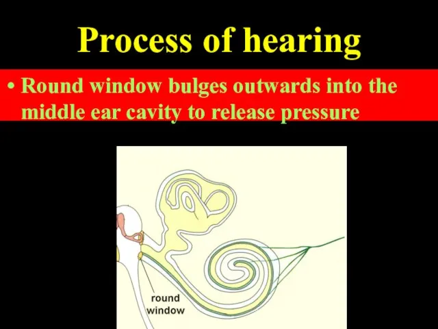
The vibrations of perilymph are transmitted to the round window
Round
Process of hearing
The vibrations of perilymph are transmitted to the round window
Round
Слайд 44All multicellular organisms have a skin composed of one or more layers.
THE
All multicellular organisms have a skin composed of one or more layers.
THE
Слайд 45Functions of Skin
It protects the inner layers of the body from physical
Functions of Skin
It protects the inner layers of the body from physical
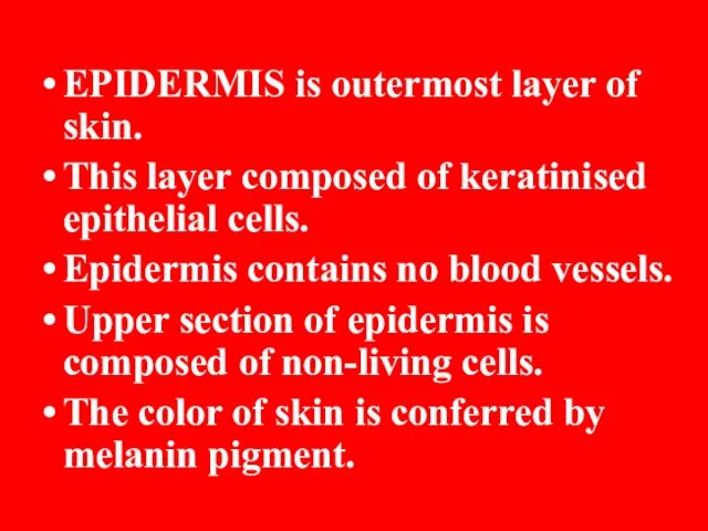
Слайд 46EPIDERMIS is outermost layer of skin.
This layer composed of keratinised epithelial cells.
Epidermis
EPIDERMIS is outermost layer of skin.
This layer composed of keratinised epithelial cells.
Epidermis
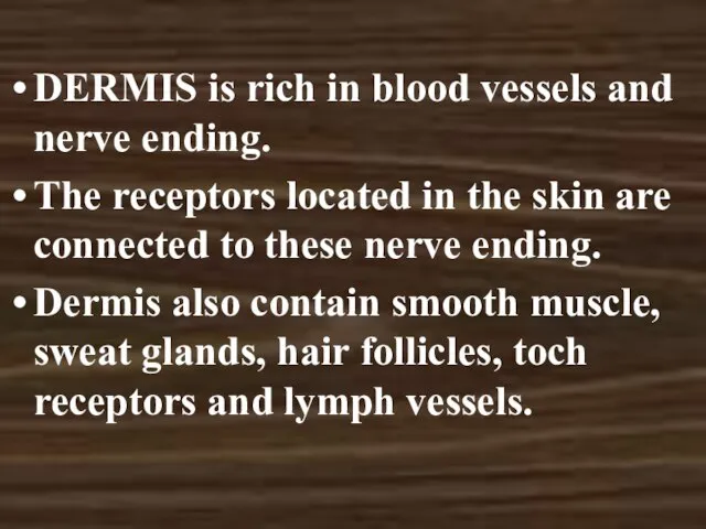
Слайд 47DERMIS is rich in blood vessels and nerve ending.
The receptors located in
DERMIS is rich in blood vessels and nerve ending.
The receptors located in
Слайд 49RECEPTORS
Meisner corpuscles: They are involved in reception of touch of the palm
RECEPTORS
Meisner corpuscles: They are involved in reception of touch of the palm
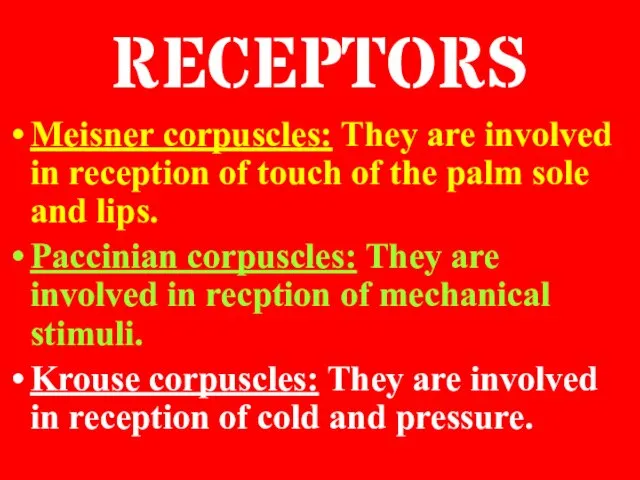
Слайд 50Ruffini corpuscles: They are involved in recption of heat, touch and pressure.
Sweat
Ruffini corpuscles: They are involved in recption of heat, touch and pressure.
Sweat
Слайд 52They are involved in removal of water, minerals, urea and other substances.
The
They are involved in removal of water, minerals, urea and other substances.
The
Слайд 53Nose is the organ of the body involved in botdh respiration and
Nose is the organ of the body involved in botdh respiration and
Слайд 55Smelling is fundemantal in the detection of food, maintenance of relationship, reproduction
Smelling is fundemantal in the detection of food, maintenance of relationship, reproduction
Слайд 57The surface of the tongue is covered with small projections called papillae.
There
The surface of the tongue is covered with small projections called papillae.
There
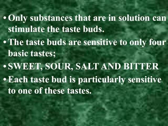
Слайд 58Only substances that are in solution can stimulate the taste buds.
The taste
Only substances that are in solution can stimulate the taste buds.
The taste
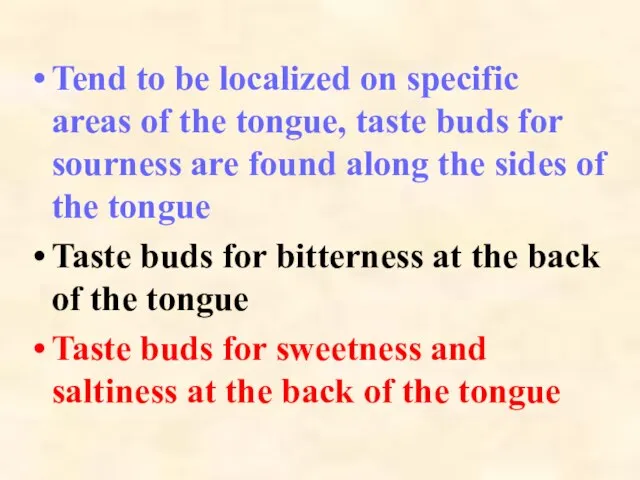
Слайд 59Tend to be localized on specific areas of the tongue, taste buds
Tend to be localized on specific areas of the tongue, taste buds
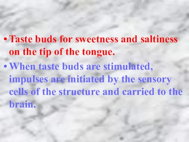
Слайд 60Taste buds for sweetness and saltiness on the tip of the tongue.
When
Taste buds for sweetness and saltiness on the tip of the tongue.
When
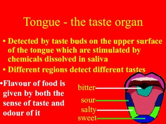
Слайд 61Tongue - the taste organ
Detected by taste buds on the upper surface
Tongue - the taste organ
Detected by taste buds on the upper surface
 Геном про и эукариот
 Поурочные планы
 Система построения уроков Иностранного языка на базе ИКТ Царькова О.А. Учитель английского языка МБОУ Новослободской основной
 ОБЪЕДИНЕННЫЕ КОММУНИКАЦИИ В УКРАИНЕСОСТОЯНИЕ И ПЕРСПЕКТИВЫ Национальная комиссия по вопросам регулирования связи УкраиныДепа
 Новое поколение RAID-контроллеров и HBA Unified Serial SATA/SAS и решенияна их основе
 Население Земли (7 класс)
 Церковь Покрова на Нерли
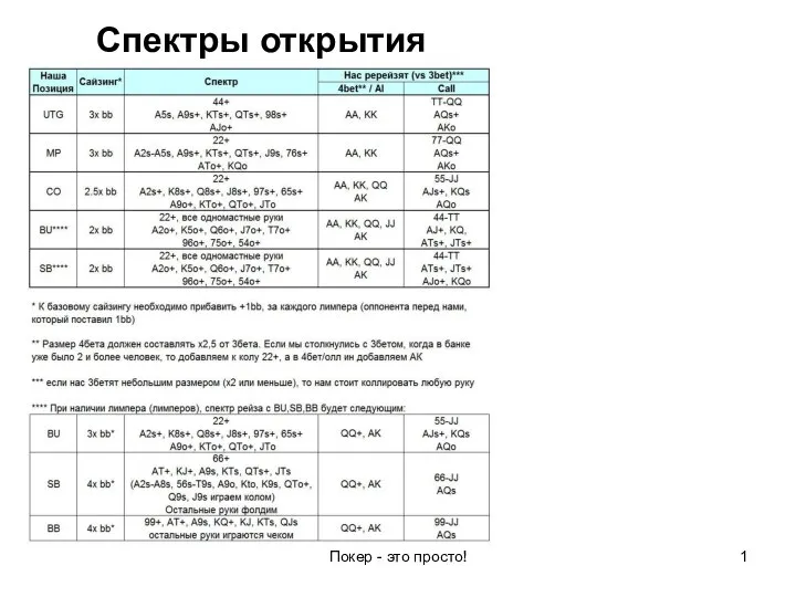 Спектры открытия. Покер
 Структура уголовного права РФ
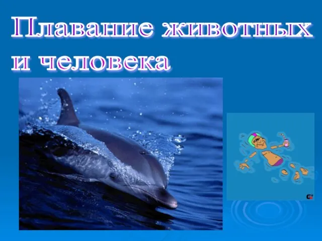 Плавание животных и человека
 Robert Koch
 Презентация на тему Танцы народов мира
 Источники финансирования. Особенности законодательства РФ. Виды инвесторов
 Алкогольные напитки
 Психика_Развитие_психики_Мозг_как_материальная_основа_психики_Сознание
 Травматизм: причины, профилактика, первая помощь
 4. Qui, je parle. Francais. 6 класс
 Презентация Серия He (lit-ion)
 Опыт использования ИКТ на уроках и во внеурочное время в начальной школе Волкова Татьяна Се
 Презентация на тему Опасные погодные явления
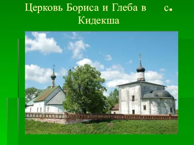 Церковь Бориса и Глеба (Кидекша)
 Foreign languages in our life
 Англия: сложный путь к величию и процветанию
 Что такое CDIO
 Презентация на тему Работаем с папой
 Л Кривые второго порядка
 Dance Heads
 Федор Иванович Тютчев