- Воспалительном процессе. Внутричерепной гипертензии Сосудистой патологии. Врожденной патологии

Содержание
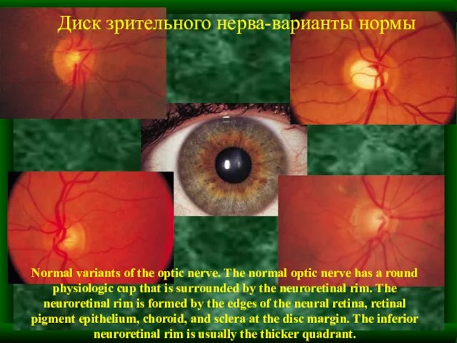
- 2. Normal variants of the optic nerve. The normal optic nerve has a round physiologic cup that
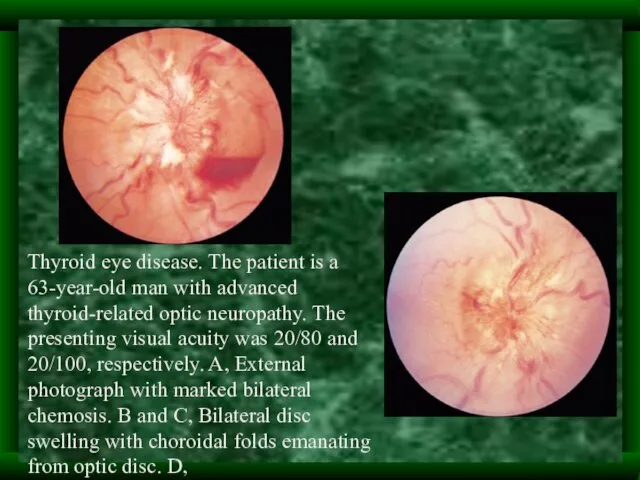
- 3. Thyroid eye disease. The patient is a 63-year-old man with advanced thyroid-related optic neuropathy. The presenting
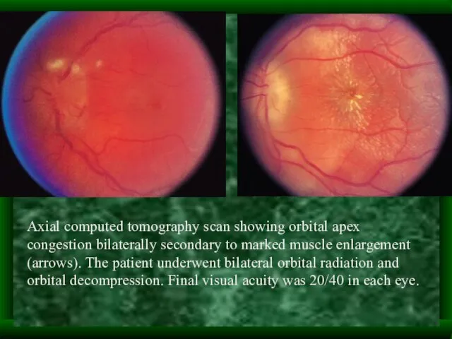
- 4. Axial computed tomography scan showing orbital apex congestion bilaterally secondary to marked muscle enlargement (arrows). The
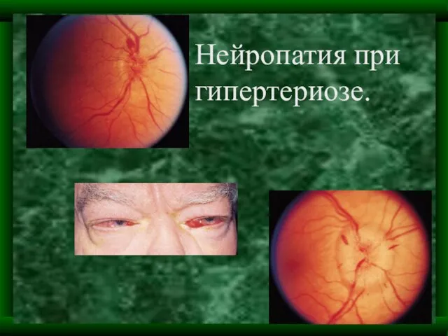
- 5. Нейропатия при гипертериозе.
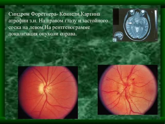
- 6. Синдром Форстнера- Кеннеди.Картина атрофии з.н. На правом глазу и застойного соска на левом.На рентгенограмме локализация опухоли
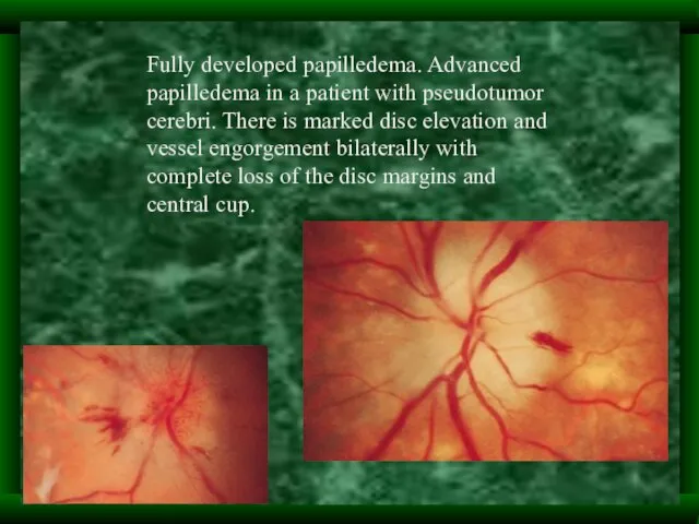
- 8. Fully developed papilledema. Advanced papilledema in a patient with pseudotumor cerebri. There is marked disc elevation
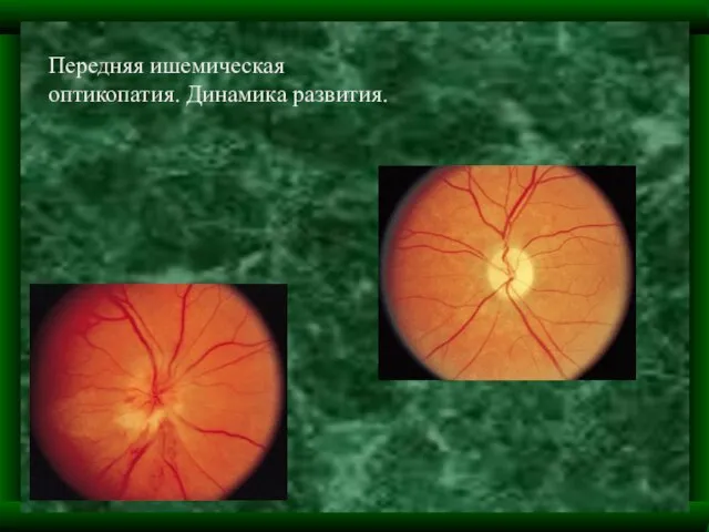
- 9. Передняя ишемическая оптикопатия. Динамика развития.
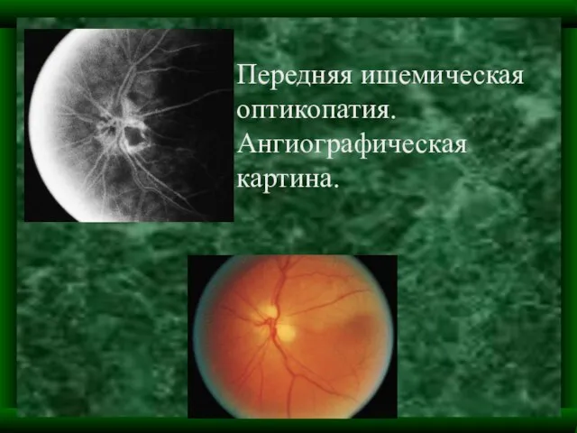
- 10. Передняя ишемическая оптикопатия. Ангиографическая картина.
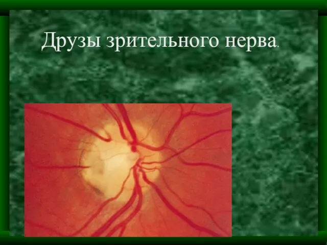
- 11. Друзы зрительного нерва.
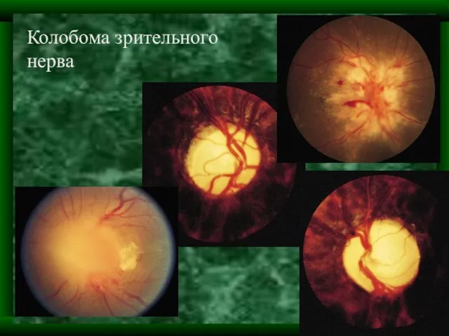
- 12. Колобома зрительного нерва
- 13. Благодарим за внимание. Надеемся, что дальнейшие выступления будут оформлены по образцу настоящей презентац или лутше. С
- 15. Скачать презентацию
Слайд 3Thyroid eye disease. The patient is a 63-year-old man with advanced thyroid-related
Thyroid eye disease. The patient is a 63-year-old man with advanced thyroid-related
Слайд 4Axial computed tomography scan showing orbital apex congestion bilaterally secondary to marked
Axial computed tomography scan showing orbital apex congestion bilaterally secondary to marked
Слайд 5Нейропатия при гипертериозе.
Нейропатия при гипертериозе.
Слайд 6Синдром Форстнера- Кеннеди.Картина атрофии з.н. На правом глазу и застойного соска на
Синдром Форстнера- Кеннеди.Картина атрофии з.н. На правом глазу и застойного соска на
Слайд 8Fully developed papilledema. Advanced papilledema in a patient with pseudotumor cerebri. There
Fully developed papilledema. Advanced papilledema in a patient with pseudotumor cerebri. There
Слайд 9Передняя ишемическая оптикопатия. Динамика развития.
Передняя ишемическая оптикопатия. Динамика развития.
Слайд 10Передняя ишемическая оптикопатия. Ангиографическая картина.
Передняя ишемическая оптикопатия. Ангиографическая картина.
Слайд 11Друзы зрительного нерва.
Друзы зрительного нерва.
Слайд 12Колобома зрительного нерва
Колобома зрительного нерва
Слайд 13Благодарим за внимание.
Надеемся, что дальнейшие выступления будут оформлены по образцу настоящей презентац
Благодарим за внимание.
Надеемся, что дальнейшие выступления будут оформлены по образцу настоящей презентац
 Научно-исследовательский проект
 Учреждения социального обслуживания населения, их виды и специфика деятельности
 эконом-класс
 Анализ целевых показателей ФЦП за 2009 год(научно-образовательные центры)
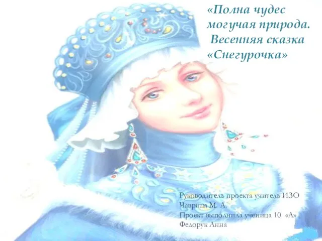 Полна чудес могучая природа. Весенняя сказка «Снегурочка»
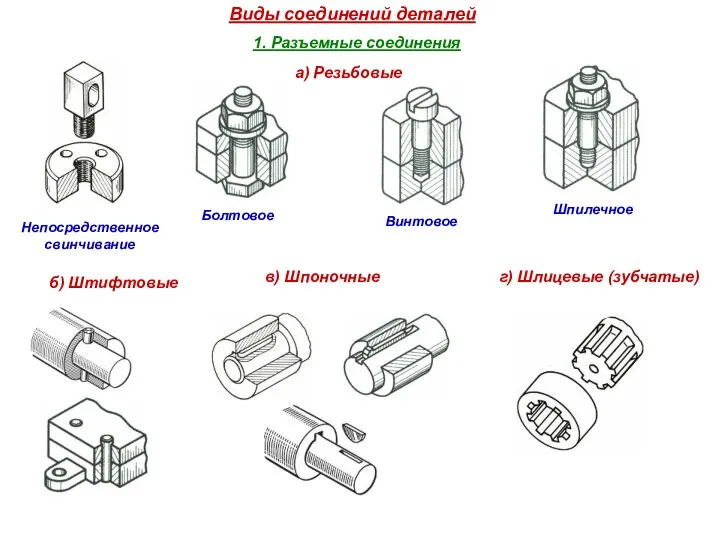 Виды соединений деталей
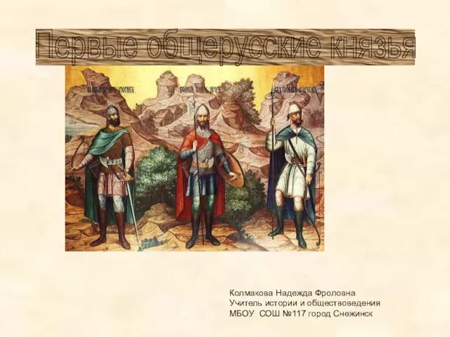 Первые общерусские князья
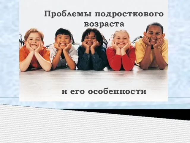 Презентация на тему: Проблемы подросткового возраста и его особенности
 Сказка про осень
 Открытый классный час на тему:
 Сытнее овоща нет в мире
 Презентация на тему Герои древней Руси
 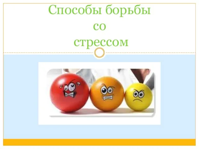 Способы борьбы со стрессом
 Презентация на тему Обитатели морей и океанов
  Динамично развивающаяся компания по продаже автозапчастей А-Detal.ru
 "Темперамент"
 Кишечные инфекции
 styles
 Материалы для рисования. Вопросы
 Класс элементарных функций и их графики
 Презентация на тему Разделительные Ъ и Ь знаки
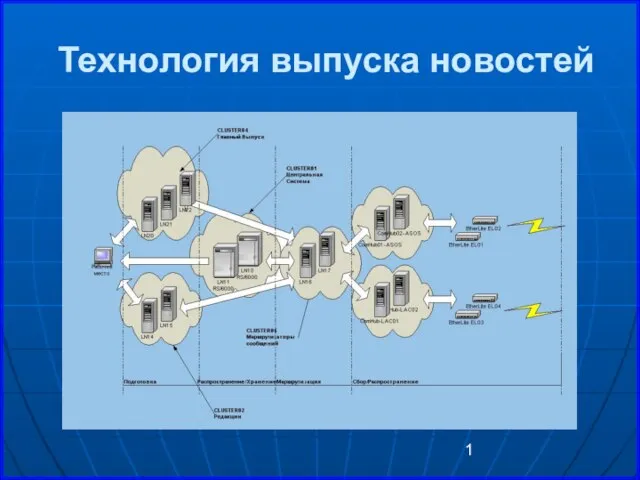 Технология выпуска новостей
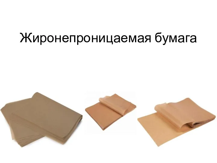 Жиронепроницаемая бумага. Пергамент
 1п екон
 We like the place we live
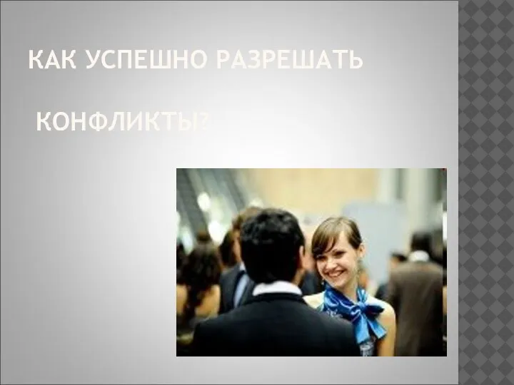 Как успешно разрешать конфликты?
 Правильная осанка - залог здоровья
 Государство в политической системе (11 класс)