- Animal Development

Содержание
- 2. Overview: A Body-Building Plan It is difficult to imagine that each of us began life as
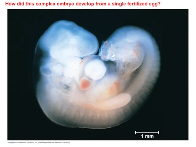
- 3. How did this complex embryo develop from a single fertilized egg? 1 mm
- 4. Development is determined by the zygote’s genome and molecules in the egg cytoplasm called Cytoplasmic determinants.
- 5. After fertilization, embryonic development proceeds through cleavage, gastrulation, and organogenesis Important events regulating development occur during
- 6. Fertilization: sperm + egg = zygote n + n = 2n Fertilization brings the haploid nuclei
- 7. The Acrosomal Reaction The acrosomal reaction is triggered when the sperm meets the egg. The acrosome
- 8. The acrosomal and cortical reactions during sea urchin fertilization Basal body (centriole) Sperm head Sperm-binding receptors
- 9. The Cortical Reaction Fusion of egg and sperm also initiates the cortical reaction: This reaction induces
- 10. Activation of the Egg The sharp rise in Ca2+ in the egg’s cytosol increases the rates
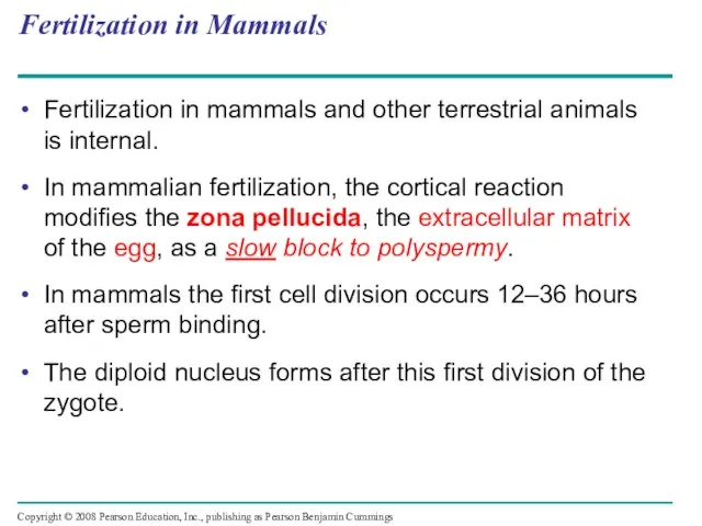
- 11. Fertilization in Mammals Fertilization in mammals and other terrestrial animals is internal. In mammalian fertilization, the
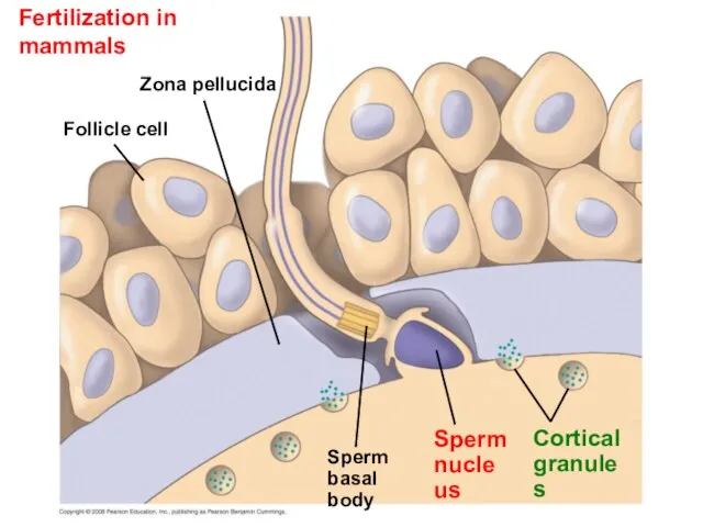
- 12. Fertilization in mammals Follicle cell Zona pellucida Cortical granules Sperm nucleus Sperm basal body
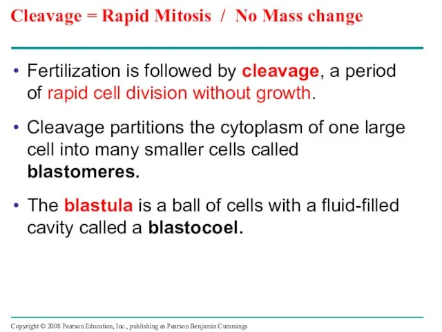
- 13. Cleavage = Rapid Mitosis / No Mass change Fertilization is followed by cleavage, a period of
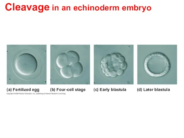
- 14. Cleavage in an echinoderm embryo (a) Fertilized egg (b) Four-cell stage (c) Early blastula (d) Later
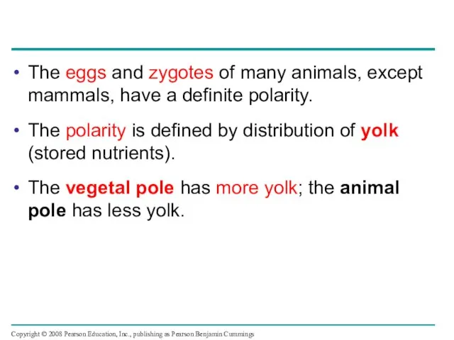
- 15. The eggs and zygotes of many animals, except mammals, have a definite polarity. The polarity is
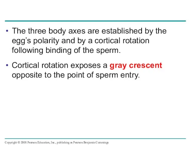
- 16. The three body axes are established by the egg’s polarity and by a cortical rotation following
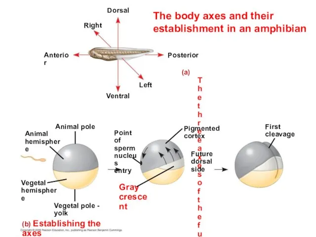
- 17. The body axes and their establishment in an amphibian (a) The three axes of the fully
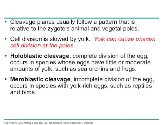
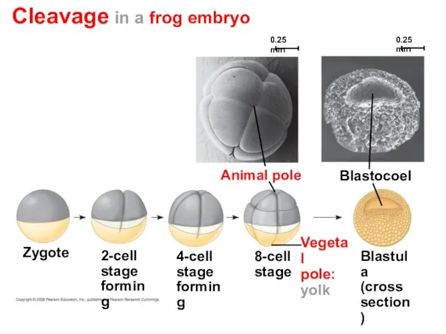
- 18. Cleavage planes usually follow a pattern that is relative to the zygote’s animal and vegetal poles.
- 19. Cleavage in a frog embryo Blastula (cross section) Blastocoel Animal pole 4-cell stage forming 2-cell stage
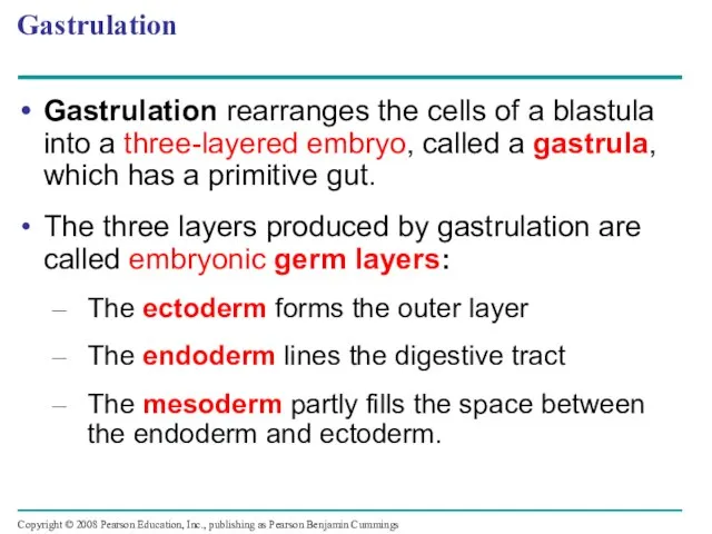
- 20. Gastrulation Gastrulation rearranges the cells of a blastula into a three-layered embryo, called a gastrula, which
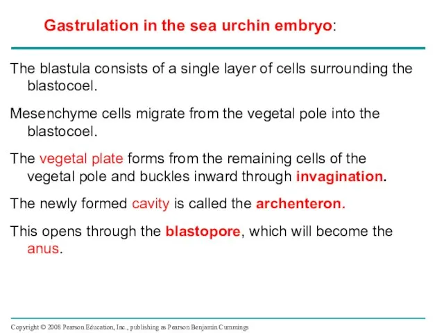
- 21. The blastula consists of a single layer of cells surrounding the blastocoel. Mesenchyme cells migrate from
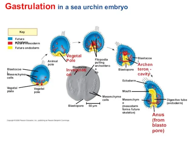
- 22. Gastrulation in a sea urchin embryo Future ectoderm Key Future endoderm Digestive tube (endoderm) Mouth Ectoderm
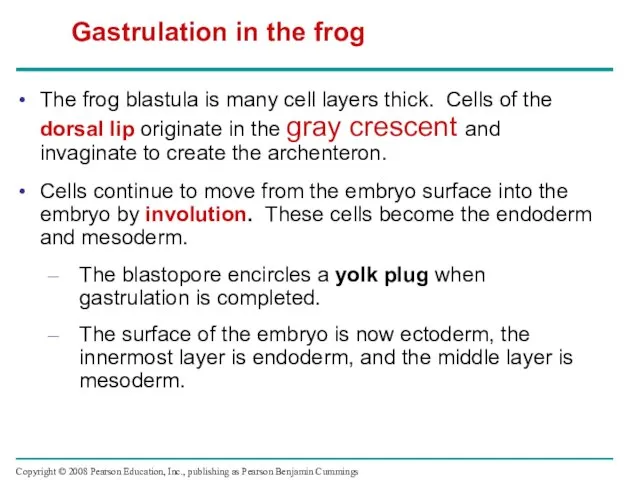
- 23. The frog blastula is many cell layers thick. Cells of the dorsal lip originate in the
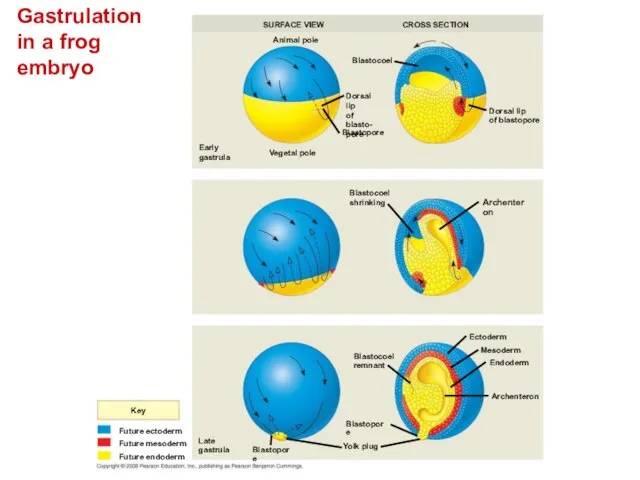
- 24. Gastrulation in a frog embryo Future ectoderm Key Future endoderm Future mesoderm SURFACE VIEW Animal pole
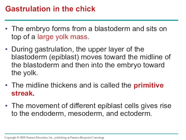
- 25. The embryo forms from a blastoderm and sits on top of a large yolk mass. During
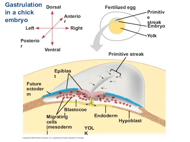
- 26. Gastrulation in a chick embryo Endoderm Future ectoderm Migrating cells (mesoderm) Hypoblast Dorsal Fertilized egg Blastocoel
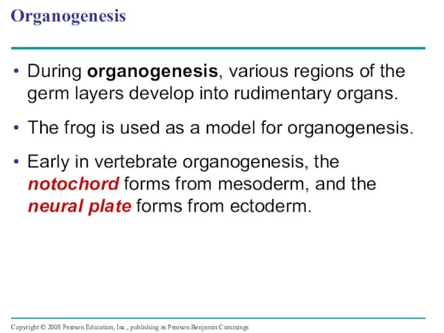
- 27. Organogenesis During organogenesis, various regions of the germ layers develop into rudimentary organs. The frog is
- 28. Early organogenesis in a frog embryo Neural folds Tail bud Neural tube (b) Neural tube formation
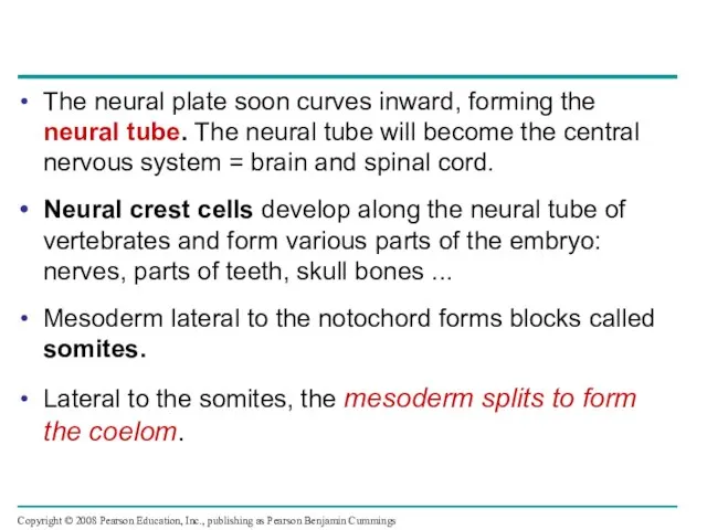
- 29. The neural plate soon curves inward, forming the neural tube. The neural tube will become the
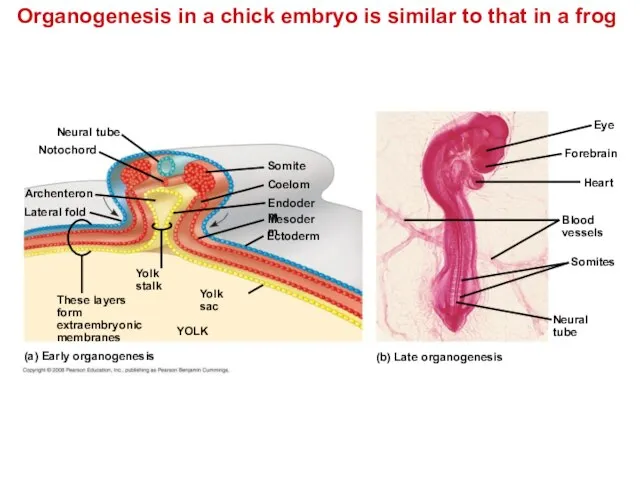
- 30. Organogenesis in a chick embryo is similar to that in a frog Endoderm (a) Early organogenesis
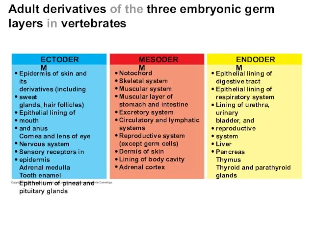
- 31. Adult derivatives of the three embryonic germ layers in vertebrates ECTODERM MESODERM ENDODERM Epidermis of skin
- 32. Developmental Adaptations of Amniotes Embryos of birds, other reptiles, and mammals develop in a fluid-filled sac
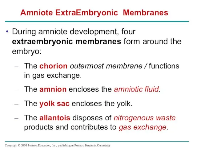
- 33. During amniote development, four extraembryonic membranes form around the embryo: The chorion outermost membrane / functions
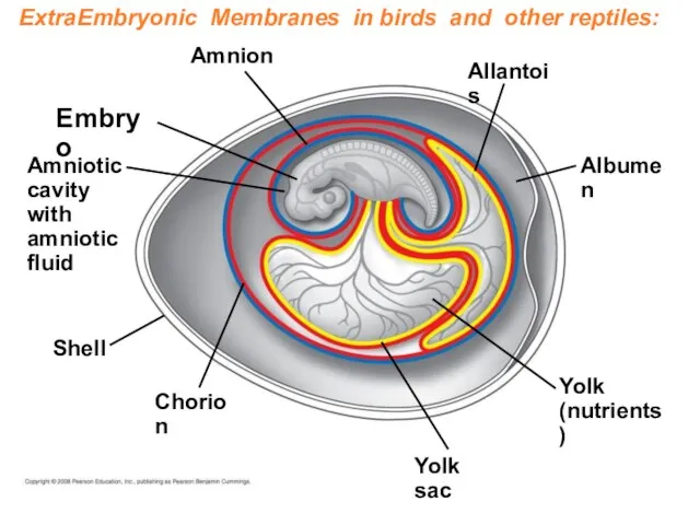
- 34. ExtraEmbryonic Membranes in birds and other reptiles: Embryo Amnion Amniotic cavity with amniotic fluid Shell Chorion
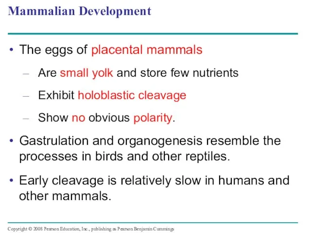
- 35. Mammalian Development The eggs of placental mammals Are small yolk and store few nutrients Exhibit holoblastic
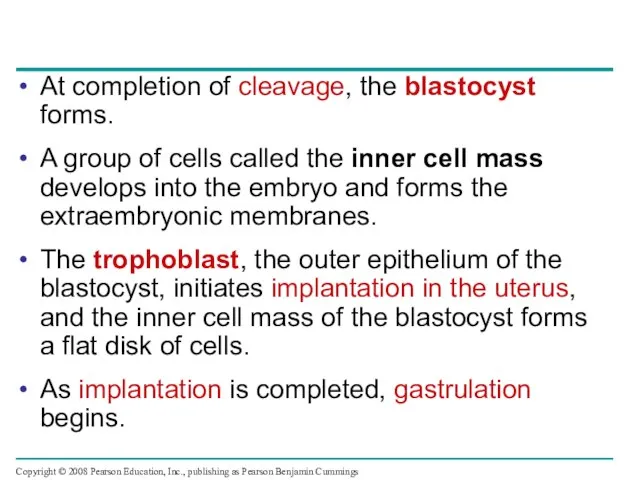
- 36. At completion of cleavage, the blastocyst forms. A group of cells called the inner cell mass
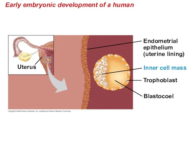
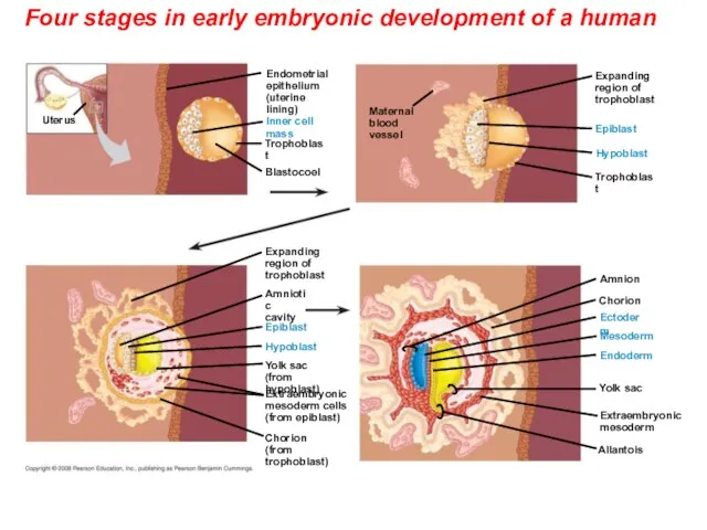
- 37. Early embryonic development of a human Blastocoel Trophoblast Uterus Endometrial epithelium (uterine lining) Inner cell mass
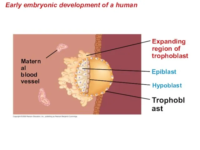
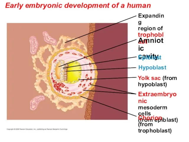
- 38. Early embryonic development of a human Trophoblast Hypoblast Maternal blood vessel Expanding region of trophoblast Epiblast
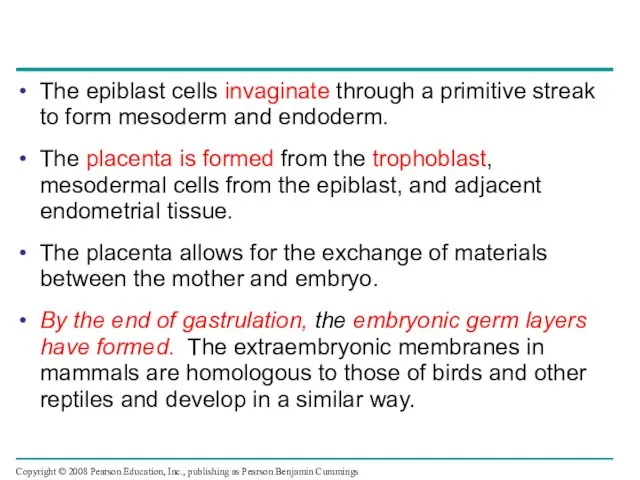
- 39. The epiblast cells invaginate through a primitive streak to form mesoderm and endoderm. The placenta is
- 40. Early embryonic development of a human Yolk sac (from hypoblast) Hypoblast Expanding region of trophoblast Amniotic
- 41. Early embryonic development of a human Yolk sac Mesoderm Amnion Chorion Ectoderm Extraembryonic mesoderm Atlantois Endoderm
- 42. Four stages in early embryonic development of a human Yolk sac Mesoderm Amnion Chorion Ectoderm Extraembryonic
- 43. Morphogenesis in animals involves specific changes in cell shape, position, and adhesion Morphogenesis is a major
- 44. The Cytoskeleton, Cell Motility, and Convergent Extension Changes in cell shape usually involve reorganization of the
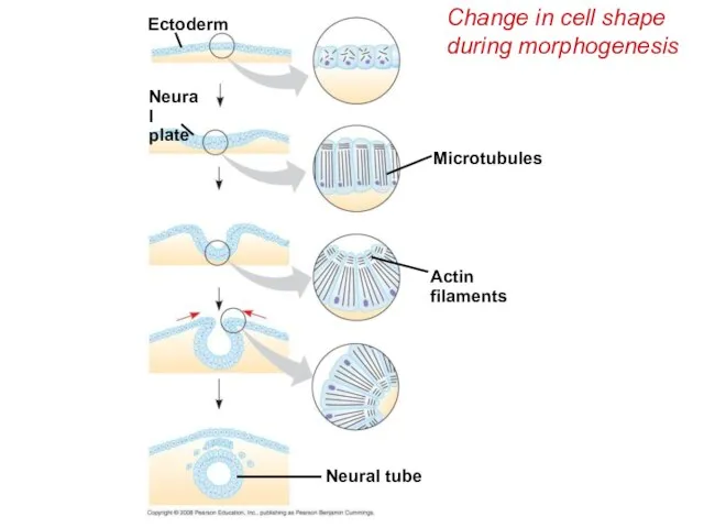
- 45. Change in cell shape during morphogenesis Neural tube Actin filaments Microtubules Ectoderm Neural plate
- 46. The cytoskeleton also drives cell migration, or cell crawling, the active movement of cells. In gastrulation,
- 47. Role of Cell Adhesion Molecules and the Extracellular Matrix Cell adhesion molecules located on cell surfaces
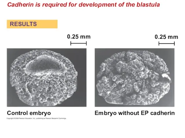
- 48. Cadherin is required for development of the blastula Control embryo Embryo without EP cadherin 0.25 mm
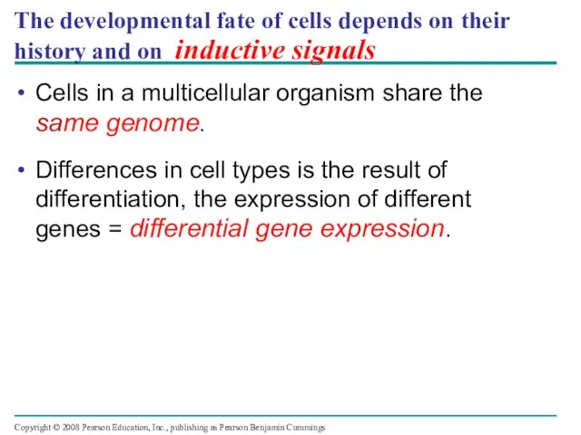
- 49. The developmental fate of cells depends on their history and on inductive signals Cells in a
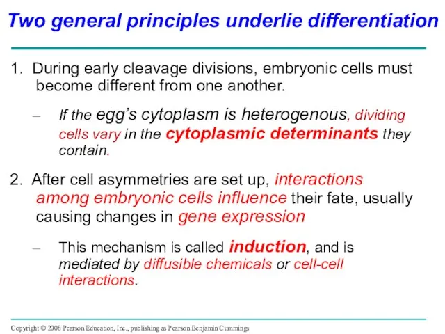
- 50. 1. During early cleavage divisions, embryonic cells must become different from one another. If the egg’s
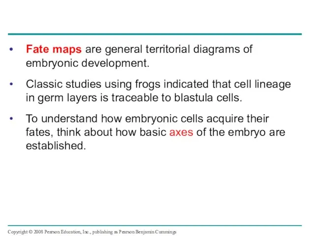
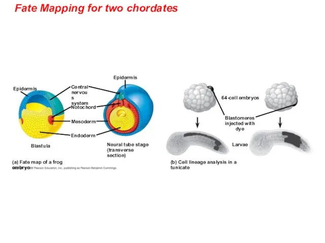
- 51. Fate maps are general territorial diagrams of embryonic development. Classic studies using frogs indicated that cell
- 52. Fate Mapping for two chordates Epidermis (b) Cell lineage analysis in a tunicate (a) Fate map
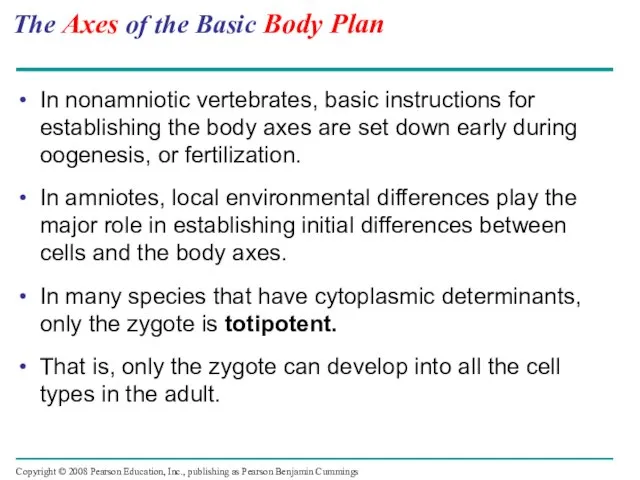
- 53. The Axes of the Basic Body Plan In nonamniotic vertebrates, basic instructions for establishing the body
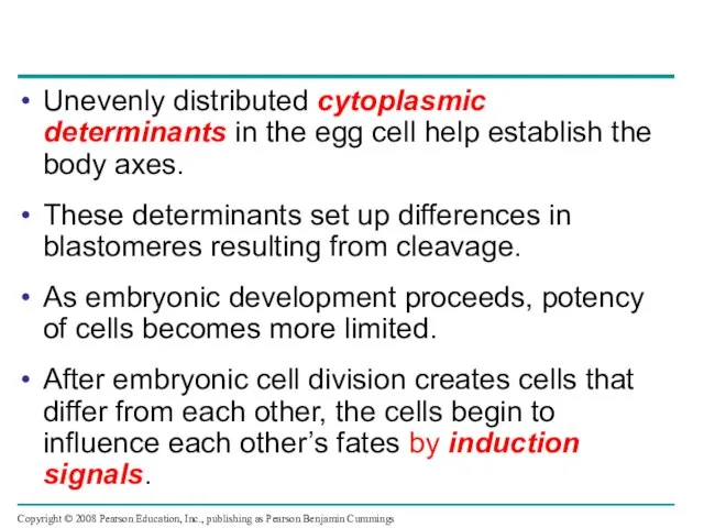
- 54. Unevenly distributed cytoplasmic determinants in the egg cell help establish the body axes. These determinants set
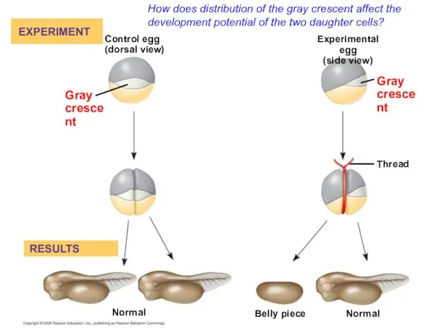
- 55. How does distribution of the gray crescent affect the development potential of the two daughter cells?
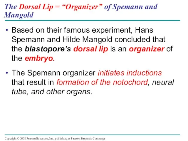
- 56. The Dorsal Lip = “Organizer” of Spemann and Mangold Based on their famous experiment, Hans Spemann
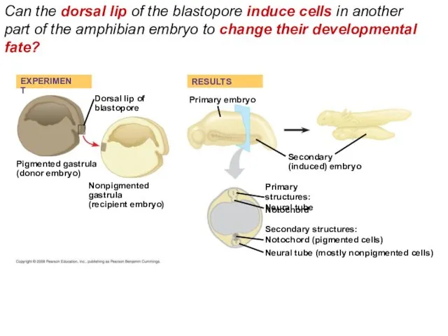
- 57. Can the dorsal lip of the blastopore induce cells in another part of the amphibian embryo
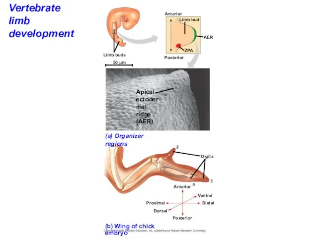
- 58. Formation of the Vertebrate Limb Inductive signals play a major role in pattern formation, development of
- 59. The wings and legs of chicks, like all vertebrate limbs, begin as bumps of tissue called
- 60. Vertebrate limb development (a) Organizer regions Apical ectodermal ridge (AER) Digits Limb buds (b) Wing of
- 61. Signal molecules produced by inducing cells influence gene expression in cells receiving them. Signal molecules lead
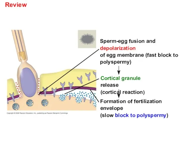
- 62. Review Sperm-egg fusion and depolarization of egg membrane (fast block to polyspermy) Cortical granule release (cortical
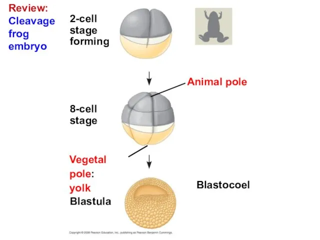
- 63. Review: Cleavage frog embryo Blastocoel Animal pole 2-cell stage forming 8-cell stage Blastula Vegetal pole: yolk
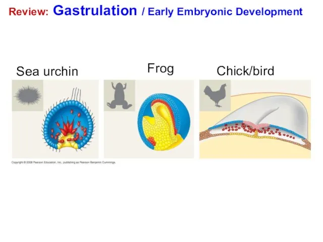
- 64. Review: Gastrulation / Early Embryonic Development Sea urchin Frog Chick/bird
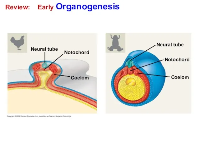
- 65. Review: Early Organogenesis Neural tube Coelom Notochord Coelom Notochord Neural tube
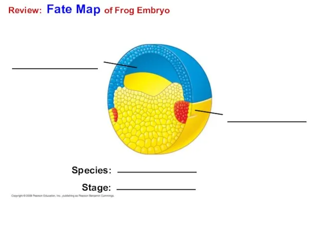
- 66. Review: Fate Map of Frog Embryo Species: Stage:
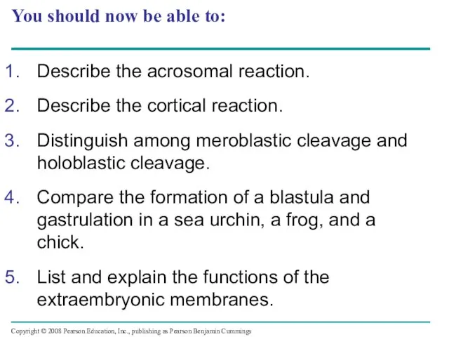
- 67. You should now be able to: Describe the acrosomal reaction. Describe the cortical reaction. Distinguish among
- 69. Скачать презентацию
Слайд 3How did this complex embryo develop from a single fertilized egg?
1 mm
How did this complex embryo develop from a single fertilized egg?
1 mm
Слайд 4Development is determined by the zygote’s genome and molecules in the egg
Development is determined by the zygote’s genome and molecules in the egg
Слайд 5After fertilization, embryonic development proceeds through cleavage, gastrulation, and organogenesis
Important events regulating
After fertilization, embryonic development proceeds through cleavage, gastrulation, and organogenesis
Important events regulating
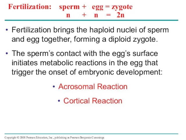
Слайд 6Fertilization: sperm + egg = zygote
n + n = 2n
Fertilization brings
Fertilization: sperm + egg = zygote
n + n = 2n
Fertilization brings
Слайд 7The Acrosomal Reaction
The acrosomal reaction is triggered when the sperm meets the
The Acrosomal Reaction
The acrosomal reaction is triggered when the sperm meets the

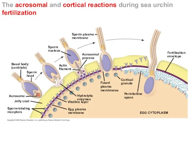
Слайд 8The acrosomal and cortical reactions during sea urchin fertilization
Basal body
(centriole)
Sperm
head
Sperm-binding
receptors
Acrosome
Jelly coat
Vitelline layer
Egg
The acrosomal and cortical reactions during sea urchin fertilization
Basal body
(centriole)
Sperm
head
Sperm-binding
receptors
Acrosome
Jelly coat
Vitelline layer
Egg
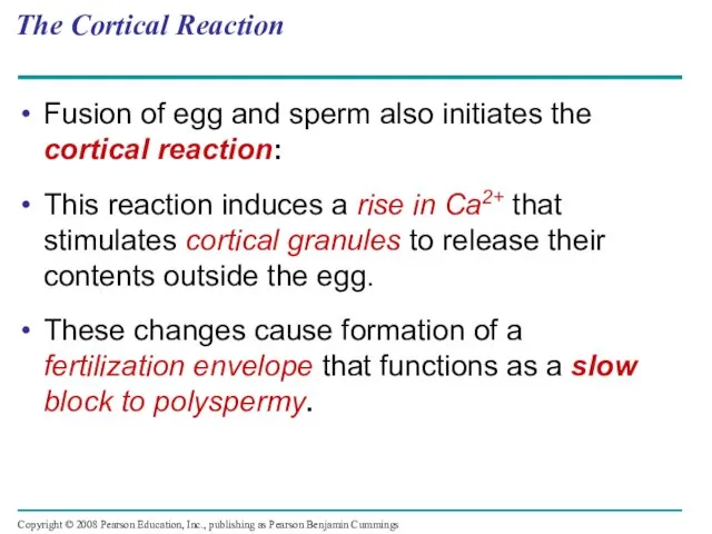
Слайд 9The Cortical Reaction
Fusion of egg and sperm also initiates the cortical reaction:
This
The Cortical Reaction
Fusion of egg and sperm also initiates the cortical reaction:
This
Слайд 10Activation of the Egg
The sharp rise in Ca2+ in the egg’s cytosol
Activation of the Egg
The sharp rise in Ca2+ in the egg’s cytosol
Слайд 11Fertilization in Mammals
Fertilization in mammals and other terrestrial animals is internal.
In mammalian
Fertilization in Mammals
Fertilization in mammals and other terrestrial animals is internal.
In mammalian
Слайд 12Fertilization in mammals
Follicle cell
Zona pellucida
Cortical
granules
Sperm
nucleus
Sperm
basal body
Fertilization in mammals
Follicle cell
Zona pellucida
Cortical
granules
Sperm
nucleus
Sperm
basal body
Слайд 13Cleavage = Rapid Mitosis / No Mass change
Fertilization is followed by cleavage,
Cleavage = Rapid Mitosis / No Mass change
Fertilization is followed by cleavage,
Слайд 14Cleavage in an echinoderm embryo
(a) Fertilized egg
(b) Four-cell stage
(c) Early blastula
(d) Later
Cleavage in an echinoderm embryo
(a) Fertilized egg
(b) Four-cell stage
(c) Early blastula
(d) Later
Слайд 15The eggs and zygotes of many animals, except mammals, have a definite
The eggs and zygotes of many animals, except mammals, have a definite
Слайд 16The three body axes are established by the egg’s polarity and by
The three body axes are established by the egg’s polarity and by
Слайд 17The body axes and their establishment in an amphibian
(a) The three axes
The body axes and their establishment in an amphibian
(a) The three axes
Слайд 18Cleavage planes usually follow a pattern that is relative to the zygote’s
Cleavage planes usually follow a pattern that is relative to the zygote’s
Слайд 19Cleavage in a frog embryo
Blastula
(cross
section)
Blastocoel
Animal pole
4-cell
stage
forming
2-cell
stage
forming
Zygote
8-cell
stage
Vegetal
pole:
yolk
0.25 mm
0.25 mm
Cleavage in a frog embryo
Blastula
(cross
section)
Blastocoel
Animal pole
4-cell
stage
forming
2-cell
stage
forming
Zygote
8-cell
stage
Vegetal
pole:
yolk
0.25 mm
0.25 mm
Слайд 20Gastrulation
Gastrulation rearranges the cells of a blastula into a three-layered embryo, called
Gastrulation
Gastrulation rearranges the cells of a blastula into a three-layered embryo, called
Слайд 21The blastula consists of a single layer of cells surrounding the blastocoel.
Mesenchyme
The blastula consists of a single layer of cells surrounding the blastocoel.
Mesenchyme
Слайд 22Gastrulation in a sea urchin embryo
Future ectoderm
Key
Future endoderm
Digestive tube
(endoderm)
Mouth
Ectoderm
Mesenchyme
(mesoderm
forms future
skeleton)
Anus (from
Gastrulation in a sea urchin embryo
Future ectoderm
Key
Future endoderm
Digestive tube
(endoderm)
Mouth
Ectoderm
Mesenchyme
(mesoderm
forms future
skeleton)
Anus (from
Слайд 23The frog blastula is many cell layers thick. Cells of the dorsal
The frog blastula is many cell layers thick. Cells of the dorsal
Слайд 24Gastrulation in a frog embryo
Future ectoderm
Key
Future endoderm
Future mesoderm
SURFACE VIEW
Animal pole
Vegetal pole
Early
gastrula
Blastopore
Blastocoel
Dorsal lip
of
Gastrulation in a frog embryo
Future ectoderm
Key
Future endoderm
Future mesoderm
SURFACE VIEW
Animal pole
Vegetal pole
Early
gastrula
Blastopore
Blastocoel
Dorsal lip of
Слайд 25The embryo forms from a blastoderm and sits on top of a
The embryo forms from a blastoderm and sits on top of a
Слайд 26Gastrulation in a chick embryo
Endoderm
Future
ectoderm
Migrating
cells
(mesoderm)
Hypoblast
Dorsal
Fertilized egg
Blastocoel
YOLK
Anterior
Right
Ventral
Posterior
Left
Epiblast
Primitive streak
Embryo
Yolk
Primitive
streak
Gastrulation in a chick embryo
Endoderm
Future
ectoderm
Migrating
cells
(mesoderm)
Hypoblast
Dorsal
Fertilized egg
Blastocoel
YOLK
Anterior
Right
Ventral
Posterior
Left
Epiblast
Primitive streak
Embryo
Yolk
Primitive
streak
Слайд 27Organogenesis
During organogenesis, various regions of the germ layers develop into rudimentary organs.
The
Organogenesis
During organogenesis, various regions of the germ layers develop into rudimentary organs.
The
Слайд 28Early organogenesis in a frog embryo
Neural folds
Tail bud
Neural tube
(b) Neural tube formation
Neural
fold
Neural
Early organogenesis in a frog embryo
Neural folds
Tail bud
Neural tube
(b) Neural tube formation
Neural
fold
Neural
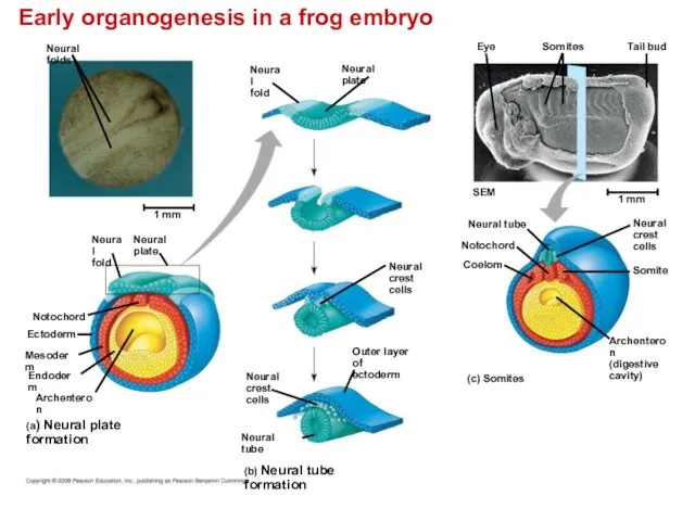
Слайд 29The neural plate soon curves inward, forming the neural tube. The neural
The neural plate soon curves inward, forming the neural tube. The neural
Слайд 30Organogenesis in a chick embryo is similar to that in a frog
Endoderm
(a)
Organogenesis in a chick embryo is similar to that in a frog
Endoderm
(a)
Слайд 31Adult derivatives of the three embryonic germ layers in vertebrates
ECTODERM
MESODERM
ENDODERM
Epidermis of skin
Adult derivatives of the three embryonic germ layers in vertebrates
ECTODERM
MESODERM
ENDODERM
Epidermis of skin
Слайд 32Developmental Adaptations of Amniotes
Embryos of birds, other reptiles, and mammals develop in
Developmental Adaptations of Amniotes
Embryos of birds, other reptiles, and mammals develop in
Слайд 33During amniote development, four extraembryonic membranes form around the embryo:
The chorion outermost
During amniote development, four extraembryonic membranes form around the embryo:
The chorion outermost
Слайд 34ExtraEmbryonic Membranes in birds and other reptiles:
Embryo
Amnion
Amniotic
cavity
with
amniotic
fluid
Shell
Chorion
Yolk sac
Yolk
(nutrients)
Allantois
Albumen
ExtraEmbryonic Membranes in birds and other reptiles:
Embryo
Amnion
Amniotic
cavity
with
amniotic
fluid
Shell
Chorion
Yolk sac
Yolk
(nutrients)
Allantois
Albumen
Слайд 35Mammalian Development
The eggs of placental mammals
Are small yolk and store few nutrients
Exhibit
Mammalian Development
The eggs of placental mammals
Are small yolk and store few nutrients
Exhibit
Слайд 36At completion of cleavage, the blastocyst forms.
A group of cells called the
At completion of cleavage, the blastocyst forms.
A group of cells called the
Слайд 37Early embryonic development of a human
Blastocoel
Trophoblast
Uterus
Endometrial
epithelium
(uterine lining)
Inner cell mass
Early embryonic development of a human
Blastocoel
Trophoblast
Uterus
Endometrial
epithelium
(uterine lining)
Inner cell mass
Слайд 38Early embryonic development of a human
Trophoblast
Hypoblast
Maternal
blood
vessel
Expanding
region of
trophoblast
Epiblast
Early embryonic development of a human
Trophoblast
Hypoblast
Maternal
blood
vessel
Expanding
region of
trophoblast
Epiblast
Слайд 39The epiblast cells invaginate through a primitive streak to form mesoderm and
The epiblast cells invaginate through a primitive streak to form mesoderm and
Слайд 40Early embryonic development of a human
Yolk sac (from
hypoblast)
Hypoblast
Expanding
region of
trophoblast
Amniotic
cavity
Epiblast
Extraembryonic
mesoderm cells
(from epiblast)
Chorion (from
trophoblast)
Early embryonic development of a human
Yolk sac (from
hypoblast)
Hypoblast
Expanding
region of
trophoblast
Amniotic
cavity
Epiblast
Extraembryonic
mesoderm cells
(from epiblast)
Chorion (from
trophoblast)
Слайд 41 Early embryonic development of a human
Yolk sac
Mesoderm
Amnion
Chorion
Ectoderm
Extraembryonic
mesoderm
Atlantois
Endoderm
Early embryonic development of a human
Yolk sac
Mesoderm
Amnion
Chorion
Ectoderm
Extraembryonic
mesoderm
Atlantois
Endoderm

Слайд 42Four stages in early embryonic development of a human
Yolk sac
Mesoderm
Amnion
Chorion
Ectoderm
Extraembryonic
mesoderm
Trophoblast
Endoderm
Hypoblast
Expanding
region of
trophoblast
Epiblast
Maternal
blood
vessel
Allantois
Trophoblast
Hypoblast
Endometrial
epithelium
(uterine
Four stages in early embryonic development of a human
Yolk sac
Mesoderm
Amnion
Chorion
Ectoderm
Extraembryonic
mesoderm
Trophoblast
Endoderm
Hypoblast
Expanding
region of
trophoblast
Epiblast
Maternal
blood
vessel
Allantois
Trophoblast
Hypoblast
Endometrial epithelium (uterine
Слайд 43Morphogenesis in animals involves specific changes in cell shape, position, and adhesion
Morphogenesis
Morphogenesis in animals involves specific changes in cell shape, position, and adhesion
Morphogenesis
Слайд 44The Cytoskeleton, Cell Motility, and Convergent Extension
Changes in cell shape usually involve
The Cytoskeleton, Cell Motility, and Convergent Extension
Changes in cell shape usually involve
Слайд 45Change in cell shape during morphogenesis
Neural tube
Actin filaments
Microtubules
Ectoderm
Neural
plate
Change in cell shape during morphogenesis
Neural tube
Actin filaments
Microtubules
Ectoderm
Neural
plate
Слайд 46The cytoskeleton also drives cell migration, or cell crawling, the active movement
The cytoskeleton also drives cell migration, or cell crawling, the active movement
Слайд 47Role of Cell Adhesion Molecules and the Extracellular Matrix
Cell adhesion molecules located
Role of Cell Adhesion Molecules and the Extracellular Matrix
Cell adhesion molecules located
Слайд 48 Cadherin is required for development of the blastula
Control embryo
Embryo without
Cadherin is required for development of the blastula
Control embryo
Embryo without
Слайд 49The developmental fate of cells depends on their history and on inductive
The developmental fate of cells depends on their history and on inductive
Слайд 501. During early cleavage divisions, embryonic cells must become different from one
1. During early cleavage divisions, embryonic cells must become different from one
Слайд 51Fate maps are general territorial diagrams of embryonic development.
Classic studies using frogs
Fate maps are general territorial diagrams of embryonic development.
Classic studies using frogs
Слайд 52 Fate Mapping for two chordates
Epidermis
(b) Cell lineage analysis in a tunicate
(a)
Fate Mapping for two chordates
Epidermis
(b) Cell lineage analysis in a tunicate
(a)
Слайд 53The Axes of the Basic Body Plan
In nonamniotic vertebrates, basic instructions for
The Axes of the Basic Body Plan
In nonamniotic vertebrates, basic instructions for
Слайд 54Unevenly distributed cytoplasmic determinants in the egg cell help establish the body
Unevenly distributed cytoplasmic determinants in the egg cell help establish the body
Слайд 55How does distribution of the gray crescent affect the development potential of
How does distribution of the gray crescent affect the development potential of
Слайд 56The Dorsal Lip = “Organizer” of Spemann and Mangold
Based on their famous
The Dorsal Lip = “Organizer” of Spemann and Mangold
Based on their famous
Слайд 57Can the dorsal lip of the blastopore induce cells in another part
Can the dorsal lip of the blastopore induce cells in another part
Слайд 58Formation of the Vertebrate Limb
Inductive signals play a major role in
Formation of the Vertebrate Limb
Inductive signals play a major role in
Слайд 59The wings and legs of chicks, like all vertebrate limbs, begin as
The wings and legs of chicks, like all vertebrate limbs, begin as
Слайд 60Vertebrate limb development
(a) Organizer regions
Apical
ectodermal
ridge (AER)
Digits
Limb buds
(b) Wing of chick embryo
Posterior
Anterior
Limb bud
AER
ZPA
50
Vertebrate limb development
(a) Organizer regions
Apical
ectodermal
ridge (AER)
Digits
Limb buds
(b) Wing of chick embryo
Posterior
Anterior
Limb bud
AER
ZPA
50
Слайд 61Signal molecules produced by inducing cells influence gene expression in cells receiving
Signal molecules produced by inducing cells influence gene expression in cells receiving
Слайд 62Review
Sperm-egg fusion and
depolarization
of egg membrane (fast block to
polyspermy)
Cortical granule release
(cortical reaction)
Formation
Review
Sperm-egg fusion and
depolarization
of egg membrane (fast block to
polyspermy)
Cortical granule release
(cortical reaction)
Formation
Слайд 63Review:
Cleavage
frog
embryo
Blastocoel
Animal pole
2-cell
stage
forming
8-cell
stage
Blastula
Vegetal pole:
yolk
Review:
Cleavage
frog
embryo
Blastocoel
Animal pole
2-cell
stage
forming
8-cell
stage
Blastula
Vegetal pole:
yolk
Слайд 64Review: Gastrulation / Early Embryonic Development
Sea urchin
Frog
Chick/bird
Review: Gastrulation / Early Embryonic Development
Sea urchin
Frog
Chick/bird
Слайд 65Review: Early Organogenesis
Neural tube
Coelom
Notochord
Coelom
Notochord
Neural tube
Review: Early Organogenesis
Neural tube
Coelom
Notochord
Coelom
Notochord
Neural tube
Слайд 66Review: Fate Map of Frog Embryo
Species:
Stage:
Review: Fate Map of Frog Embryo
Species:
Stage:
Слайд 67You should now be able to:
Describe the acrosomal reaction.
Describe the cortical reaction.
Distinguish
You should now be able to:
Describe the acrosomal reaction.
Describe the cortical reaction.
Distinguish
 Презентация на тему Половое размножение
  Отруйні гриби
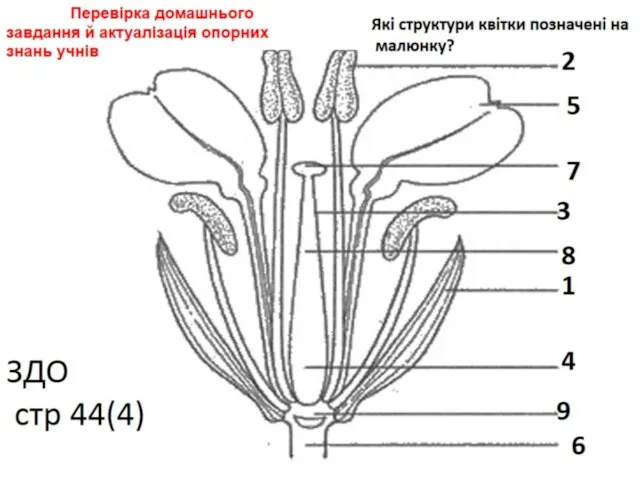 Структура квітки
 Зачем человеку растения
 Простые и сложные методы окрашивания микроорганизмов: окраска по Граму, окраска спор, капсул, жгутиков
 Многообразие земноводных
 Опыты с растениями. Банан. 7 класс
 Клуб любителей кошек
 Членистоногие обитающие в Республике Крым
 Презентация на тему Цитология
  Презентация на тему Рациональное питание залог здоровья
  pril
pril Нервная система человека
 Ткани
Ткани Виороиды. Инфекционные молекулы РНК, способные вызывать заболевания высших растений
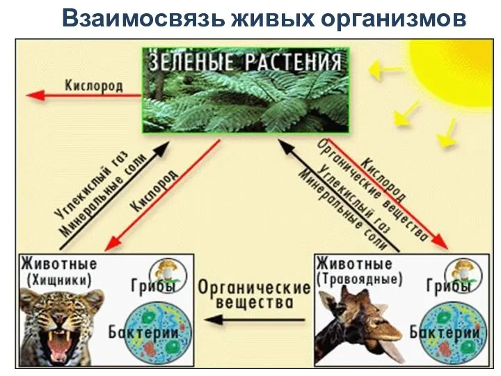 Биосфера. Часть 3. Взаимосвязь живых организмов
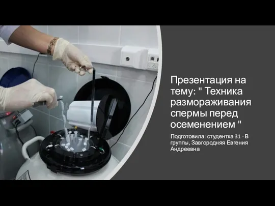 Техника размораживания спермы перед осеменением
 Царство Грибы
 Синица. Внешний вид
 Zheludochno-kishechny_trakt_Prezentatsia_3
 Красная книга Карелии
 Царство грибы (часть 1)
 Гигиена сенсорных систем
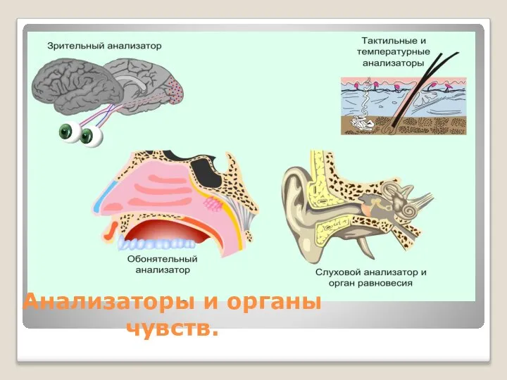 Анализаторы и органы чувств
 Анатомия человека. Понятие о костях
 Тема 1
 Низшие растения. Водоросли
 Презентация на тему МИКРООРГАНИЗМЫ-ХУДОЖНИКИ