- Concept about connections of bones

Содержание
- 2. The plan of lecture: 1. Introduction. 2. Development connection of bones. 3. Continuous connections of bones.
- 3. JOINTS Joints are part of the support and locomotion apparatus. They retain bones close to each
- 4. Synostosis and semi-joint. A — syndesmosis: 1 — interosseus membrane of forearm; В — synchondrosis: 2
- 7. They can be fibrous, cartilaginous and bony. Fibrous joints (junctura fibrosa) include sutures, gomphoses and syndesmoses.
- 8. Transverse section of articular cartilage. 1 — superficial layer; 2 — cartilaginous basic substance; 3 —
- 9. The articular capsule (capsula articularis) is attached to the edges of the articular cartilage or at
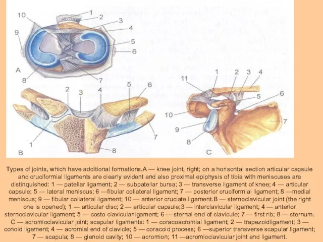
- 10. Types of joints, which have additional formations.A — knee joint, right; on a horisontal section articular
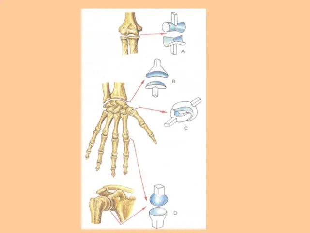
- 11. The articular cavity (cavum articulare) is a closed fissure-like space confined by the articular surfaces and
- 13. AGE CHARACTERISTICS OF JOINTS In embryogenesis all articulations are at first formed as continuous joints. Later,
- 14. General myology. A muscle as body. Development of muscles in ontogenesis. Classification of muscles. The auxiliary
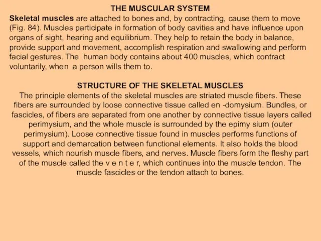
- 15. THE MUSCULAR SYSTEM Skeletal muscles are attached to bones and, by contracting, cause them to move
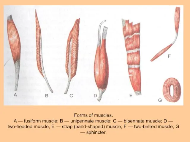
- 17. CLASSIFICATION OF MUSCLES There are several ways of classification of skeletal muscles. Muscles can be classified
- 18. Forms of muscles. A — fusiform muscle; В — unipennate muscle; С — bipennate muscle; D
- 19. Many muscles of the body are named according to their shape. Thus, / there is a
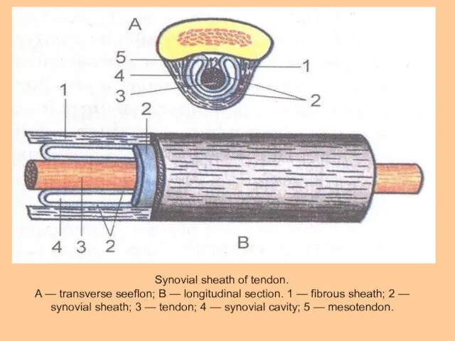
- 20. Synovial sheath of tendon. A — transverse seeflon; В — longitudinal section. 1 — fibrous sheath;
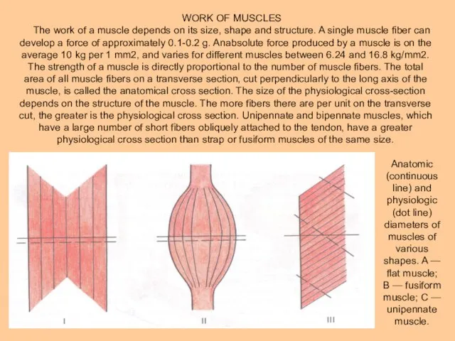
- 21. WORK OF MUSCLES The work of a muscle depends on its size, shape and structure. A
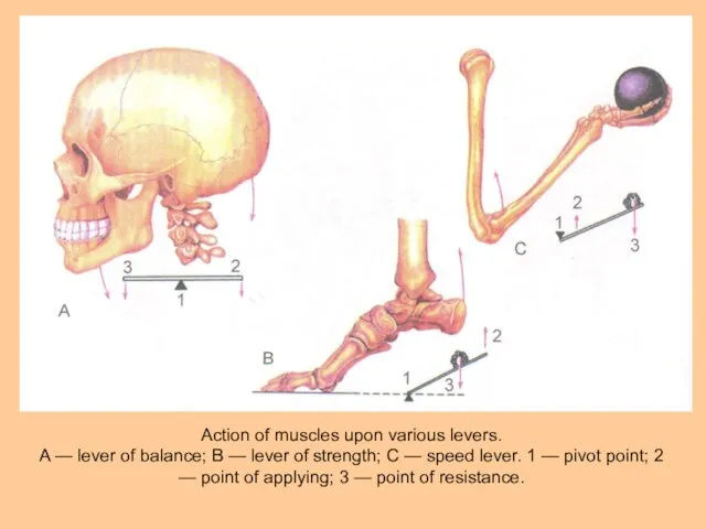
- 22. Action of muscles upon various levers. A — lever of balance; В — lever of strength;
- 23. Overcoming work is carried out when muscle contraction changes the positions of the body or its
- 24. This lever system has an advantage in power, but a disadvantage in speed. The other type
- 26. Скачать презентацию
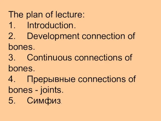
Слайд 2The plan of lecture:
1. Introduction.
2. Development connection of bones.
3. Continuous connections of
The plan of lecture:
1. Introduction.
2. Development connection of bones.
3. Continuous connections of
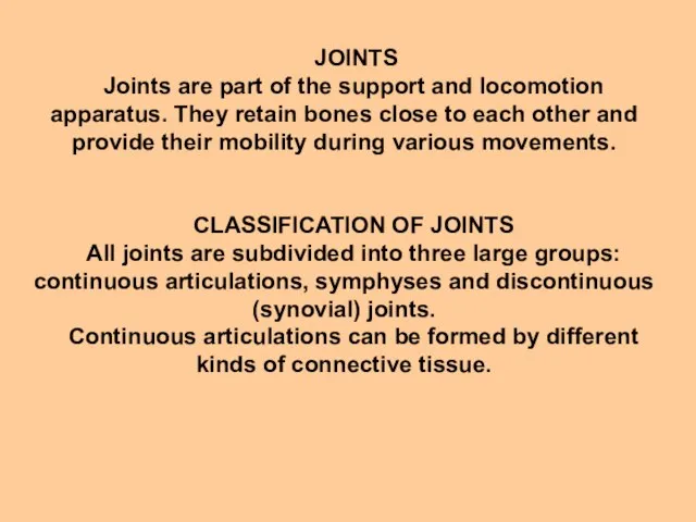
Слайд 3 JOINTS
Joints are part of the support and locomotion apparatus. They retain
JOINTS
Joints are part of the support and locomotion apparatus. They retain
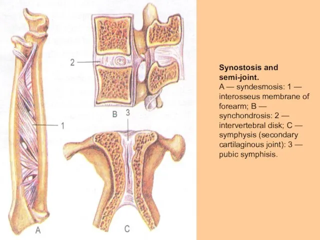
Слайд 4Synostosis and semi-joint.
A — syndesmosis: 1 — interosseus membrane of forearm; В
Synostosis and semi-joint.
A — syndesmosis: 1 — interosseus membrane of forearm; В
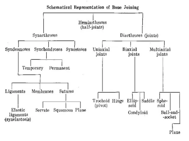
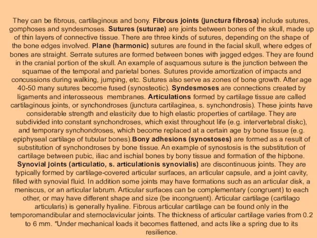
Слайд 7They can be fibrous, cartilaginous and bony. Fibrous joints (junctura fibrosa) include
They can be fibrous, cartilaginous and bony. Fibrous joints (junctura fibrosa) include
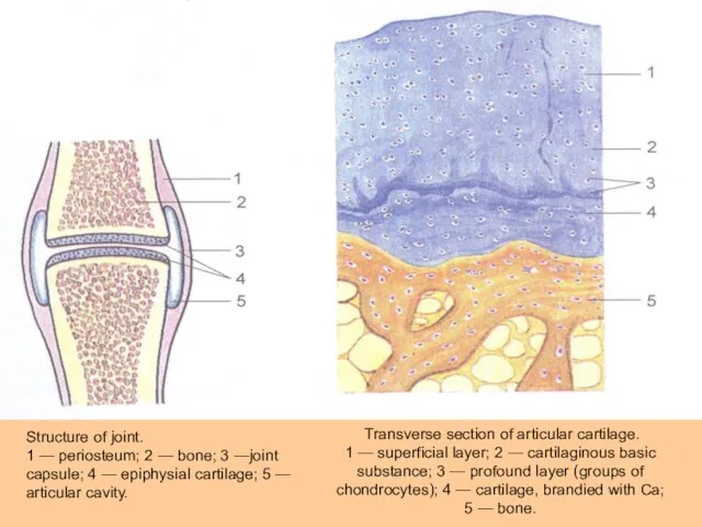
Слайд 8 Transverse section of articular cartilage.
1 — superficial layer; 2 — cartilaginous
Transverse section of articular cartilage.
1 — superficial layer; 2 — cartilaginous
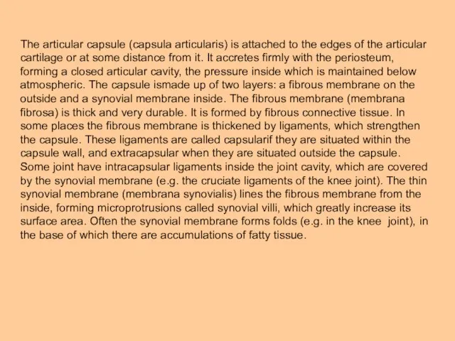
Слайд 9The articular capsule (capsula articularis) is attached to the edges of the
The articular capsule (capsula articularis) is attached to the edges of the
Слайд 10Types of joints, which have additional formations.A — knee joint, right; on
Types of joints, which have additional formations.A — knee joint, right; on
Слайд 11The articular cavity (cavum articulare) is a closed fissure-like space confined by
The articular cavity (cavum articulare) is a closed fissure-like space confined by
Слайд 13AGE CHARACTERISTICS OF JOINTS
In embryogenesis all articulations are at first formed as
AGE CHARACTERISTICS OF JOINTS
In embryogenesis all articulations are at first formed as
Слайд 14General myology. A muscle as body. Development of muscles in ontogenesis. Classification
General myology. A muscle as body. Development of muscles in ontogenesis. Classification
Слайд 15THE MUSCULAR SYSTEM
Skeletal muscles are attached to bones and, by contracting, cause
THE MUSCULAR SYSTEM
Skeletal muscles are attached to bones and, by contracting, cause

Слайд 17CLASSIFICATION OF MUSCLES
There are several ways of classification of skeletal muscles. Muscles
CLASSIFICATION OF MUSCLES
There are several ways of classification of skeletal muscles. Muscles
Слайд 18Forms of muscles.
A — fusiform muscle; В — unipennate muscle; С —
Forms of muscles.
A — fusiform muscle; В — unipennate muscle; С —
Слайд 19Many muscles of the body are named according to their shape. Thus,
Many muscles of the body are named according to their shape. Thus,
Слайд 20Synovial sheath of tendon.
A — transverse seeflon; В — longitudinal section. 1
Synovial sheath of tendon.
A — transverse seeflon; В — longitudinal section. 1
Слайд 21WORK OF MUSCLES
The work of a muscle depends on its size, shape
WORK OF MUSCLES
The work of a muscle depends on its size, shape
Слайд 22Action of muscles upon various levers.
A — lever of balance; В —
Action of muscles upon various levers.
A — lever of balance; В —
Слайд 23Overcoming work is carried out when muscle contraction changes the positions of
Overcoming work is carried out when muscle contraction changes the positions of
Слайд 24This lever system has an advantage in power, but a disadvantage in
This lever system has an advantage in power, but a disadvantage in
 Этикет
 Сплавы металлов
 Презентация на тему Труд как нравственность
 Растения и животные
 Хозяин судьбы
 Органическая химия в быту
 Блюда из овощей. 5 класс
 Презентация на тему Своя игра ("Путь к грамотности")
 Фото Близнецы. Курсовая работа
 Эмоции человека
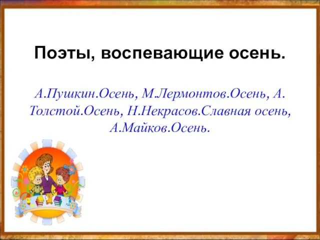 Поэты, воспевающие осень.А.Пушкин.Осень, М.Лермонтов.Осень, А.Толстой.Осень, Н.Некрасов.Славная осень, А.Майков.Осень.
 Презентация на тему Как размножаются живые организмы
 ОНТОЛОГИИ: формальное и неформальное
 Строительно-монтажные работы при восстановлении объектов ж/д транспорта при возникновении ЧС
 Интегрированная концепция и уровни абстракции
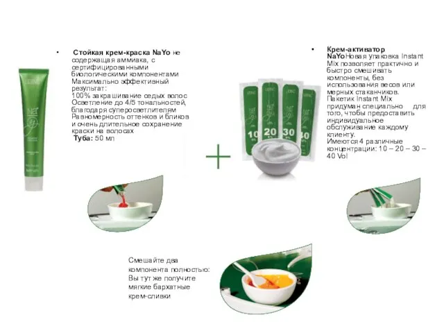 Стойкая крем-краска NaYo не содержащая аммиака, с сертифицированными биологическими компонентамиМаксимально эффективный результа
 Презентация на тему Реалистическая живопись Голландии
  Lektsia_1
 Документация компании
 Лига супервайзеров. Плиточный шоколад Lacmi
 Презентация на тему Путешествие по сказкам А.С. Пушкина
 Пустыня Сахара
 Тест по окружающему миру
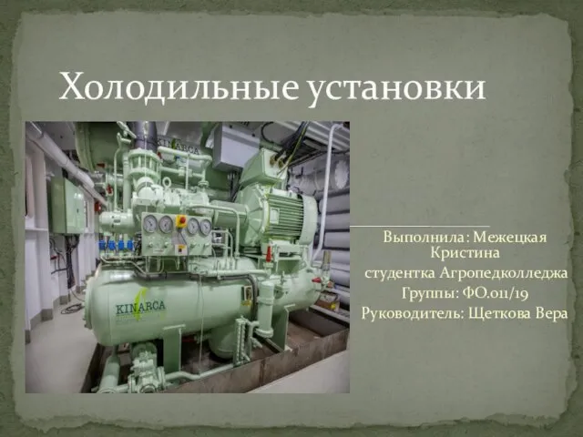 Холодильные установки
 Екатеринбургскому Энергетическому-90!
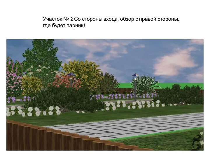 Проекты участков
 Детская организация Тенгрин герл
 Самопрезентация2