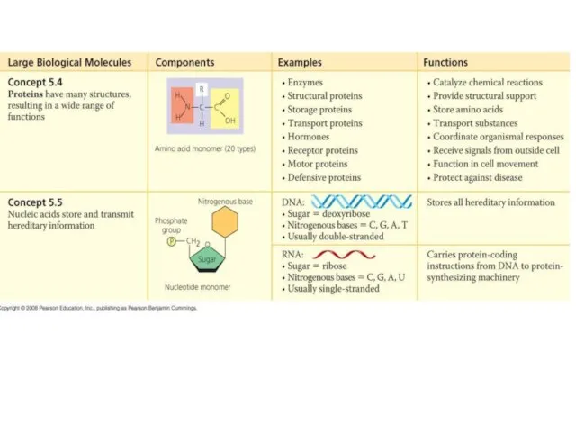
- Cтруктурно-функциональная организация генов и белков

Содержание
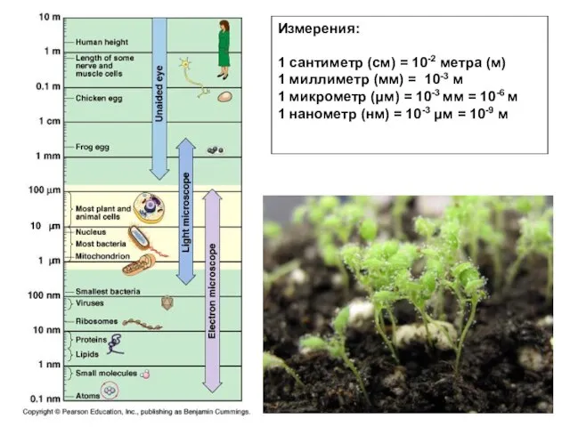
- 2. Измерения: 1 сантиметр (cм) = 10-2 метра (м) 1 миллиметр (мм) = 10-3 м 1 микрометр
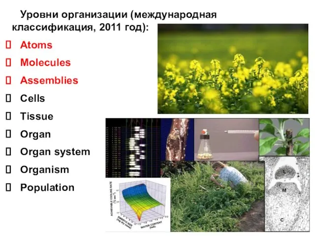
- 3. Уровни организации (международная классификация, 2011 год): Atoms Molecules Assemblies Cells Tissue Organ Organ system Organism Population
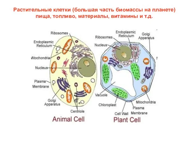
- 4. Растительные клетки (большая часть биомассы на планете) пища, топливо, материалы, витамины и т.д.
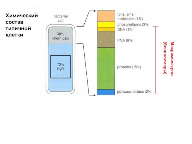
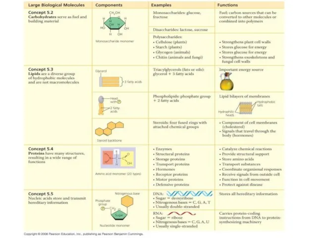
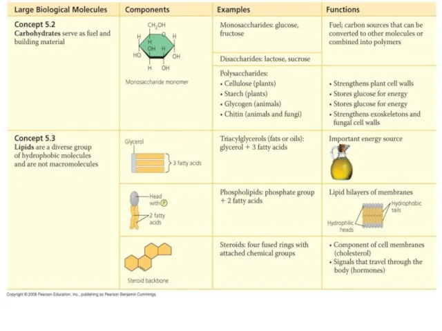
- 5. Химический состав типичной клетки Макромолекулы (биополимеры)
- 6. Общий принцип образования и распада биополимеров синтез……… распад.............
- 7. Всего 20 аминокислот участвует в образовании белков и других реакциях (международные названия и классификация):
- 8. Аденозинтрифосфат - АТФ АТФ – нуклеотид предшественник биосинтеза нуклеиновых кислот источник энергии медиатор, нейротрансмиттер, регулятор
- 9. Строение нуклеиновых кислот:
- 10. Строение липидов:
- 11. Строение липидов:
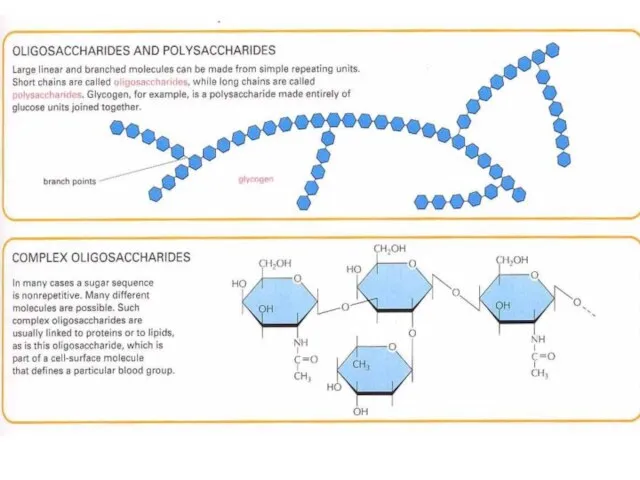
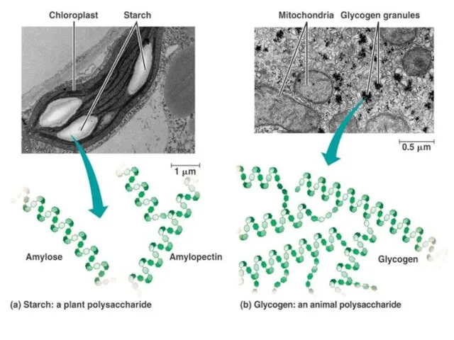
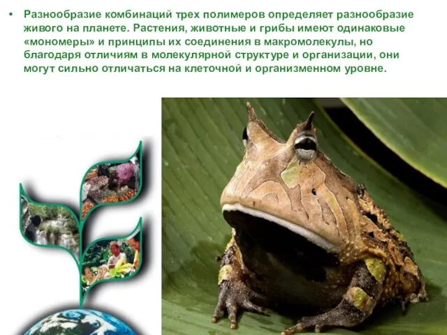
- 19. Разнообразие комбинаций трех полимеров определяет разнообразие живого на планете. Растения, животные и грибы имеют одинаковые «мономеры»
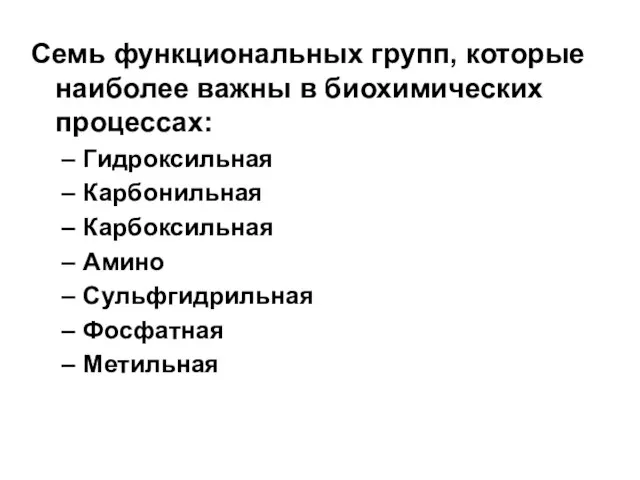
- 20. Семь функциональных групп, которые наиболее важны в биохимических процессах: Гидроксильная Карбонильная Карбоксильная Амино Сульфгидрильная Фосфатная Метильная
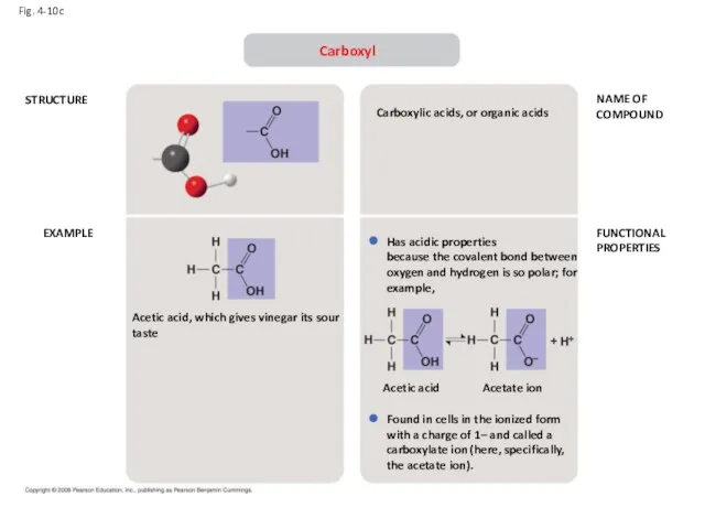
- 21. Fig. 4-10c STRUCTURE EXAMPLE NAME OF COMPOUND FUNCTIONAL PROPERTIES Carboxyl Acetic acid, which gives vinegar its
- 22. Fig. 4-10d STRUCTURE EXAMPLE NAME OF COMPOUND FUNCTIONAL PROPERTIES Amino Because it also has a carboxyl
- 23. Fig. 4-10e STRUCTURE EXAMPLE NAME OF COMPOUND FUNCTIONAL PROPERTIES Sulfhydryl (may be written HS—) Cysteine Cysteine
- 24. Fig. 4-10f STRUCTURE EXAMPLE NAME OF COMPOUND FUNCTIONAL PROPERTIES Phosphate In addition to taking part in
- 25. Fig. 4-10g STRUCTURE EXAMPLE NAME OF COMPOUND FUNCTIONAL PROPERTIES Methyl 5-Methyl cytidine is a component of
- 27. James Watson and Francis Crick, UK Erwin Chargaff, Ukraine Linus Pauling, USA Maurice Wilkins, NZ Rosalind
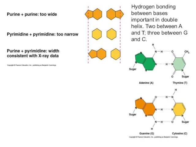
- 28. Первое правило Чаргафа: В природной ДНК количество единиц гуанина равно количеству единиц цитозина, тогда как количество
- 29. Pyrimidines: uracil, cytosine, thiamine Purines : Adenine, guanosine
- 30. Структура оснований в ДНК и РНК: пиримидины: урацил, цитозин, тиамин пурины: аденин, гуанозин
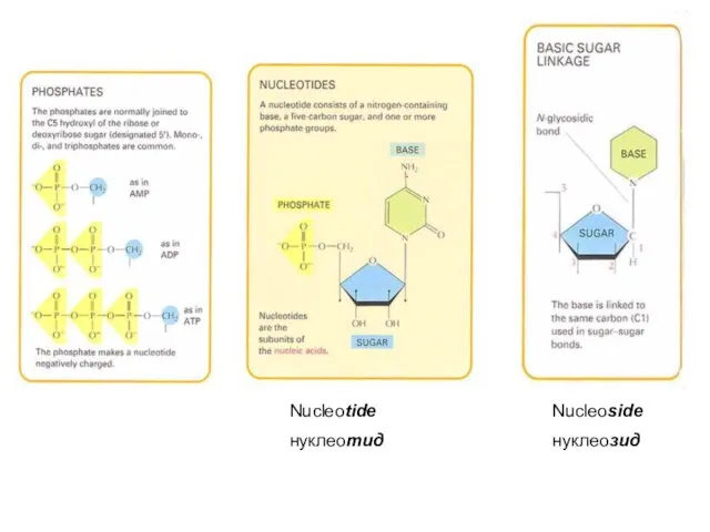
- 31. Nucleoside нуклеозид Nucleotide нуклеотид
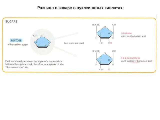
- 32. Разница в сахаре в нуклеиновых кислотах:
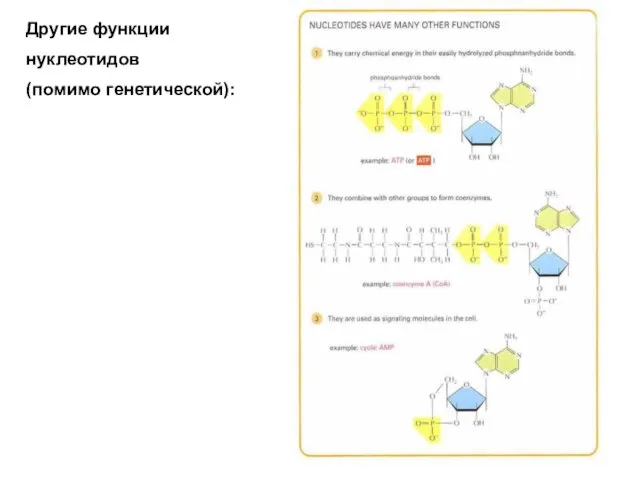
- 33. Другие функции нуклеотидов (помимо генетической):
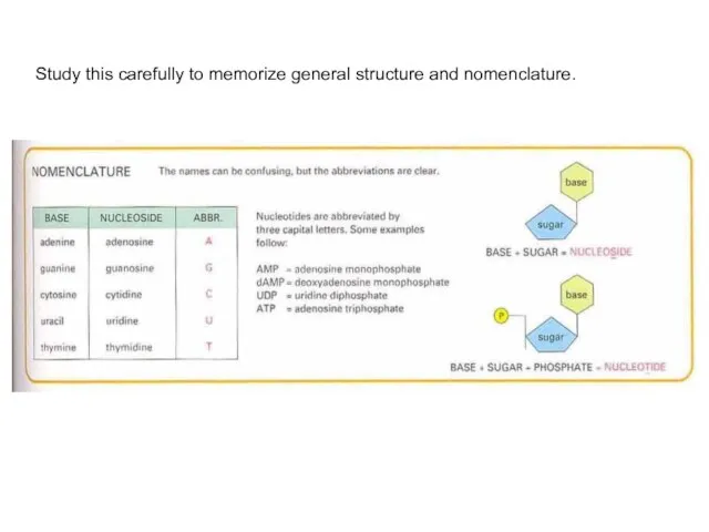
- 34. Study this carefully to memorize general structure and nomenclature.
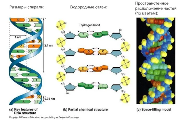
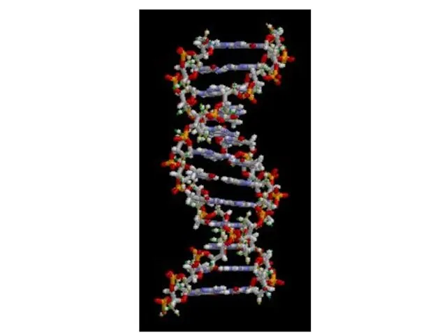
- 35. Размеры спирали: Водородные связи: Пространстенное расположение частей (по цветам):
- 37. Hydrogen bonding between bases important in double helix. Two between A and T; three between G
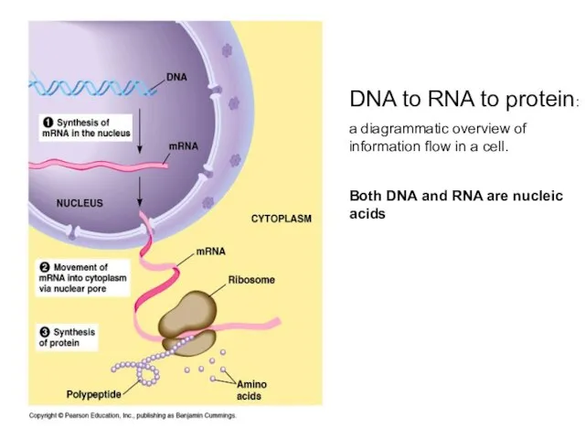
- 38. DNA to RNA to protein: a diagrammatic overview of information flow in a cell. Both DNA
- 39. Campbell and Reece 8 Figure 17.20 Polyribosomes mRNA is translated into a polypeptide/protein in the cytoplasm.
- 40. Campbell and Reece Figure 17.14 The structure of transfer RNA (tRNA). Transfer RNA is an adaptor
- 41. Campbell and Reece 8 Chapter 17 These first three are the types of RNA that you
- 43. Making a polypeptide chain Amino acids are linked by the formation of a peptide bond Note
- 44. The primary structure of a protein is the sequence of amino acids This is the enzyme
- 45. Campbell and Reece Figure 5.19 Conformation of the enzyme lysozyme. Two types of model, the ribbon
- 46. Campbell and Reece 8 Figure 5.21 Exploring levels of protein structure.
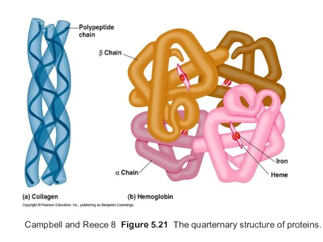
- 47. Campbell and Reece 8 Figure 5.21 The quarternary structure of proteins.
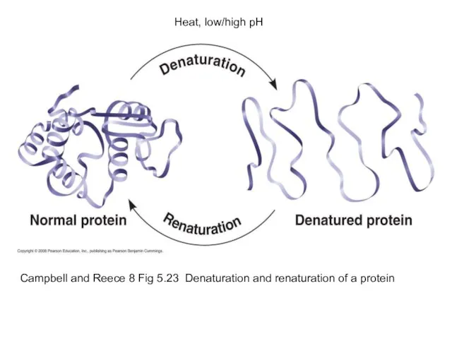
- 48. Campbell and Reece 8 Fig 5.23 Denaturation and renaturation of a protein Heat, low/high pH
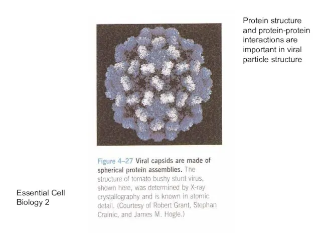
- 49. Essential Cell Biology 2 Protein structure and protein-protein interactions are important in viral particle structure
- 51. Скачать презентацию
Слайд 3 Уровни организации (международная классификация, 2011 год):
Atoms
Molecules
Assemblies
Cells
Tissue
Уровни организации (международная классификация, 2011 год):
Atoms
Molecules
Assemblies
Cells
Tissue
Слайд 4Растительные клетки (большая часть биомассы на планете) пища, топливо, материалы, витамины и
Растительные клетки (большая часть биомассы на планете) пища, топливо, материалы, витамины и
Слайд 5Химический состав типичной клетки
Макромолекулы
(биополимеры)
Химический состав типичной клетки
Макромолекулы
(биополимеры)
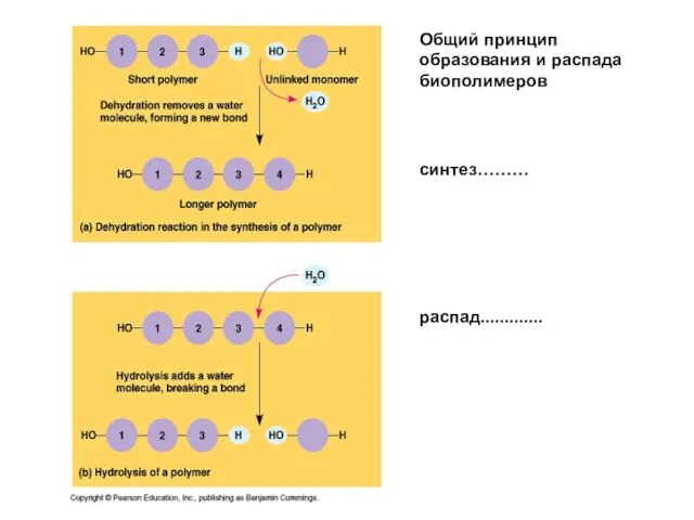
Слайд 6Общий принцип образования и распада биополимеров
синтез………
распад.............
Общий принцип образования и распада биополимеров
синтез………
распад.............
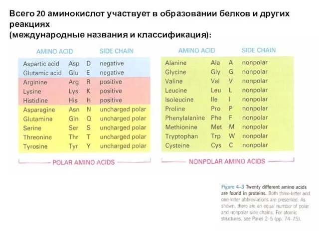
Слайд 7Всего 20 аминокислот участвует в образовании белков и других реакциях
(международные названия и
Всего 20 аминокислот участвует в образовании белков и других реакциях
(международные названия и
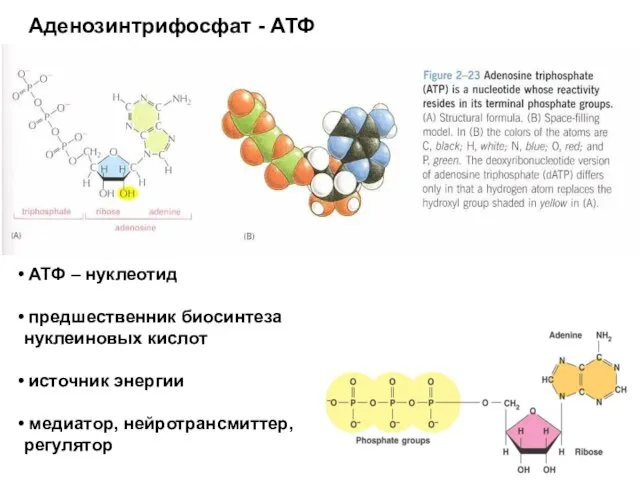
Слайд 8Аденозинтрифосфат - АТФ
АТФ – нуклеотид
предшественник биосинтеза нуклеиновых кислот
источник
Аденозинтрифосфат - АТФ
АТФ – нуклеотид
предшественник биосинтеза нуклеиновых кислот
источник
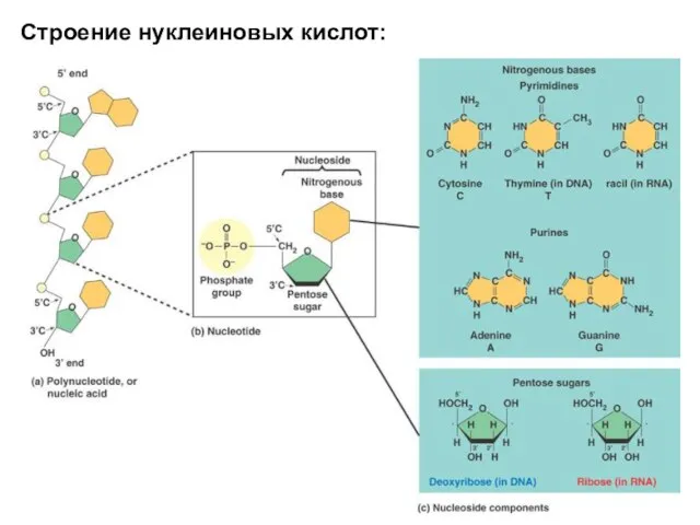
Слайд 9Строение нуклеиновых кислот:
Строение нуклеиновых кислот:
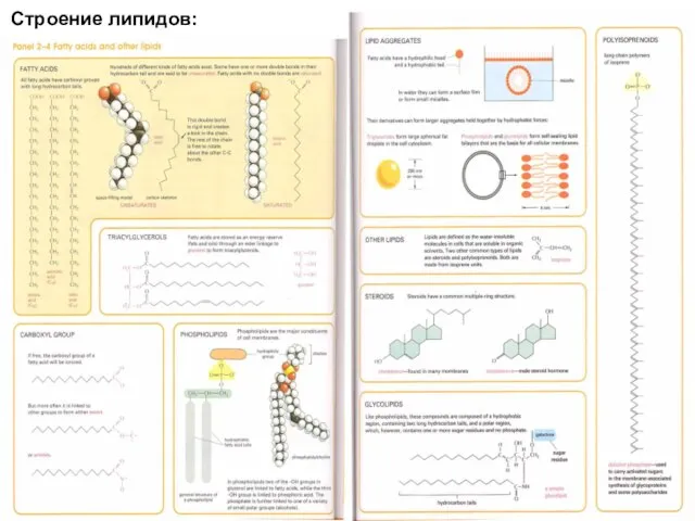
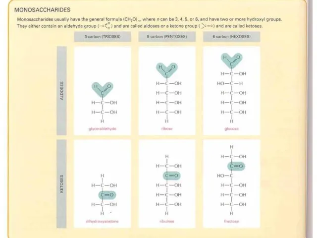
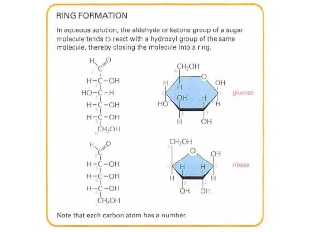
Слайд 10Строение липидов:
Строение липидов:
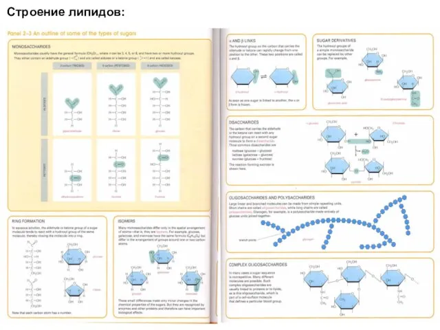
Слайд 11Строение липидов:
Строение липидов:
Слайд 19Разнообразие комбинаций трех полимеров определяет разнообразие живого на планете. Растения, животные и
Разнообразие комбинаций трех полимеров определяет разнообразие живого на планете. Растения, животные и
Слайд 20Семь функциональных групп, которые наиболее важны в биохимических процессах:
Гидроксильная
Карбонильная
Карбоксильная
Амино
Сульфгидрильная
Фосфатная
Метильная
Семь функциональных групп, которые наиболее важны в биохимических процессах:
Гидроксильная
Карбонильная
Карбоксильная
Амино
Сульфгидрильная
Фосфатная
Метильная
Слайд 21Fig. 4-10c
STRUCTURE
EXAMPLE
NAME OF
COMPOUND
FUNCTIONAL
PROPERTIES
Carboxyl
Acetic acid, which gives vinegar its sour taste
Carboxylic acids, or
Fig. 4-10c
STRUCTURE
EXAMPLE
NAME OF
COMPOUND
FUNCTIONAL
PROPERTIES
Carboxyl
Acetic acid, which gives vinegar its sour taste
Carboxylic acids, or
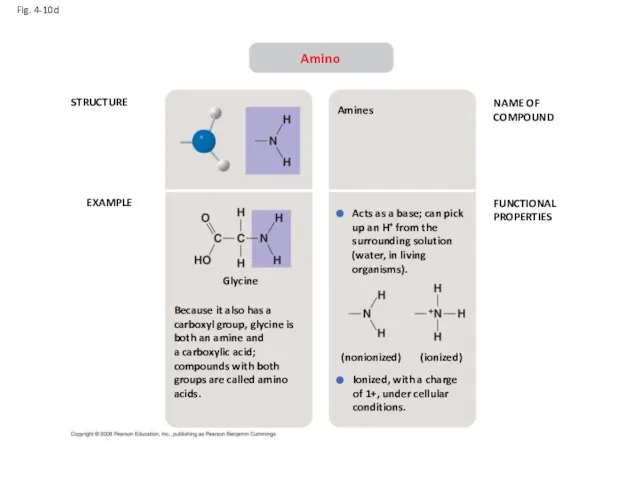
Слайд 22Fig. 4-10d
STRUCTURE
EXAMPLE
NAME OF
COMPOUND
FUNCTIONAL
PROPERTIES
Amino
Because it also has a carboxyl group, glycine is both
Fig. 4-10d
STRUCTURE
EXAMPLE
NAME OF
COMPOUND
FUNCTIONAL
PROPERTIES
Amino
Because it also has a carboxyl group, glycine is both
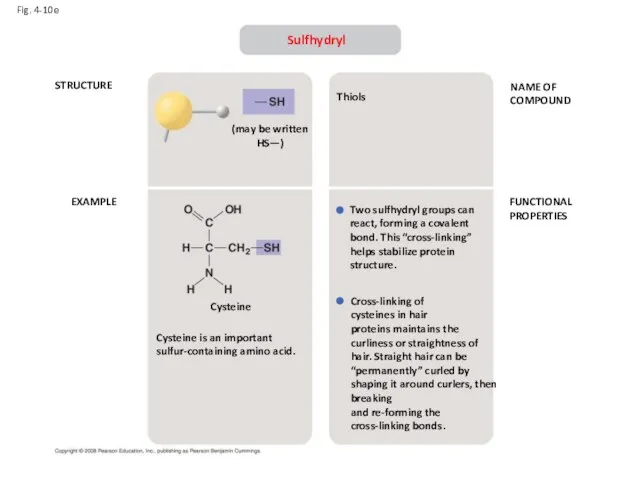
Слайд 23Fig. 4-10e
STRUCTURE
EXAMPLE
NAME OF
COMPOUND
FUNCTIONAL
PROPERTIES
Sulfhydryl
(may be written HS—)
Cysteine
Cysteine is an important sulfur-containing amino acid.
Thiols
Two
Fig. 4-10e
STRUCTURE
EXAMPLE
NAME OF
COMPOUND
FUNCTIONAL
PROPERTIES
Sulfhydryl
(may be written HS—)
Cysteine
Cysteine is an important sulfur-containing amino acid.
Thiols
Two
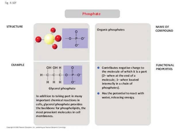
Слайд 24Fig. 4-10f
STRUCTURE
EXAMPLE
NAME OF
COMPOUND
FUNCTIONAL
PROPERTIES
Phosphate
In addition to taking part in many important chemical reactions
Fig. 4-10f
STRUCTURE
EXAMPLE
NAME OF
COMPOUND
FUNCTIONAL
PROPERTIES
Phosphate
In addition to taking part in many important chemical reactions
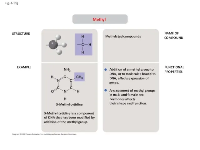
Слайд 25Fig. 4-10g
STRUCTURE
EXAMPLE
NAME OF
COMPOUND
FUNCTIONAL
PROPERTIES
Methyl
5-Methyl cytidine is a component of DNA that has been
Fig. 4-10g
STRUCTURE
EXAMPLE
NAME OF
COMPOUND
FUNCTIONAL
PROPERTIES
Methyl
5-Methyl cytidine is a component of DNA that has been
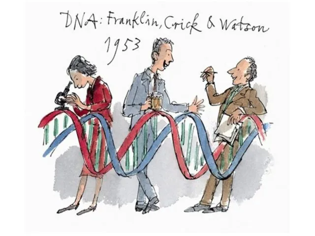
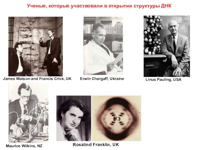
Слайд 27James Watson and Francis Crick, UK
Erwin Chargaff, Ukraine
Linus Pauling, USA
Maurice Wilkins, NZ
Rosalind
James Watson and Francis Crick, UK
Erwin Chargaff, Ukraine
Linus Pauling, USA
Maurice Wilkins, NZ
Rosalind
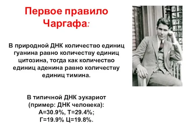
Слайд 28Первое правило Чаргафа:
В природной ДНК количество единиц гуанина равно количеству единиц цитозина,
Первое правило Чаргафа:
В природной ДНК количество единиц гуанина равно количеству единиц цитозина,
Слайд 29Pyrimidines:
uracil, cytosine, thiamine
Purines :
Adenine, guanosine
Pyrimidines:
uracil, cytosine, thiamine
Purines :
Adenine, guanosine

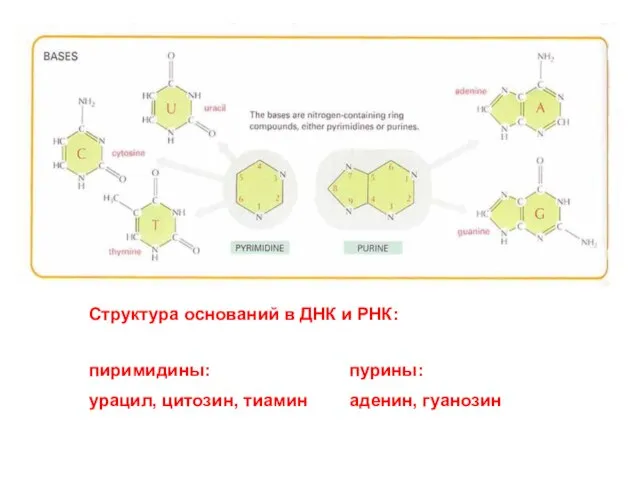
Слайд 30Структура оснований в ДНК и РНК:
пиримидины:
урацил, цитозин, тиамин
пурины:
аденин, гуанозин
Структура оснований в ДНК и РНК:
пиримидины:
урацил, цитозин, тиамин
пурины:
аденин, гуанозин
Слайд 31Nucleoside
нуклеозид
Nucleotide
нуклеотид
Nucleoside
нуклеозид
Nucleotide
нуклеотид
Слайд 32Разница в сахаре в нуклеиновых кислотах:
Разница в сахаре в нуклеиновых кислотах:
Слайд 33Другие функции
нуклеотидов
(помимо генетической):
Другие функции
нуклеотидов
(помимо генетической):
Слайд 34Study this carefully to memorize general structure and nomenclature.
Study this carefully to memorize general structure and nomenclature.
Слайд 35Размеры спирали:
Водородные связи:
Пространстенное расположение частей (по цветам):
Размеры спирали:
Водородные связи:
Пространстенное расположение частей (по цветам):
Слайд 37Hydrogen bonding between bases important in double helix. Two between A and
Hydrogen bonding between bases important in double helix. Two between A and
Слайд 38DNA to RNA to protein:
a diagrammatic overview of information flow in
a diagrammatic overview of information flow in
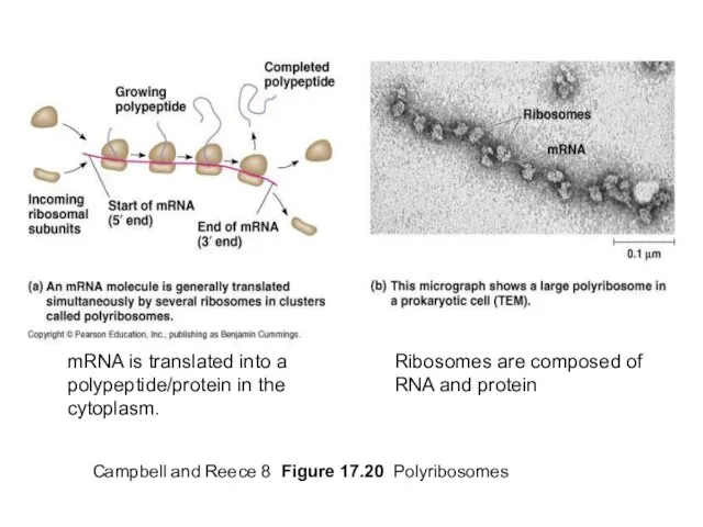
Слайд 39Campbell and Reece 8 Figure 17.20 Polyribosomes
mRNA is translated into a polypeptide/protein
Campbell and Reece 8 Figure 17.20 Polyribosomes
mRNA is translated into a polypeptide/protein
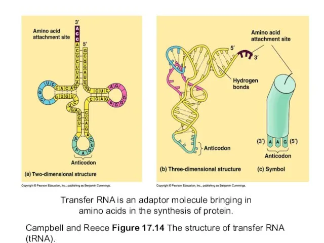
Слайд 40Campbell and Reece Figure 17.14 The structure of transfer RNA (tRNA).
Transfer RNA
Campbell and Reece Figure 17.14 The structure of transfer RNA (tRNA).
Transfer RNA
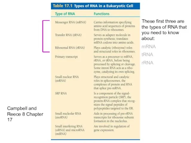
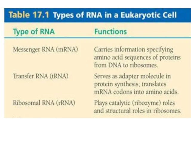
Слайд 41Campbell and Reece 8 Chapter 17
These first three are the types of
Campbell and Reece 8 Chapter 17
These first three are the types of
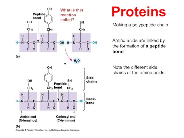
Слайд 43Making a polypeptide chain
Amino acids are linked by the formation of a
Making a polypeptide chain
Amino acids are linked by the formation of a
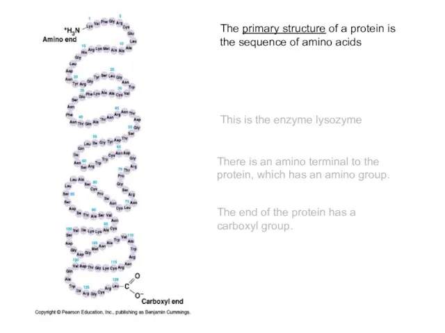
Слайд 44The primary structure of a protein is the sequence of amino acids
This
The primary structure of a protein is the sequence of amino acids
This
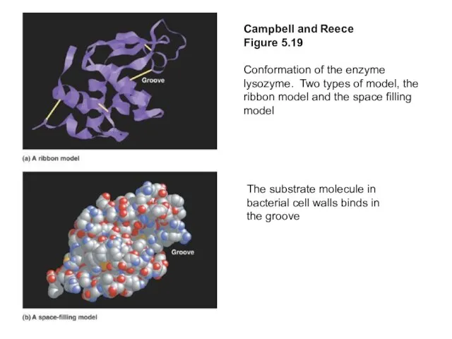
Слайд 45Campbell and Reece
Figure 5.19
Conformation of the enzyme lysozyme. Two types of
Campbell and Reece
Figure 5.19
Conformation of the enzyme lysozyme. Two types of
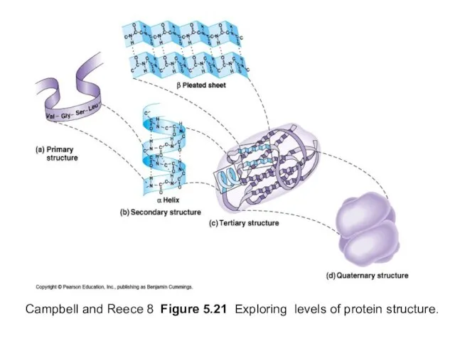
Слайд 46Campbell and Reece 8 Figure 5.21 Exploring levels of protein structure.
Campbell and Reece 8 Figure 5.21 Exploring levels of protein structure.
Слайд 47Campbell and Reece 8 Figure 5.21 The quarternary structure of proteins.
Campbell and Reece 8 Figure 5.21 The quarternary structure of proteins.
Слайд 48Campbell and Reece 8 Fig 5.23 Denaturation and renaturation of a protein
Heat,
Campbell and Reece 8 Fig 5.23 Denaturation and renaturation of a protein
Heat,
Слайд 49Essential Cell Biology 2
Protein structure and protein-protein interactions are important in viral
Essential Cell Biology 2
Protein structure and protein-protein interactions are important in viral
 Формирование умения решать уравнения в начальной школе
 Программы кредитования субъектов малого и среднего бизнеса
 Что такое дружба
 Весна море 3-бклассx
 Развитие школьной библиотеки
 Джедайское искуство боя
 Предложения по формированию корпоративной культуры Департамента культуры и архивного дела Ульяновской области
 Отдел Мохообразные. Общая характеристика
 Изобразительно- выразительные средства языка Сравнение урок литературы, 5 класс
 Плавный пуск электродвигателей
 Реализм во Франции
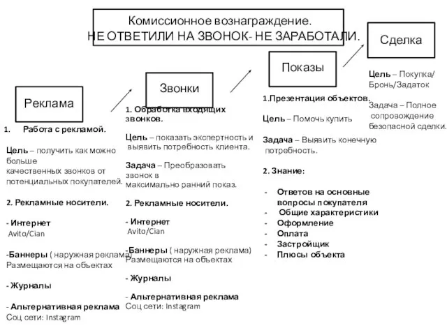 Комиссионное вознаграждение
 Мотивация
 Мастер группа Хранители снов
 Восстание декабристов (8 класс)
 «Мир начинается с тебя»рабочая программакурса внеурочной деятельностидля учащихся 1-4 классов
 Верста значение
 Органическое управление(методология эффективной управленческойдеятельности в информационном обществе)
 Правила работы в группе: 1. Уважение к говорящему, 1. Уважение к говорящему, не перебивать говорящего, не шуметь, когда кто-то говорит;
 Новые рекламные возможности торговой сети «Молния»
 Финансовые результаты деятельности предприятия ОАО Лазмурь
 Кузьма Минин и Дмитрий Пожарский
 Кафе “Унесенные кошками”
 Теорема Пифагора
 Основы теории мотивации
  Торговля на новостях. Фундаментальный анализ
 Трудовые организации. Системы и формы оплаты труда. Тема 3
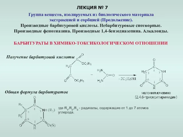 ЛЕКЦИЯ № 7