- Human heart

Содержание
- 2. Plan of work Heart(structure) The projection of the heart valves and large vessels The topography of
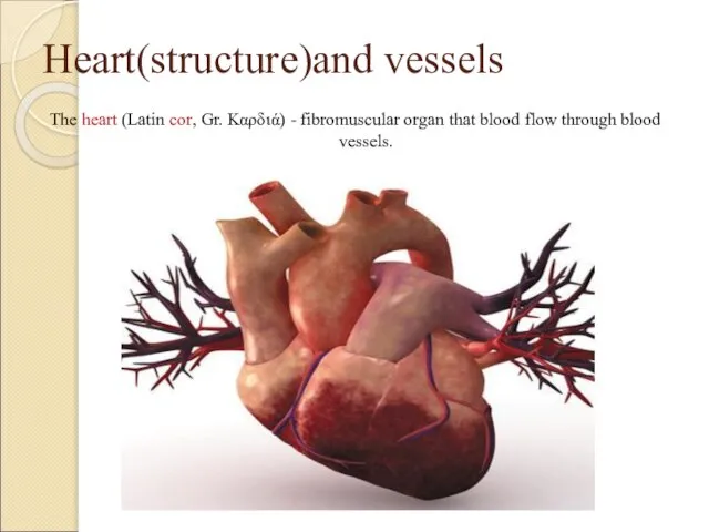
- 3. Heart(structure)and vessels The heart (Latin сor, Gr. Καρδιά) - fibromuscular organ that blood flow through blood
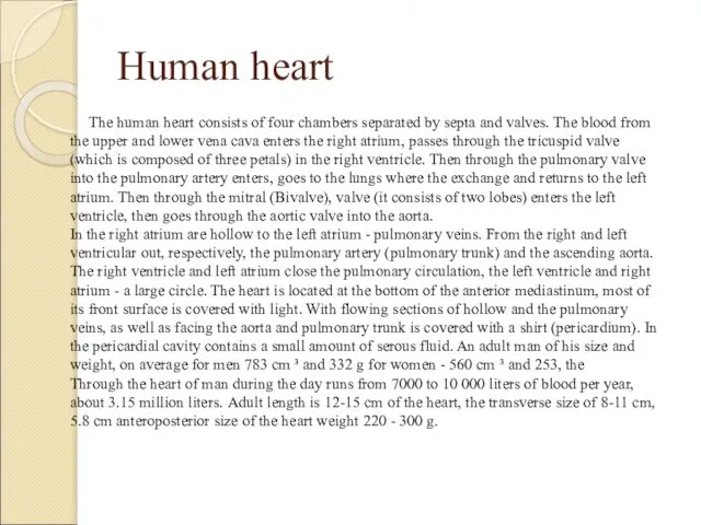
- 4. Human heart The human heart consists of four chambers separated by septa and valves. The blood
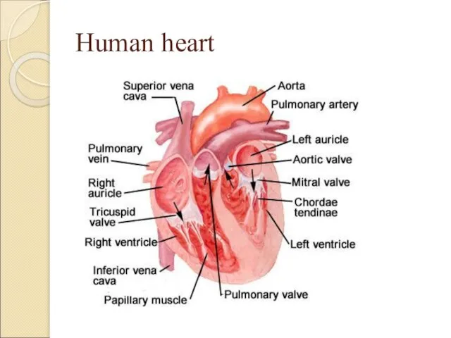
- 5. Human heart
- 6. The projection of the heart valves and large vessels In Fig. 1. The projection of the
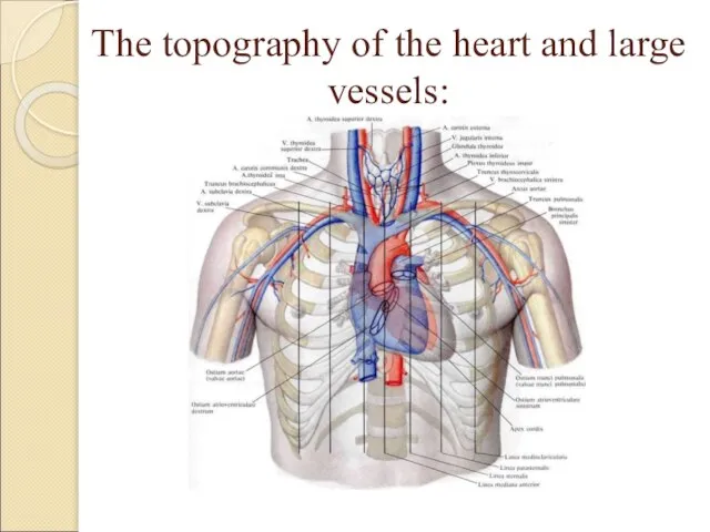
- 7. The topography of the heart and large vessels: The heart is located behind the lower half
- 8. The topography of the heart and large vessels: To the right of the median plane of
- 9. The topography of the heart and large vessels:
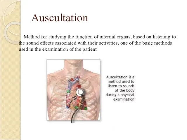
- 10. Auscultation Method for studying the function of internal organs, based on listening to the sound effects
- 11. The 1st point of auscultation the apex of the heart, i.e, apex beat area or if
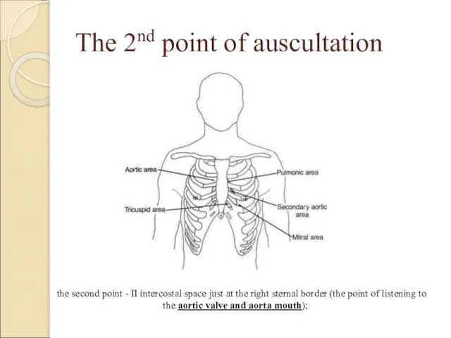
- 12. The 2nd point of auscultation the second point - II intercostal space just at the right
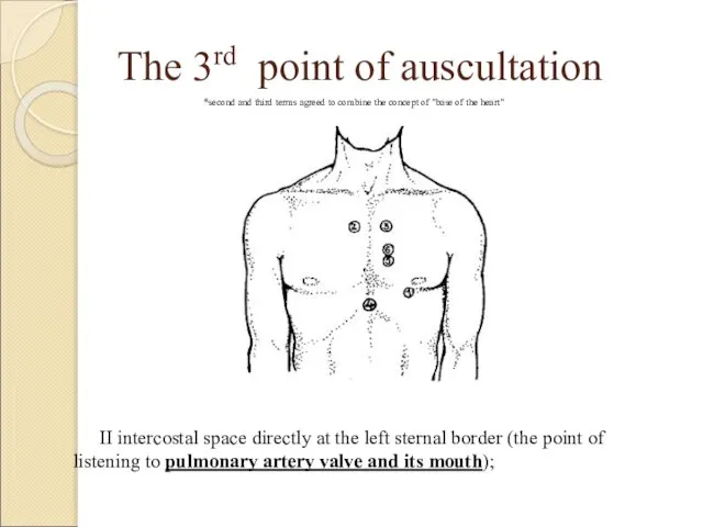
- 13. The 3rd point of auscultation II intercostal space directly at the left sternal border (the point
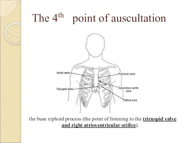
- 14. The 4th point of auscultation the base xiphoid process (the point of listening to the tricuspid
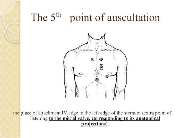
- 15. The 5th point of auscultation the place of attachment IV edge to the left edge of
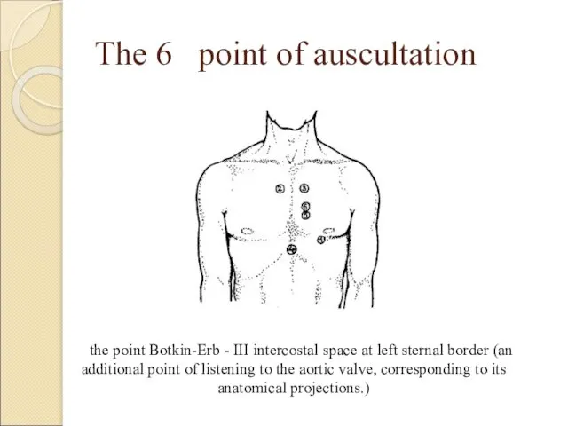
- 16. The 6 point of auscultation the point Botkin-Erb - III intercostal space at left sternal border
- 18. Скачать презентацию
Слайд 2Plan of work
Heart(structure)
The projection of the heart valves and large vessels
The topography
Plan of work
Heart(structure)
The projection of the heart valves and large vessels
The topography
Слайд 3Heart(structure)and vessels
The heart (Latin сor, Gr. Καρδιά) - fibromuscular organ that blood
Heart(structure)and vessels
The heart (Latin сor, Gr. Καρδιά) - fibromuscular organ that blood
Слайд 4Human heart
The human heart consists of four chambers separated by septa and
Human heart
The human heart consists of four chambers separated by septa and
Слайд 5Human heart
Human heart
Слайд 6The projection of the heart valves and large vessels
In Fig. 1. The
The projection of the heart valves and large vessels
In Fig. 1. The

Слайд 7The topography of the heart and large vessels:
The heart is located behind the lower half
The topography of the heart and large vessels:
The heart is located behind the lower half

Слайд 8The topography of the heart and large vessels:
To the right of the
The topography of the heart and large vessels:
To the right of the
Слайд 9The topography of the heart and large vessels:
The topography of the heart and large vessels:
Слайд 10Auscultation
Method for studying the function of internal organs, based on listening to
Auscultation
Method for studying the function of internal organs, based on listening to
Слайд 11The 1st point of auscultation
the apex of the heart, i.e, apex beat area or if it is not
The 1st point of auscultation
the apex of the heart, i.e, apex beat area or if it is not

Слайд 12The 2nd point of auscultation
the second point - II intercostal space just
The 2nd point of auscultation
the second point - II intercostal space just
Слайд 13The 3rd point of auscultation
II intercostal space directly at the left sternal
The 3rd point of auscultation
II intercostal space directly at the left sternal
Слайд 14The 4th point of auscultation
the base xiphoid process (the point of listening
The 4th point of auscultation
the base xiphoid process (the point of listening
Слайд 15The 5th point of auscultation
the place of attachment IV edge to the
The 5th point of auscultation
the place of attachment IV edge to the
Слайд 16The 6 point of auscultation
the point Botkin-Erb - III intercostal space at
The 6 point of auscultation
the point Botkin-Erb - III intercostal space at
 ИТС ПРОФ - профессиональная информационная система для бухгалтераи руководителя
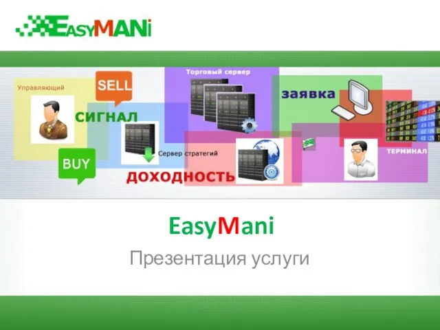 EasyMani
 Pepsi - участник фестиваля Пикник Афиша
 Именины Загоскина М.Н
 Исаак Бабель
 В некотором царстве,В некотором государстве,вернее в ТюрлемеЖивет своей жизнью
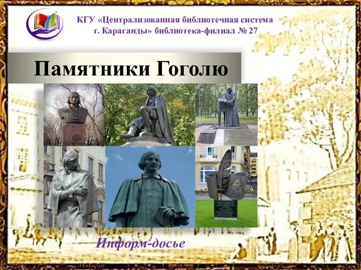 Памятники Гоголю
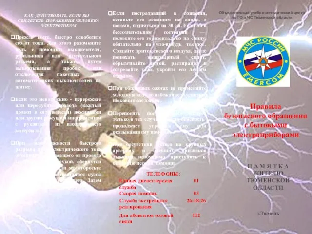 Правила безопасного обращения с бытовыми электроприборами
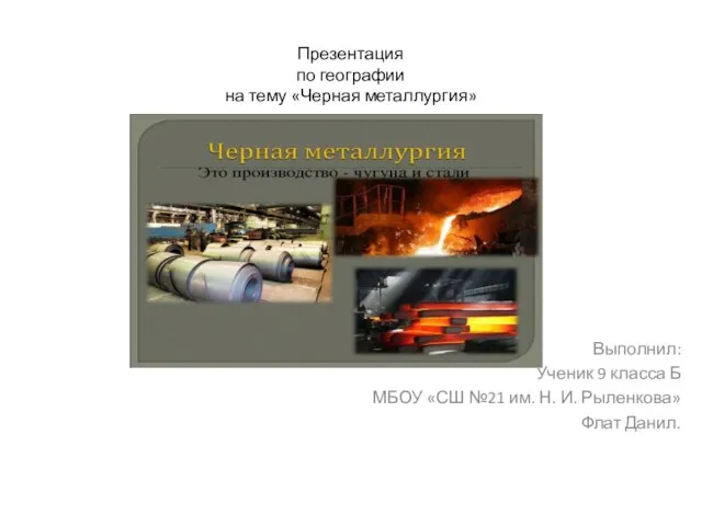 Черная металлургия
 Психолого-педагогическая диагностика
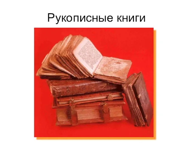 Рукописные книги
 Использование приёмов технологии развития критического мышления при написании части «С».
 МЕДИЦИНСКАЯ И БИОЛОГИЧЕСКАЯ ФИЗИКА
 Передача жилых домов в управление
 Aleksander Sergeevich PUSHKIN 1799-1837
 Сергей Александрович Есенин.(1895-1925)
 Презентация на тему "Будни и праздники 5 класса" - скачать презентации по Педагогике
 Эпоха возрождения
 Интуитивные решения. Интуитивное мышление
 Ведение реестра организаций отдыха детей и их оздоровления , расположенных на территории Свердловской области
 экстр пси ДПО
 Профессии в области хореографии
 Изменение величин
 Поведение во время грозы
 Тайна бумажного листа
 Воспитательная программа «Любознайки»
 Век Просвещения
 Проблемный метод обученияв преподавании истории