- Manual of Structural Kinesiology

Содержание
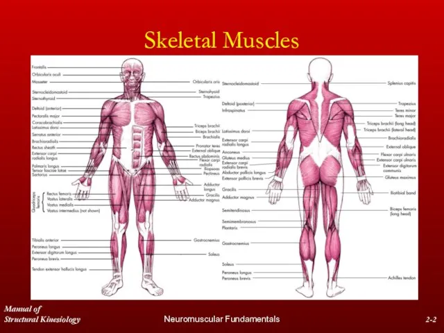
- 2. Manual of Structural Kinesiology Neuromuscular Fundamentals 2- Skeletal Muscles
- 3. Manual of Structural Kinesiology Neuromuscular Fundamentals 2- Skeletal Muscles Over 600 skeletal muscles comprise approximately 40
- 4. Manual of Structural Kinesiology Neuromuscular Fundamentals 2- Muscle Nomenclature Muscles are usually named due to visual
- 5. Manual of Structural Kinesiology Neuromuscular Fundamentals 2- Muscle Nomenclature Action & size – adductor magnus Shape
- 6. Manual of Structural Kinesiology Neuromuscular Fundamentals 2- Muscle Tissue Properties Skeletal muscle tissue has 4 properties
- 7. Manual of Structural Kinesiology Neuromuscular Fundamentals 2- Muscle Tissue Properties Irritability - property of muscle being
- 8. Manual of Structural Kinesiology Neuromuscular Fundamentals 2- Muscle Tissue Properties Extensibility - ability of muscle to
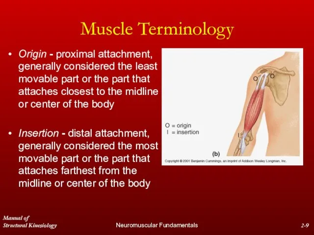
- 9. Manual of Structural Kinesiology Neuromuscular Fundamentals 2- Muscle Terminology Origin - proximal attachment, generally considered the
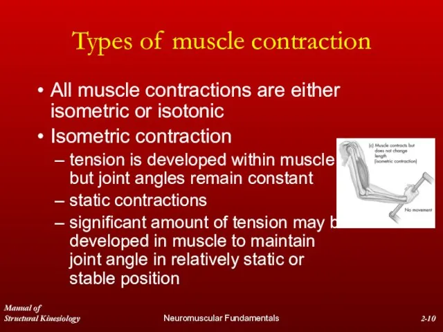
- 10. Manual of Structural Kinesiology Neuromuscular Fundamentals 2- Types of muscle contraction All muscle contractions are either
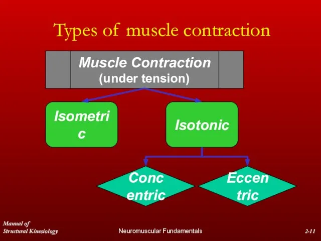
- 11. Manual of Structural Kinesiology Neuromuscular Fundamentals 2- Types of muscle contraction Muscle Contraction (under tension)
- 12. Manual of Structural Kinesiology Neuromuscular Fundamentals 2- Types of muscle contraction Isotonic contractions involve muscle developing
- 13. Manual of Structural Kinesiology Neuromuscular Fundamentals 2- Types of muscle contraction Concentric contractions involve muscle developing
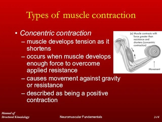
- 14. Manual of Structural Kinesiology Neuromuscular Fundamentals 2- Types of muscle contraction Concentric contraction muscle develops tension
- 15. Manual of Structural Kinesiology Neuromuscular Fundamentals 2- Types of muscle contraction Concentric contraction force developed by
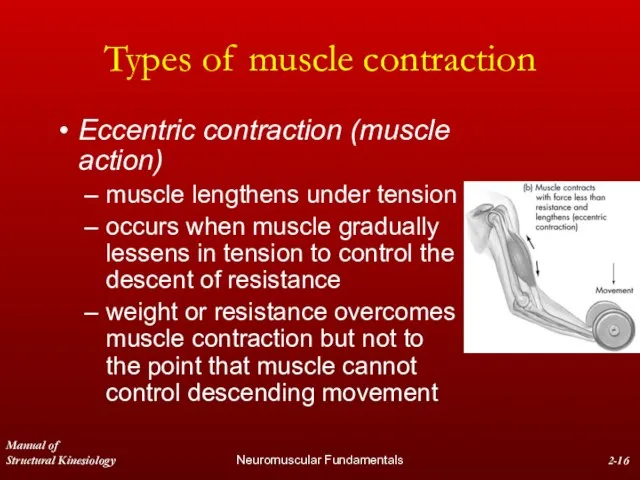
- 16. Manual of Structural Kinesiology Neuromuscular Fundamentals 2- Types of muscle contraction Eccentric contraction (muscle action) muscle
- 17. Manual of Structural Kinesiology Neuromuscular Fundamentals 2- Types of muscle contraction Eccentric contraction (muscle action) controls
- 18. Manual of Structural Kinesiology Neuromuscular Fundamentals 2- Types of muscle contraction Eccentric contraction (muscle action) Some
- 19. Manual of Structural Kinesiology Neuromuscular Fundamentals 2- Types of muscle contraction Isokinetics - a type of
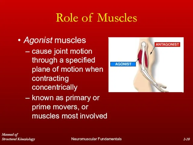
- 20. Manual of Structural Kinesiology Neuromuscular Fundamentals 2- Role of Muscles Agonist muscles cause joint motion through
- 21. Manual of Structural Kinesiology Neuromuscular Fundamentals 2- Role of Muscles Antagonist muscles located on opposite side
- 22. Manual of Structural Kinesiology Neuromuscular Fundamentals 2- Role of Muscles Stabilizers surround joint or body part
- 23. Manual of Structural Kinesiology Neuromuscular Fundamentals 2- Role of Muscles Synergist assist in action of agonists
- 24. Manual of Structural Kinesiology Neuromuscular Fundamentals 2- Role of Muscles Neutralizers Counteract or neutralize the action
- 25. Manual of Structural Kinesiology Neuromuscular Fundamentals 2- Tying Roles of Muscles All Together Muscles with multiple
- 26. Manual of Structural Kinesiology Neuromuscular Fundamentals 2- Tying Roles of Muscles All Together Two muscles may
- 27. Manual of Structural Kinesiology Neuromuscular Fundamentals 2- Tying Roles of Muscles All Together Example of muscle
- 28. Manual of Structural Kinesiology Neuromuscular Fundamentals 2- Tying Roles of Muscles All Together Example of muscle
- 29. Manual of Structural Kinesiology Neuromuscular Fundamentals 2- Tying Roles of Muscles All Together Example of muscle
- 30. Manual of Structural Kinesiology Neuromuscular Fundamentals 2- Tying Roles of Muscles All Together Example of muscle
- 31. Manual of Structural Kinesiology Neuromuscular Fundamentals 2- Tying Roles of Muscles All Together Example of muscle
- 32. Manual of Structural Kinesiology Neuromuscular Fundamentals 2- Tying Roles of Muscles All Together Antagonistic muscles produce
- 33. Manual of Structural Kinesiology Neuromuscular Fundamentals 2- Tying Roles of Muscles All Together Antagonistic muscles produce
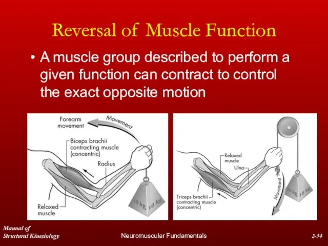
- 34. Manual of Structural Kinesiology Neuromuscular Fundamentals 2- Reversal of Muscle Function A muscle group described to
- 35. Manual of Structural Kinesiology Neuromuscular Fundamentals 2- Neural control of voluntary movement Muscle contraction result from
- 36. Manual of Structural Kinesiology Neuromuscular Fundamentals 2- Neural control of voluntary movement Sensory neurons transmit impulses
- 37. Manual of Structural Kinesiology Neuromuscular Fundamentals 2- Proprioception & Kinesthesis Activity performance is significantly dependent upon
- 38. Manual of Structural Kinesiology Neuromuscular Fundamentals 2- Proprioception & Kinesthesis Taken for granted are sensations associated
- 39. Manual of Structural Kinesiology Neuromuscular Fundamentals 2- Proprioception & Kinesthesis Proprioceptors work in combination with other
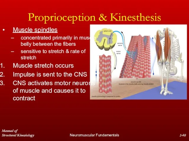
- 40. Manual of Structural Kinesiology Neuromuscular Fundamentals 2- Proprioception & Kinesthesis Muscle spindles concentrated primarily in muscle
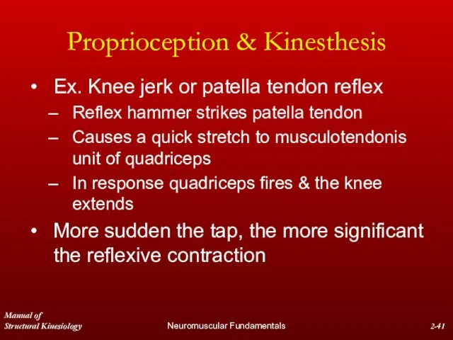
- 41. Manual of Structural Kinesiology Neuromuscular Fundamentals 2- Proprioception & Kinesthesis Ex. Knee jerk or patella tendon
- 42. Manual of Structural Kinesiology Neuromuscular Fundamentals 2- Proprioception & Kinesthesis Stretch reflex may be utilized to
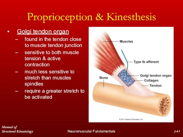
- 43. Manual of Structural Kinesiology Neuromuscular Fundamentals 2- Proprioception & Kinesthesis Golgi tendon organ found in the
- 44. Manual of Structural Kinesiology Neuromuscular Fundamentals 2- Proprioception & Kinesthesis Tension in tendons & GTO increases
- 45. Manual of Structural Kinesiology Neuromuscular Fundamentals 2- All or None Principle All or None Principle -
- 46. Manual of Structural Kinesiology Neuromuscular Fundamentals 2- All or None Principle The number of muscle fibers
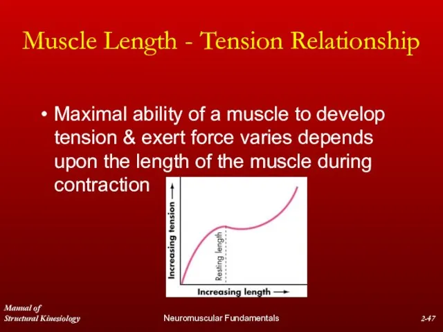
- 47. Manual of Structural Kinesiology Neuromuscular Fundamentals 2- Muscle Length - Tension Relationship Maximal ability of a
- 48. Manual of Structural Kinesiology Neuromuscular Fundamentals 2- Muscle Length - Tension Relationship Generally, depending upon muscle
- 49. Manual of Structural Kinesiology Neuromuscular Fundamentals 2- Muscle Length - Tension Relationship Generally, depending upon muscle
- 50. Manual of Structural Kinesiology Neuromuscular Fundamentals 2- Muscle Length - Tension Relationship Ex. 1 Increasing ability
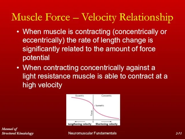
- 51. Manual of Structural Kinesiology Neuromuscular Fundamentals 2- Muscle Force – Velocity Relationship When muscle is contracting
- 52. Manual of Structural Kinesiology Neuromuscular Fundamentals 2- Muscle Force – Velocity Relationship As resistance increases, the
- 53. Manual of Structural Kinesiology Neuromuscular Fundamentals 2- Muscle Force – Velocity Relationship Slight increases in load
- 54. Manual of Structural Kinesiology Neuromuscular Fundamentals 2- Muscle Force – Velocity Relationship As force needed to
- 55. Manual of Structural Kinesiology Neuromuscular Fundamentals 2- Angle of pull Angle between the line of pull
- 56. Manual of Structural Kinesiology Neuromuscular Fundamentals 2- Angle of pull Angle of pull decreases as bone
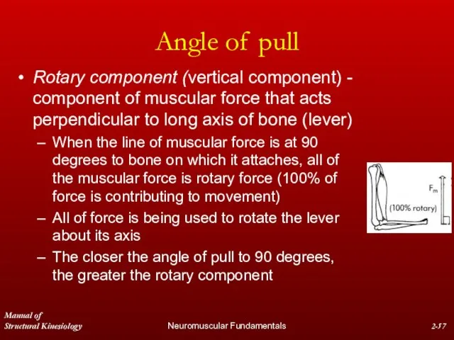
- 57. Manual of Structural Kinesiology Neuromuscular Fundamentals 2- Angle of pull Rotary component (vertical component) - component
- 58. Manual of Structural Kinesiology Neuromuscular Fundamentals 2- Angle of pull At all other degrees of the
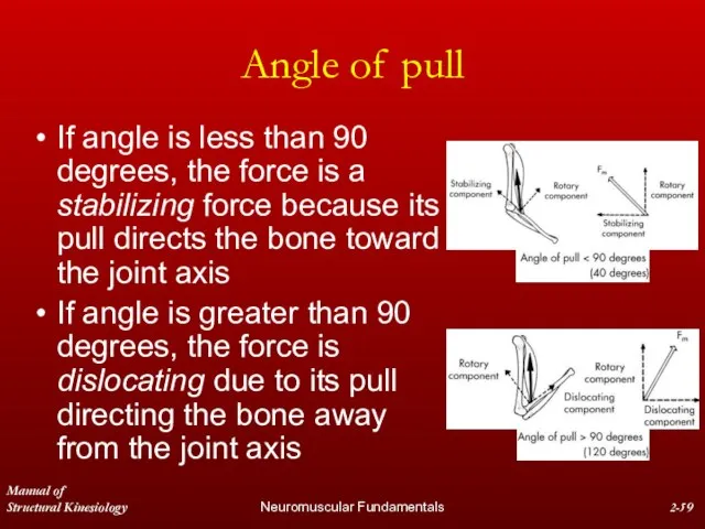
- 59. Manual of Structural Kinesiology Neuromuscular Fundamentals 2- Angle of pull If angle is less than 90
- 60. Manual of Structural Kinesiology Neuromuscular Fundamentals 2- Angle of pull Sometimes desirable to begin with the
- 61. Manual of Structural Kinesiology Neuromuscular Fundamentals 2- Biarticular or Multiarticular Muscles Biarticular muscles – cross &
- 62. Manual of Structural Kinesiology Neuromuscular Fundamentals 2- Biarticular or Multiarticular Muscles Muscle does not actually shorten
- 63. Manual of Structural Kinesiology Neuromuscular Fundamentals 2- Biarticular or Multiarticular Muscles Ex.1 Hip & knee biarticular
- 64. Manual of Structural Kinesiology Neuromuscular Fundamentals 2- Biarticular or Multiarticular Muscles Ex. 2 Hip & knee
- 65. Manual of Structural Kinesiology Neuromuscular Fundamentals 2- Biarticular or Multiarticular Muscles Multiarticular muscles act on three
- 66. Manual of Structural Kinesiology Neuromuscular Fundamentals 2- Reciprocal Inhibition or Innervation Antagonist muscles groups must relax
- 67. Manual of Structural Kinesiology Neuromuscular Fundamentals 2- Reciprocal Inhibition or Innervation Ex. Compare the ease of
- 68. Manual of Structural Kinesiology Neuromuscular Fundamentals 2- Active & Passive Insufficiency As muscle shortens its ability
- 69. Manual of Structural Kinesiology Neuromuscular Fundamentals 2- Active & Passive Insufficiency Easily observed in either biarticular
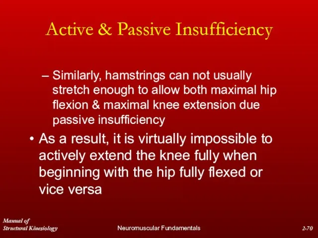
- 70. Manual of Structural Kinesiology Neuromuscular Fundamentals 2- Active & Passive Insufficiency Similarly, hamstrings can not usually
- 71. Manual of Structural Kinesiology Neuromuscular Fundamentals 2- Web Sites Neurologic Exam: An anatomical approach http://medlib.med.utah.edu/neurologicexam/home_exam.html A
- 72. Manual of Structural Kinesiology Neuromuscular Fundamentals 2- Web Sites Dermatomes www.meddean.luc.edu/lumen/MedEd/GrossAnatomy/learnem/dermat/main_der.htm An interactive review of the
- 74. Скачать презентацию
Слайд 3Manual of
Structural Kinesiology
Neuromuscular Fundamentals
2-
Skeletal Muscles
Over 600 skeletal muscles comprise approximately 40
Manual of
Structural Kinesiology
Neuromuscular Fundamentals
2-
Skeletal Muscles
Over 600 skeletal muscles comprise approximately 40
Слайд 4Manual of
Structural Kinesiology
Neuromuscular Fundamentals
2-
Muscle Nomenclature
Muscles are usually named due to
visual appearance
anatomical
Manual of
Structural Kinesiology
Neuromuscular Fundamentals
2-
Muscle Nomenclature
Muscles are usually named due to
visual appearance
anatomical
Слайд 5Manual of
Structural Kinesiology
Neuromuscular Fundamentals
2-
Muscle Nomenclature
Action & size – adductor magnus
Shape &
Manual of
Structural Kinesiology
Neuromuscular Fundamentals
2-
Muscle Nomenclature
Action & size – adductor magnus
Shape &
Слайд 6Manual of
Structural Kinesiology
Neuromuscular Fundamentals
2-
Muscle Tissue Properties
Skeletal muscle tissue has 4 properties
Manual of
Structural Kinesiology
Neuromuscular Fundamentals
2-
Muscle Tissue Properties
Skeletal muscle tissue has 4 properties
Слайд 7Manual of
Structural Kinesiology
Neuromuscular Fundamentals
2-
Muscle Tissue Properties
Irritability - property of muscle being
Manual of
Structural Kinesiology
Neuromuscular Fundamentals
2-
Muscle Tissue Properties
Irritability - property of muscle being
Слайд 8Manual of
Structural Kinesiology
Neuromuscular Fundamentals
2-
Muscle Tissue Properties
Extensibility - ability of muscle to
Manual of
Structural Kinesiology
Neuromuscular Fundamentals
2-
Muscle Tissue Properties
Extensibility - ability of muscle to
Слайд 9Manual of
Structural Kinesiology
Neuromuscular Fundamentals
2-
Muscle Terminology
Origin - proximal attachment, generally considered the
Manual of
Structural Kinesiology
Neuromuscular Fundamentals
2-
Muscle Terminology
Origin - proximal attachment, generally considered the
Слайд 10Manual of
Structural Kinesiology
Neuromuscular Fundamentals
2-
Types of muscle contraction
All muscle contractions are either
Manual of
Structural Kinesiology
Neuromuscular Fundamentals
2-
Types of muscle contraction
All muscle contractions are either
Слайд 11Manual of
Structural Kinesiology
Neuromuscular Fundamentals
2-
Types of muscle contraction
Muscle Contraction
(under tension)
Manual of
Structural Kinesiology
Neuromuscular Fundamentals
2-
Types of muscle contraction
Muscle Contraction
(under tension)
Слайд 12Manual of
Structural Kinesiology
Neuromuscular Fundamentals
2-
Types of muscle contraction
Isotonic contractions involve muscle developing
Manual of
Structural Kinesiology
Neuromuscular Fundamentals
2-
Types of muscle contraction
Isotonic contractions involve muscle developing
Слайд 13Manual of
Structural Kinesiology
Neuromuscular Fundamentals
2-
Types of muscle contraction
Concentric contractions involve muscle developing
Manual of
Structural Kinesiology
Neuromuscular Fundamentals
2-
Types of muscle contraction
Concentric contractions involve muscle developing
Слайд 14Manual of
Structural Kinesiology
Neuromuscular Fundamentals
2-
Types of muscle contraction
Concentric contraction
muscle develops tension as
Manual of
Structural Kinesiology
Neuromuscular Fundamentals
2-
Types of muscle contraction
Concentric contraction
muscle develops tension as
Слайд 15Manual of
Structural Kinesiology
Neuromuscular Fundamentals
2-
Types of muscle contraction
Concentric contraction
force developed by the
Manual of
Structural Kinesiology
Neuromuscular Fundamentals
2-
Types of muscle contraction
Concentric contraction
force developed by the
Слайд 16Manual of
Structural Kinesiology
Neuromuscular Fundamentals
2-
Types of muscle contraction
Eccentric contraction (muscle action)
muscle lengthens
Manual of
Structural Kinesiology
Neuromuscular Fundamentals
2-
Types of muscle contraction
Eccentric contraction (muscle action)
muscle lengthens
Слайд 17Manual of
Structural Kinesiology
Neuromuscular Fundamentals
2-
Types of muscle contraction
Eccentric contraction (muscle action)
controls movement
Manual of
Structural Kinesiology
Neuromuscular Fundamentals
2-
Types of muscle contraction
Eccentric contraction (muscle action)
controls movement
Слайд 18Manual of
Structural Kinesiology
Neuromuscular Fundamentals
2-
Types of muscle contraction
Eccentric contraction (muscle action)
Some refer
Manual of
Structural Kinesiology
Neuromuscular Fundamentals
2-
Types of muscle contraction
Eccentric contraction (muscle action)
Some refer
Слайд 19Manual of
Structural Kinesiology
Neuromuscular Fundamentals
2-
Types of muscle contraction
Isokinetics - a type of
Manual of
Structural Kinesiology
Neuromuscular Fundamentals
2-
Types of muscle contraction
Isokinetics - a type of
Слайд 20Manual of
Structural Kinesiology
Neuromuscular Fundamentals
2-
Role of Muscles
Agonist muscles
cause joint motion through a
Manual of
Structural Kinesiology
Neuromuscular Fundamentals
2-
Role of Muscles
Agonist muscles
cause joint motion through a
Слайд 21Manual of
Structural Kinesiology
Neuromuscular Fundamentals
2-
Role of Muscles
Antagonist muscles
located on opposite side of
Manual of
Structural Kinesiology
Neuromuscular Fundamentals
2-
Role of Muscles
Antagonist muscles
located on opposite side of
Слайд 22Manual of
Structural Kinesiology
Neuromuscular Fundamentals
2-
Role of Muscles
Stabilizers
surround joint or body part
contract to
Manual of
Structural Kinesiology
Neuromuscular Fundamentals
2-
Role of Muscles
Stabilizers
surround joint or body part
contract to
Слайд 23Manual of
Structural Kinesiology
Neuromuscular Fundamentals
2-
Role of Muscles
Synergist
assist in action of agonists
not necessarily
Manual of
Structural Kinesiology
Neuromuscular Fundamentals
2-
Role of Muscles
Synergist
assist in action of agonists
not necessarily
Слайд 24Manual of
Structural Kinesiology
Neuromuscular Fundamentals
2-
Role of Muscles
Neutralizers
Counteract or neutralize the action of
Manual of
Structural Kinesiology
Neuromuscular Fundamentals
2-
Role of Muscles
Neutralizers
Counteract or neutralize the action of
Слайд 25Manual of
Structural Kinesiology
Neuromuscular Fundamentals
2-
Tying Roles of Muscles All Together
Muscles with multiple
Manual of
Structural Kinesiology
Neuromuscular Fundamentals
2-
Tying Roles of Muscles All Together
Muscles with multiple
Слайд 26Manual of
Structural Kinesiology
Neuromuscular Fundamentals
2-
Tying Roles of Muscles All Together
Two muscles may
Manual of
Structural Kinesiology
Neuromuscular Fundamentals
2-
Tying Roles of Muscles All Together
Two muscles may
Слайд 27Manual of
Structural Kinesiology
Neuromuscular Fundamentals
2-
Tying Roles of Muscles All Together
Example of muscle
Manual of
Structural Kinesiology
Neuromuscular Fundamentals
2-
Tying Roles of Muscles All Together
Example of muscle
Слайд 28Manual of
Structural Kinesiology
Neuromuscular Fundamentals
2-
Tying Roles of Muscles All Together
Example of muscle
Manual of
Structural Kinesiology
Neuromuscular Fundamentals
2-
Tying Roles of Muscles All Together
Example of muscle
Слайд 29Manual of
Structural Kinesiology
Neuromuscular Fundamentals
2-
Tying Roles of Muscles All Together
Example of muscle
Manual of
Structural Kinesiology
Neuromuscular Fundamentals
2-
Tying Roles of Muscles All Together
Example of muscle
Слайд 30Manual of
Structural Kinesiology
Neuromuscular Fundamentals
2-
Tying Roles of Muscles All Together
Example of muscle
Manual of
Structural Kinesiology
Neuromuscular Fundamentals
2-
Tying Roles of Muscles All Together
Example of muscle
Слайд 31Manual of
Structural Kinesiology
Neuromuscular Fundamentals
2-
Tying Roles of Muscles All Together
Example of muscle
Manual of
Structural Kinesiology
Neuromuscular Fundamentals
2-
Tying Roles of Muscles All Together
Example of muscle
Слайд 32Manual of
Structural Kinesiology
Neuromuscular Fundamentals
2-
Tying Roles of Muscles All Together
Antagonistic muscles produce
Manual of
Structural Kinesiology
Neuromuscular Fundamentals
2-
Tying Roles of Muscles All Together
Antagonistic muscles produce
Слайд 33Manual of
Structural Kinesiology
Neuromuscular Fundamentals
2-
Tying Roles of Muscles All Together
Antagonistic muscles produce
Manual of
Structural Kinesiology
Neuromuscular Fundamentals
2-
Tying Roles of Muscles All Together
Antagonistic muscles produce
Слайд 34Manual of
Structural Kinesiology
Neuromuscular Fundamentals
2-
Reversal of Muscle Function
A muscle group described to
Manual of
Structural Kinesiology
Neuromuscular Fundamentals
2-
Reversal of Muscle Function
A muscle group described to
Слайд 35Manual of
Structural Kinesiology
Neuromuscular Fundamentals
2-
Neural control of voluntary movement
Muscle contraction result from
Manual of
Structural Kinesiology
Neuromuscular Fundamentals
2-
Neural control of voluntary movement
Muscle contraction result from
Слайд 36Manual of
Structural Kinesiology
Neuromuscular Fundamentals
2-
Neural control of voluntary movement
Sensory neurons transmit impulses
Manual of
Structural Kinesiology
Neuromuscular Fundamentals
2-
Neural control of voluntary movement
Sensory neurons transmit impulses
Слайд 37Manual of
Structural Kinesiology
Neuromuscular Fundamentals
2-
Proprioception & Kinesthesis
Activity performance is significantly dependent upon
Manual of
Structural Kinesiology
Neuromuscular Fundamentals
2-
Proprioception & Kinesthesis
Activity performance is significantly dependent upon
Слайд 38Manual of
Structural Kinesiology
Neuromuscular Fundamentals
2-
Proprioception & Kinesthesis
Taken for granted are sensations associated
Manual of
Structural Kinesiology
Neuromuscular Fundamentals
2-
Proprioception & Kinesthesis
Taken for granted are sensations associated
Слайд 39Manual of
Structural Kinesiology
Neuromuscular Fundamentals
2-
Proprioception & Kinesthesis
Proprioceptors work in combination with other
Manual of
Structural Kinesiology
Neuromuscular Fundamentals
2-
Proprioception & Kinesthesis
Proprioceptors work in combination with other
Слайд 40Manual of
Structural Kinesiology
Neuromuscular Fundamentals
2-
Proprioception & Kinesthesis
Muscle spindles
concentrated primarily in muscle belly
Manual of
Structural Kinesiology
Neuromuscular Fundamentals
2-
Proprioception & Kinesthesis
Muscle spindles
concentrated primarily in muscle belly
Слайд 41Manual of
Structural Kinesiology
Neuromuscular Fundamentals
2-
Proprioception & Kinesthesis
Ex. Knee jerk or patella tendon
Manual of
Structural Kinesiology
Neuromuscular Fundamentals
2-
Proprioception & Kinesthesis
Ex. Knee jerk or patella tendon
Слайд 42Manual of
Structural Kinesiology
Neuromuscular Fundamentals
2-
Proprioception & Kinesthesis
Stretch reflex may be utilized to
Manual of
Structural Kinesiology
Neuromuscular Fundamentals
2-
Proprioception & Kinesthesis
Stretch reflex may be utilized to
Слайд 43Manual of
Structural Kinesiology
Neuromuscular Fundamentals
2-
Proprioception & Kinesthesis
Golgi tendon organ
found in the tendon
Manual of
Structural Kinesiology
Neuromuscular Fundamentals
2-
Proprioception & Kinesthesis
Golgi tendon organ
found in the tendon
Слайд 44Manual of
Structural Kinesiology
Neuromuscular Fundamentals
2-
Proprioception & Kinesthesis
Tension in tendons & GTO increases
Manual of
Structural Kinesiology
Neuromuscular Fundamentals
2-
Proprioception & Kinesthesis
Tension in tendons & GTO increases
Слайд 45Manual of
Structural Kinesiology
Neuromuscular Fundamentals
2-
All or None Principle
All or None Principle -
Manual of
Structural Kinesiology
Neuromuscular Fundamentals
2-
All or None Principle
All or None Principle -
Слайд 46Manual of
Structural Kinesiology
Neuromuscular Fundamentals
2-
All or None Principle
The number of muscle fibers
Manual of
Structural Kinesiology
Neuromuscular Fundamentals
2-
All or None Principle
The number of muscle fibers
Слайд 47Manual of
Structural Kinesiology
Neuromuscular Fundamentals
2-
Muscle Length - Tension Relationship
Maximal ability of a
Manual of
Structural Kinesiology
Neuromuscular Fundamentals
2-
Muscle Length - Tension Relationship
Maximal ability of a
Слайд 48Manual of
Structural Kinesiology
Neuromuscular Fundamentals
2-
Muscle Length - Tension Relationship
Generally, depending upon muscle
Manual of
Structural Kinesiology
Neuromuscular Fundamentals
2-
Muscle Length - Tension Relationship
Generally, depending upon muscle
Слайд 49Manual of
Structural Kinesiology
Neuromuscular Fundamentals
2-
Muscle Length - Tension Relationship
Generally, depending upon muscle
Manual of
Structural Kinesiology
Neuromuscular Fundamentals
2-
Muscle Length - Tension Relationship
Generally, depending upon muscle
Слайд 50Manual of
Structural Kinesiology
Neuromuscular Fundamentals
2-
Muscle Length - Tension Relationship
Ex. 1 Increasing ability
Manual of
Structural Kinesiology
Neuromuscular Fundamentals
2-
Muscle Length - Tension Relationship
Ex. 1 Increasing ability
Слайд 51Manual of
Structural Kinesiology
Neuromuscular Fundamentals
2-
Muscle Force – Velocity Relationship
When muscle is contracting
Manual of
Structural Kinesiology
Neuromuscular Fundamentals
2-
Muscle Force – Velocity Relationship
When muscle is contracting
Слайд 52Manual of
Structural Kinesiology
Neuromuscular Fundamentals
2-
Muscle Force – Velocity Relationship
As resistance increases, the
Manual of
Structural Kinesiology
Neuromuscular Fundamentals
2-
Muscle Force – Velocity Relationship
As resistance increases, the
Слайд 53Manual of
Structural Kinesiology
Neuromuscular Fundamentals
2-
Muscle Force – Velocity Relationship
Slight increases in load
Manual of
Structural Kinesiology
Neuromuscular Fundamentals
2-
Muscle Force – Velocity Relationship
Slight increases in load
Слайд 54Manual of
Structural Kinesiology
Neuromuscular Fundamentals
2-
Muscle Force – Velocity Relationship
As force needed to
Manual of
Structural Kinesiology
Neuromuscular Fundamentals
2-
Muscle Force – Velocity Relationship
As force needed to
Слайд 55Manual of
Structural Kinesiology
Neuromuscular Fundamentals
2-
Angle of pull
Angle between the line of pull
Manual of
Structural Kinesiology
Neuromuscular Fundamentals
2-
Angle of pull
Angle between the line of pull
Слайд 56Manual of
Structural Kinesiology
Neuromuscular Fundamentals
2-
Angle of pull
Angle of pull decreases as bone
Manual of
Structural Kinesiology
Neuromuscular Fundamentals
2-
Angle of pull
Angle of pull decreases as bone
Слайд 57Manual of
Structural Kinesiology
Neuromuscular Fundamentals
2-
Angle of pull
Rotary component (vertical component) - component
Manual of
Structural Kinesiology
Neuromuscular Fundamentals
2-
Angle of pull
Rotary component (vertical component) - component
Слайд 58Manual of
Structural Kinesiology
Neuromuscular Fundamentals
2-
Angle of pull
At all other degrees of the
Manual of
Structural Kinesiology
Neuromuscular Fundamentals
2-
Angle of pull
At all other degrees of the
Слайд 59Manual of
Structural Kinesiology
Neuromuscular Fundamentals
2-
Angle of pull
If angle is less than 90
Manual of
Structural Kinesiology
Neuromuscular Fundamentals
2-
Angle of pull
If angle is less than 90
Слайд 60Manual of
Structural Kinesiology
Neuromuscular Fundamentals
2-
Angle of pull
Sometimes desirable to begin with the
Manual of
Structural Kinesiology
Neuromuscular Fundamentals
2-
Angle of pull
Sometimes desirable to begin with the
Слайд 61Manual of
Structural Kinesiology
Neuromuscular Fundamentals
2-
Biarticular or Multiarticular Muscles
Biarticular muscles – cross &
Manual of
Structural Kinesiology
Neuromuscular Fundamentals
2-
Biarticular or Multiarticular Muscles
Biarticular muscles – cross &
Слайд 62Manual of
Structural Kinesiology
Neuromuscular Fundamentals
2-
Biarticular or Multiarticular Muscles
Muscle does not actually shorten
Manual of
Structural Kinesiology
Neuromuscular Fundamentals
2-
Biarticular or Multiarticular Muscles
Muscle does not actually shorten
Слайд 63Manual of
Structural Kinesiology
Neuromuscular Fundamentals
2-
Biarticular or Multiarticular Muscles
Ex.1 Hip & knee biarticular
Manual of
Structural Kinesiology
Neuromuscular Fundamentals
2-
Biarticular or Multiarticular Muscles
Ex.1 Hip & knee biarticular
Слайд 64Manual of
Structural Kinesiology
Neuromuscular Fundamentals
2-
Biarticular or Multiarticular Muscles
Ex. 2 Hip & knee
Manual of
Structural Kinesiology
Neuromuscular Fundamentals
2-
Biarticular or Multiarticular Muscles
Ex. 2 Hip & knee
Слайд 65Manual of
Structural Kinesiology
Neuromuscular Fundamentals
2-
Biarticular or Multiarticular Muscles
Multiarticular muscles act on three
Manual of
Structural Kinesiology
Neuromuscular Fundamentals
2-
Biarticular or Multiarticular Muscles
Multiarticular muscles act on three
Слайд 66Manual of
Structural Kinesiology
Neuromuscular Fundamentals
2-
Reciprocal Inhibition or Innervation
Antagonist muscles groups must relax
Manual of
Structural Kinesiology
Neuromuscular Fundamentals
2-
Reciprocal Inhibition or Innervation
Antagonist muscles groups must relax
Слайд 67Manual of
Structural Kinesiology
Neuromuscular Fundamentals
2-
Reciprocal Inhibition or Innervation
Ex. Compare the ease of
stretching
Manual of
Structural Kinesiology
Neuromuscular Fundamentals
2-
Reciprocal Inhibition or Innervation
Ex. Compare the ease of
stretching
Слайд 68Manual of
Structural Kinesiology
Neuromuscular Fundamentals
2-
Active & Passive Insufficiency
As muscle shortens its ability
Manual of
Structural Kinesiology
Neuromuscular Fundamentals
2-
Active & Passive Insufficiency
As muscle shortens its ability
Слайд 69Manual of
Structural Kinesiology
Neuromuscular Fundamentals
2-
Active & Passive Insufficiency
Easily observed in either biarticular
Manual of
Structural Kinesiology
Neuromuscular Fundamentals
2-
Active & Passive Insufficiency
Easily observed in either biarticular
Слайд 70Manual of
Structural Kinesiology
Neuromuscular Fundamentals
2-
Active & Passive Insufficiency
Similarly, hamstrings can not usually
Manual of
Structural Kinesiology
Neuromuscular Fundamentals
2-
Active & Passive Insufficiency
Similarly, hamstrings can not usually
Слайд 71Manual of
Structural Kinesiology
Neuromuscular Fundamentals
2-
Web Sites
Neurologic Exam: An anatomical approach
http://medlib.med.utah.edu/neurologicexam/home_exam.html
A very thorough
Manual of
Structural Kinesiology
Neuromuscular Fundamentals
2-
Web Sites
Neurologic Exam: An anatomical approach
http://medlib.med.utah.edu/neurologicexam/home_exam.html
A very thorough
Слайд 72Manual of
Structural Kinesiology
Neuromuscular Fundamentals
2-
Web Sites
Dermatomes
www.meddean.luc.edu/lumen/MedEd/GrossAnatomy/learnem/dermat/main_der.htm
An interactive review of the body’s dermatomes.
Loyola
Manual of
Structural Kinesiology
Neuromuscular Fundamentals
2-
Web Sites
Dermatomes
www.meddean.luc.edu/lumen/MedEd/GrossAnatomy/learnem/dermat/main_der.htm
An interactive review of the body’s dermatomes.
Loyola
 Учение Ч.Дарвина о естественном отборе
 Die Bayer Aktiengesellschaft (Bayer AG) ist die Holding-Gesellschaft des Bayer- Konzerns
 Применение органического теплоизоляционного материала для предотвращения образования сосулек и наледи на крышах зданий
 СТАТИЧЕСКИ ОПРЕДЕЛИМЫЕ СИСТЕМЫС СТРОИТЕЛЬНАЯ МЕХАНИКА
  Настали святки
 Electronic Learning — система электронного обучения
  Презентация на тему Решение задач по нахождению величин по сумме и разности
 Что такое перспектива
 Защита от мошенников
 Восприятие россиянами товаров премиум-класса и роскоши как стиля жизни
 Архивные фотографии о первых днях ВОВ
 История шотландской реликвии
 Михаил Афанасьевич Булгаков
 Стихотворение «Дума» 1838 год
 Осевая симметрия
 London
 Историческое развитие человечества: поиски социальной макротеории
 РБД_ДЗ1
 Собственность
 Еще три дня нашей смены...
 Призыв граждан на военную службу
 Что такое информация. Виды информации
 Картины на заказ: масло, акварель, гуашь
 Анималистический жанр. 2 класс
 Московская государственная академия ветеринарной медицины и биотехнологии имени К.И. Скрябина
 Irish famous writers
  Коммерческое предложение. Бумажное шоу Снег
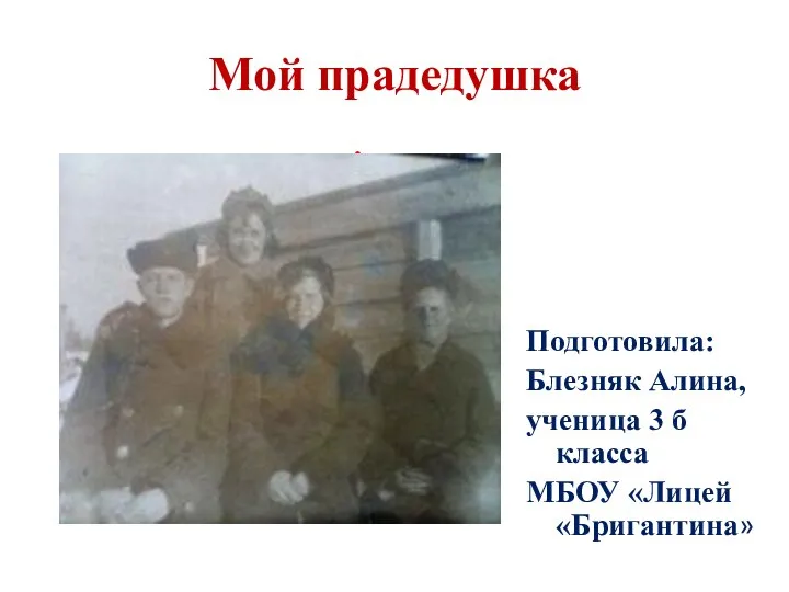 Мой прадедушка Толочко Максим Савельевич