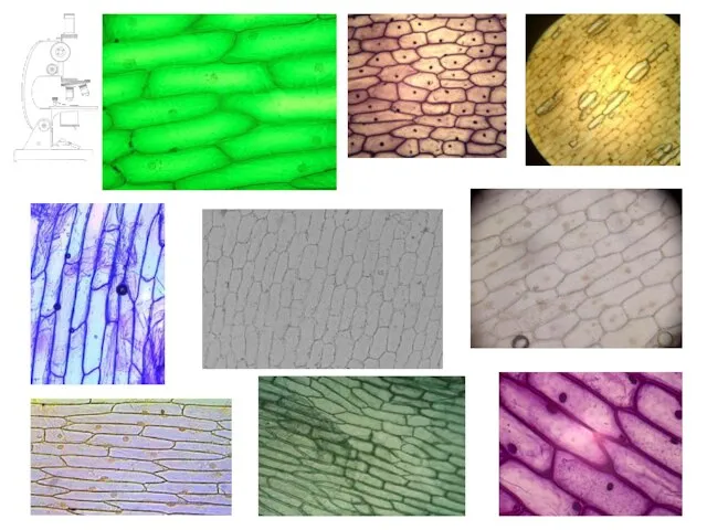
- Measuring cells

Содержание
- 2. Syllabus reference:
- 3. This symbol in the corner of a slide indicates a picture, diagram or table taken from
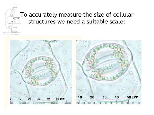
- 4. To accurately measure the size of cellular structures we need a suitable scale:
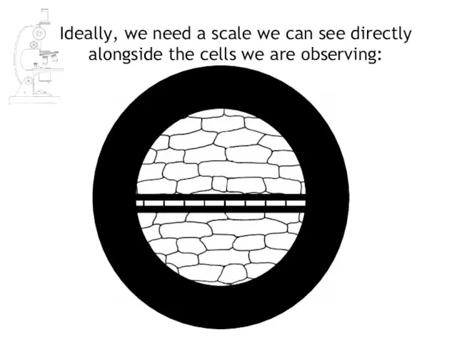
- 5. Ideally, we need a scale we can see directly alongside the cells we are observing:
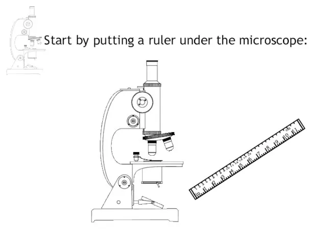
- 6. Start by putting a ruler under the microscope:
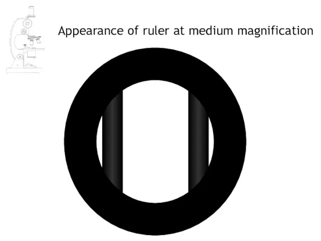
- 7. Appearance of ruler at medium magnification
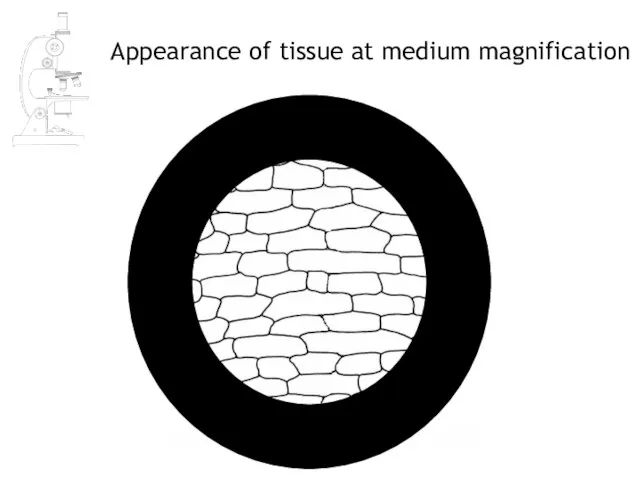
- 8. Appearance of tissue at medium magnification
- 9. Estimating cell size at medium magnification 1mm 5 1 ÷ 5 = 0.2mm 12 1 ÷
- 11. Mean length of cells = 0.2 x 1000 = 200µm 1mm = 1000µm Mean width of
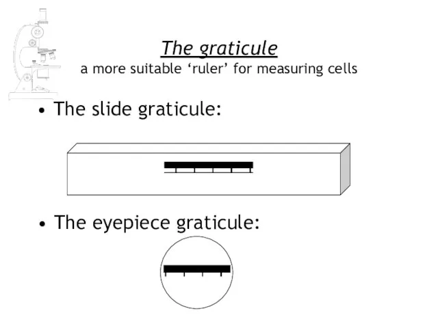
- 15. The graticule a more suitable ‘ruler’ for measuring cells The slide graticule: The eyepiece graticule:
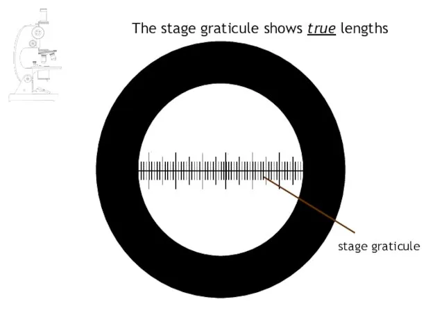
- 16. The stage graticule shows true lengths stage graticule
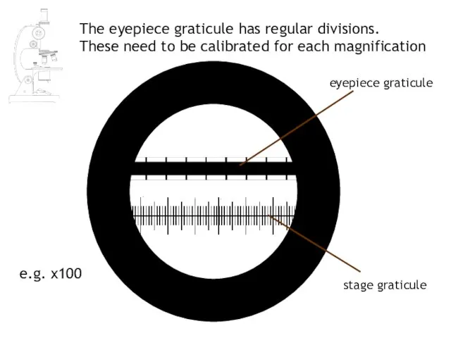
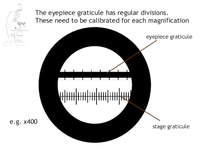
- 17. The eyepiece graticule has regular divisions. These need to be calibrated for each magnification stage graticule
- 18. stage graticule eyepiece graticule e.g. x400 The eyepiece graticule has regular divisions. These need to be
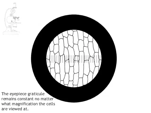
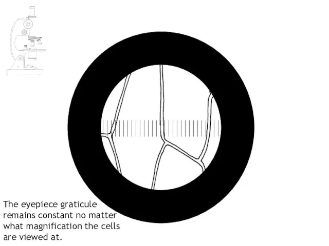
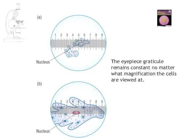
- 19. The eyepiece graticule remains constant no matter what magnification the cells are viewed at.
- 20. The eyepiece graticule remains constant no matter what magnification the cells are viewed at.
- 21. The eyepiece graticule remains constant no matter what magnification the cells are viewed at.
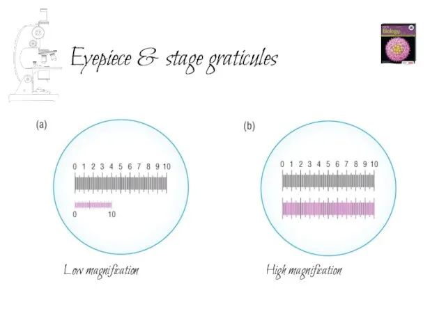
- 22. Eyepiece & stage graticules Low magnification High magnification
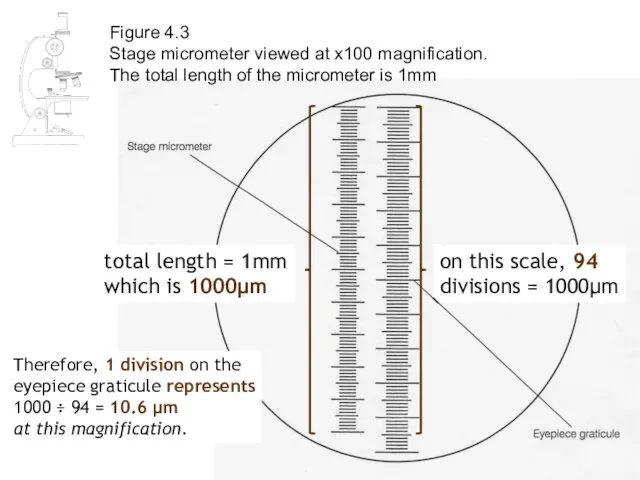
- 23. Figure 4.3 Stage micrometer viewed at x100 magnification. The total length of the micrometer is 1mm
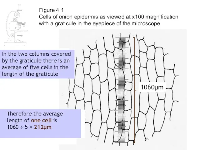
- 24. Figure 4.1 Cells of onion epidermis as viewed at x100 magnification with a graticule in the
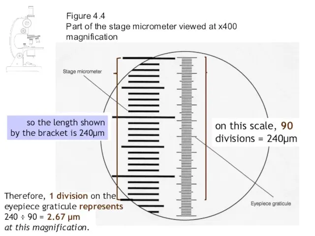
- 25. remember that each division here is 10μm Figure 4.4 Part of the stage micrometer viewed at
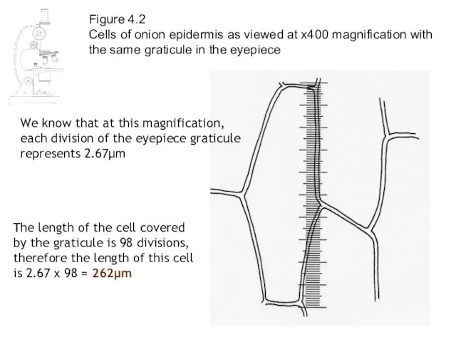
- 26. Figure 4.2 Cells of onion epidermis as viewed at x400 magnification with the same graticule in
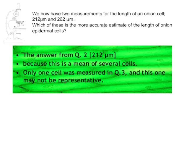
- 27. We now have two measurements for the length of an onion cell; 212μm and 262 μm.
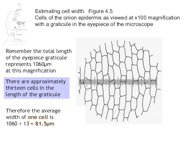
- 28. Estimating cell width. Figure 4.5. Cells of the onion epidermis as viewed at x100 magnification with
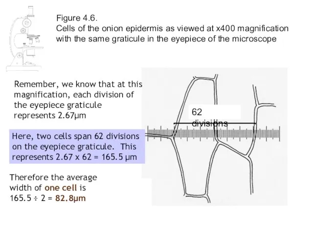
- 29. Figure 4.6. Cells of the onion epidermis as viewed at x400 magnification with the same graticule
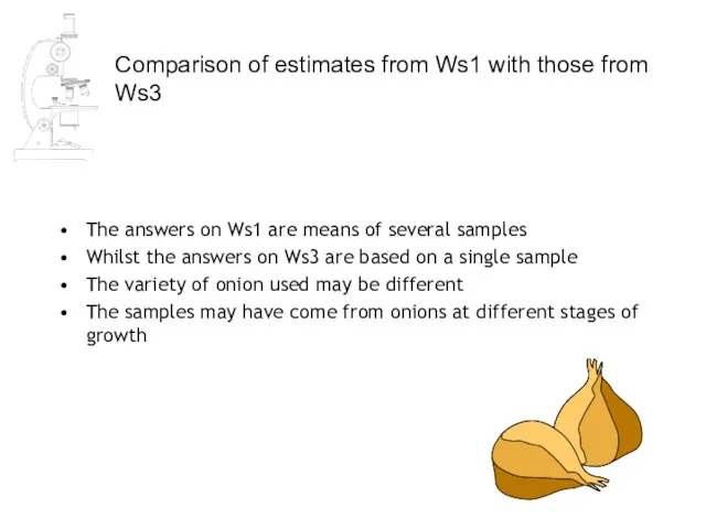
- 30. Comparison of estimates from Ws1 with those from Ws3 The answers on Ws1 are means of
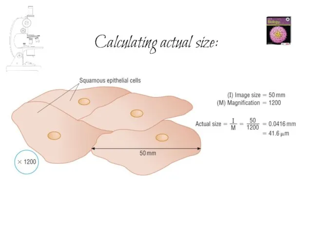
- 31. Calculating actual size:
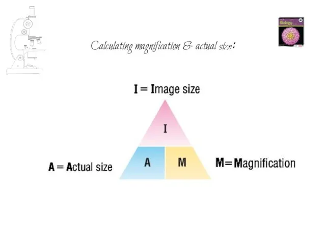
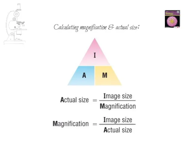
- 32. Calculating magnification & actual size:
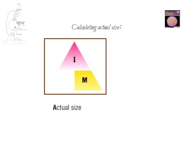
- 33. Calculating actual size:
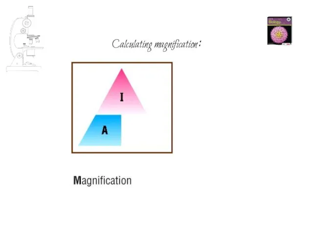
- 34. Calculating magnification:
- 35. Calculating magnification & actual size:
- 37. Скачать презентацию
Слайд 3This symbol in the corner of a slide indicates a picture, diagram
This symbol in the corner of a slide indicates a picture, diagram
Слайд 4To accurately measure the size of cellular structures we need a suitable
To accurately measure the size of cellular structures we need a suitable
Слайд 5Ideally, we need a scale we can see directly alongside the cells
Ideally, we need a scale we can see directly alongside the cells
Слайд 6Start by putting a ruler under the microscope:
Start by putting a ruler under the microscope:
Слайд 7Appearance of ruler at medium magnification
Appearance of ruler at medium magnification
Слайд 8Appearance of tissue at medium magnification
Appearance of tissue at medium magnification
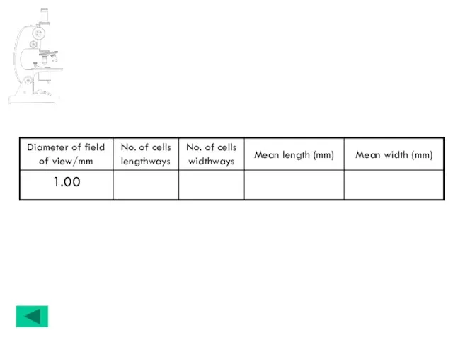
Слайд 9Estimating cell size at medium magnification
1mm
5
1 ÷ 5 = 0.2mm
12
1 ÷ 12
Estimating cell size at medium magnification
1mm
5
1 ÷ 5 = 0.2mm
12
1 ÷ 12
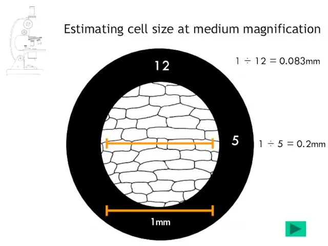
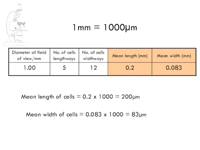
Слайд 11Mean length of cells = 0.2 x 1000 = 200µm
1mm = 1000µm
Mean
Mean length of cells = 0.2 x 1000 = 200µm
1mm = 1000µm
Mean
Слайд 15The graticule
a more suitable ‘ruler’ for measuring cells
The slide graticule:
The eyepiece
The graticule
a more suitable ‘ruler’ for measuring cells
The slide graticule:
The eyepiece
Слайд 16The stage graticule shows true lengths
stage graticule
The stage graticule shows true lengths
stage graticule
Слайд 17The eyepiece graticule has regular divisions.
These need to be calibrated for
The eyepiece graticule has regular divisions.
These need to be calibrated for
Слайд 18stage graticule
eyepiece graticule
e.g. x400
The eyepiece graticule has regular divisions.
These need to
stage graticule
eyepiece graticule
e.g. x400
The eyepiece graticule has regular divisions.
These need to
Слайд 19The eyepiece graticule
remains constant no matter
what magnification the cells
are
The eyepiece graticule
remains constant no matter
what magnification the cells
are
Слайд 20The eyepiece graticule
remains constant no matter
what magnification the cells
are
The eyepiece graticule
remains constant no matter
what magnification the cells
are
Слайд 21The eyepiece graticule
remains constant no matter
what magnification the cells
are
The eyepiece graticule
remains constant no matter
what magnification the cells
are
Слайд 22Eyepiece & stage graticules
Low magnification
High magnification
Eyepiece & stage graticules
Low magnification
High magnification
Слайд 23Figure 4.3
Stage micrometer viewed at x100 magnification.
The total length of the
Figure 4.3 Stage micrometer viewed at x100 magnification. The total length of the
Слайд 24Figure 4.1
Cells of onion epidermis as viewed at x100 magnification with a
Figure 4.1 Cells of onion epidermis as viewed at x100 magnification with a
Слайд 25remember that each
division here is 10μm
Figure 4.4
Part of the stage micrometer
remember that each
division here is 10μm
Figure 4.4 Part of the stage micrometer
Слайд 26Figure 4.2
Cells of onion epidermis as viewed at x400 magnification with the
Figure 4.2 Cells of onion epidermis as viewed at x400 magnification with the
Слайд 27We now have two measurements for the length of an onion cell;
We now have two measurements for the length of an onion cell;
Слайд 28Estimating cell width. Figure 4.5.
Cells of the onion epidermis as viewed
Estimating cell width. Figure 4.5. Cells of the onion epidermis as viewed
Слайд 29Figure 4.6.
Cells of the onion epidermis as viewed at x400 magnification
Figure 4.6. Cells of the onion epidermis as viewed at x400 magnification
Слайд 30Comparison of estimates from Ws1 with those from Ws3
The answers on Ws1
Comparison of estimates from Ws1 with those from Ws3
The answers on Ws1
Слайд 31Calculating actual size:
Calculating actual size:
Слайд 32Calculating magnification & actual size:
Calculating magnification & actual size:
Слайд 33Calculating actual size:
Calculating actual size:
Слайд 34Calculating magnification:
Calculating magnification:
Слайд 35Calculating magnification & actual size:
Calculating magnification & actual size:
 Powracam do Boga
 ПРИРОДНЫЕ СООБЩЕСТВА ВОКРУГ НАС (биология, экология)
 Uchebnaya_praktika_Golub_Maria_20PI-2
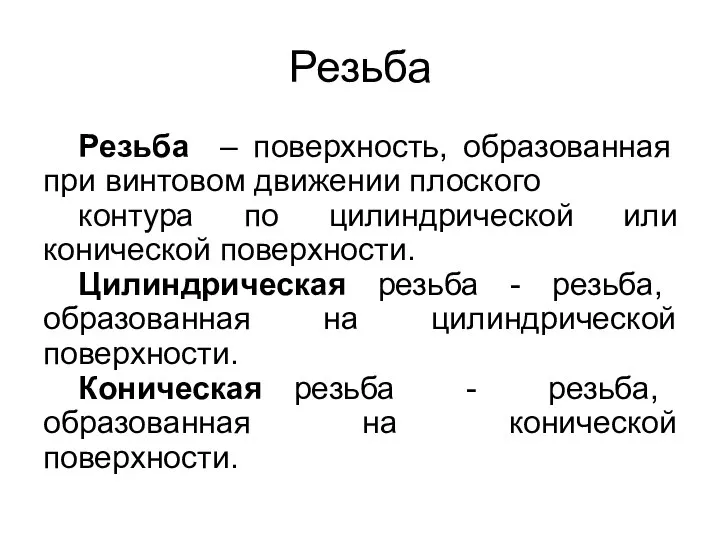 Резьба. Изображение резьбы
 Socjalizacja do kultury fizycznej
 Супер Маша
 Ahunbaev Isa Konoevich
 Системный подход к освоению месторождений с учетом природно-техногенных изменений недр – основа углеводорододобывающего промыс
 Ахиллесова пята
 Теорема о вписанном угле в окружность.
 Тема: Биотические факторы среды
 Бизнес-Менеджер АГРО Блок «Интерактивная карта»
 Архитектура барокко
 Система подготовки учащихся к ЕГЭ по обществознанию
 Конфигурация «Клиент Федерального казначейства. Администратор доходов» на платформе 1С:Предприятие 8
 Welcome to Great Britain
  SoveTshennoletniy
 Европейская экономическая комиссия ООН Подготовила: Клеутина С.А. ДС_01
 О, спорт, ты – мир!!! 6 класс
 Рождество в Великобретании
 Метод координат
 Разработка стратегии образовательного учреждения: вводные замечания
 Неиспользуемые объекты недвижимости, предлагаемые для продажи
 Научный комплекс
 Таможенное дело. Электронное декларирование
 О правилах питания
 Презентация на тему Решение задач. Закрепление (2 класс)
 ДОБРОКАЧЕСТВЕННЫЕ ОПУХОЛИ ЖЕНСКИХ ПОЛОВЫХ ОРГАНОВ (лекция)