- Sense organs

Содержание
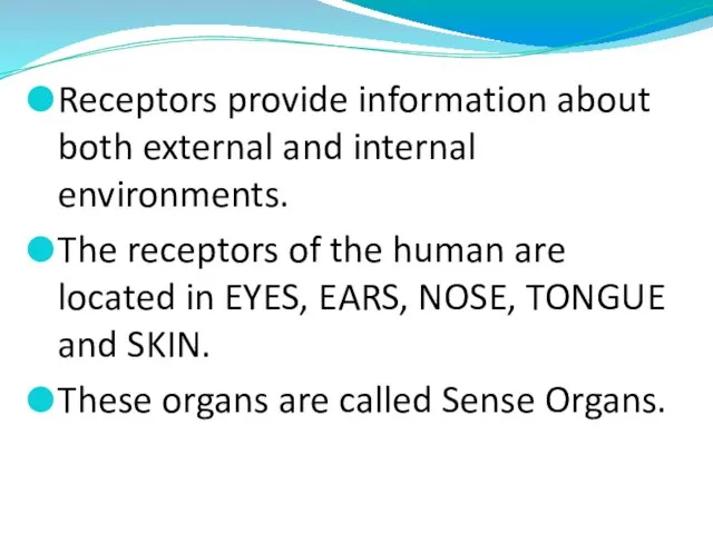
- 2. Receptors provide information about both external and internal environments. The receptors of the human are located
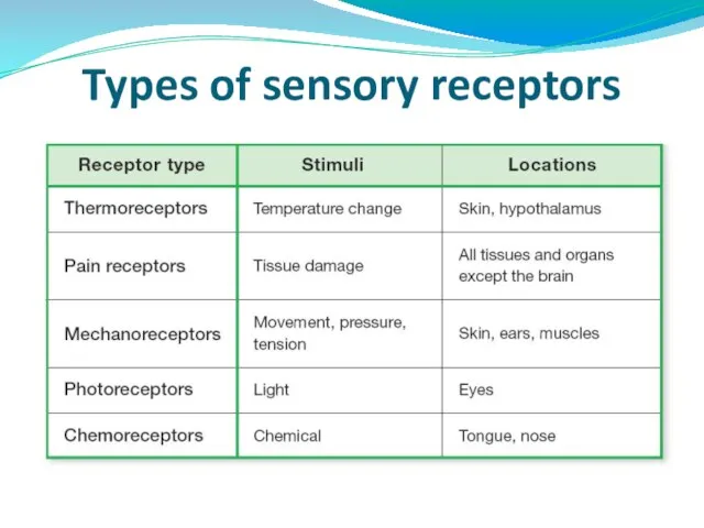
- 3. Types of sensory receptors
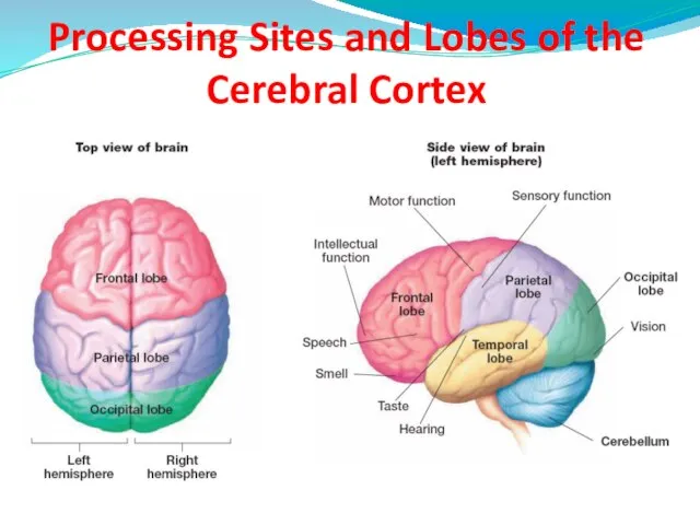
- 4. Processing Sites and Lobes of the Cerebral Cortex
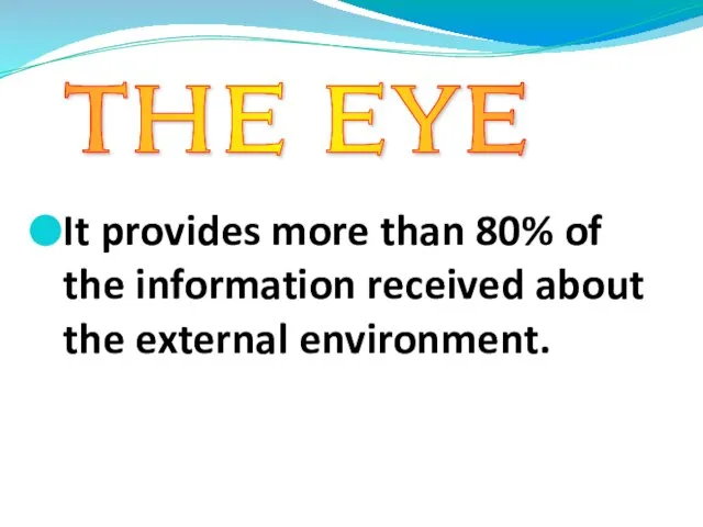
- 5. It provides more than 80% of the information received about the external environment. THE EYE
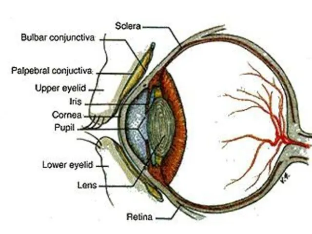
- 7. STRUCTURE OF EYE Eyes contain 3 main parts; Schlerenchyma Choroid Retina
- 8. Schlerenchyma It is supportive structure of eye that protects inner structures of the eye. In the
- 9. CHOROID Just inside the sclera is the choroid. This layer contains many blood vessels. At the
- 10. The diameter of iris is related to the amount of light. It narrows pupil under intense
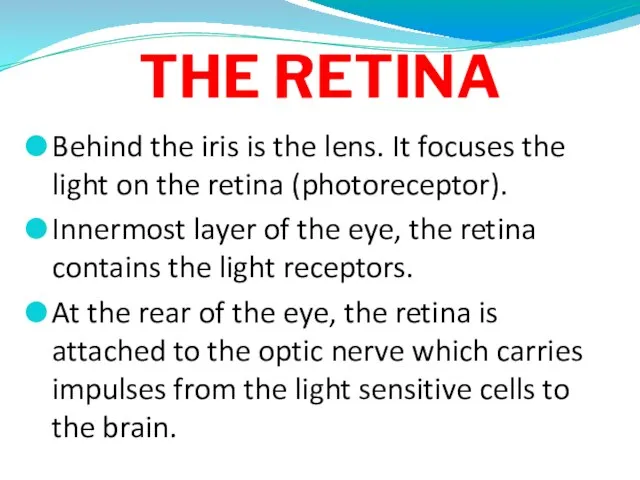
- 11. THE RETINA Behind the iris is the lens. It focuses the light on the retina (photoreceptor).
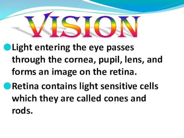
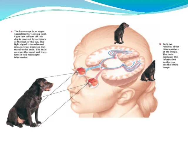
- 14. Light entering the eye passes through the cornea, pupil, lens, and forms an image on the
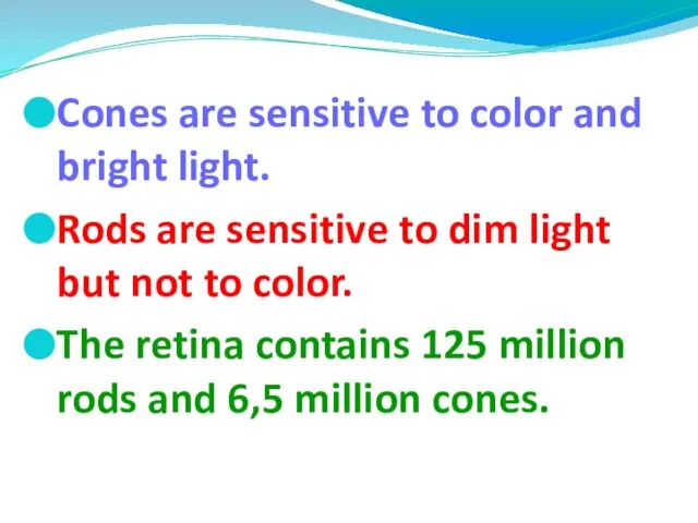
- 15. Cones are sensitive to color and bright light. Rods are sensitive to dim light but not
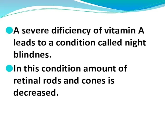
- 17. A severe dificiency of vitamin A leads to a condition called night blindnes. In this condition
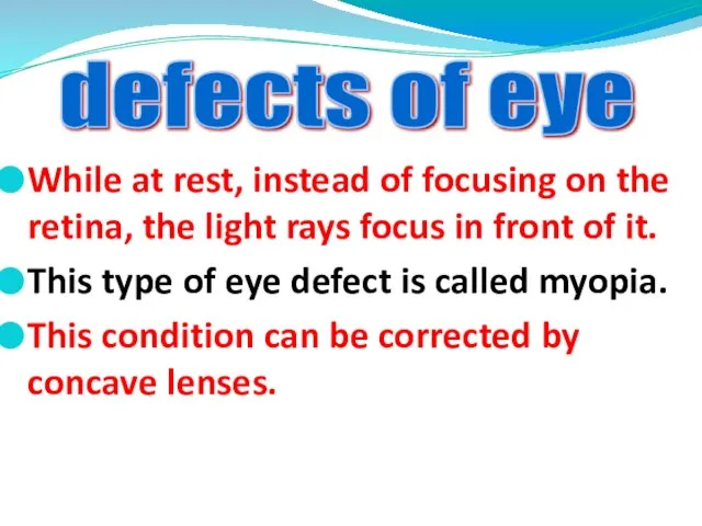
- 19. While at rest, instead of focusing on the retina, the light rays focus in front of
- 20. At rest, the light rays focus behind instead of on the retina. This type of eye
- 21. The human ear has 2 sensory functions. One of them is hearing. Other is maintaning balance
- 22. Structure of ears Ears contains 3 main parts; Outer ear The middle ear Inner ear
- 24. 1. OUTER EAR Outer ear is composed of 3 parts. These are pinna, auditory canal and
- 25. Auditory canal is a canal which is found between pinna and eardrum. It has hairs and
- 26. 2. MIDDLE EAR It contains three small bones which are called the hammer-malleus, anvil-incus and stirrup-stapes.
- 28. EUSTACHIAN TUBE It is located between pharynx and the middle ear. It equalizes air pressure between
- 29. 3. THE INNER EAR It consists of the cochlea and semicircular canals. Cochlea is organ of
- 30. They are separated from another by membranes. Lining of the membranes are specialized hair cells that
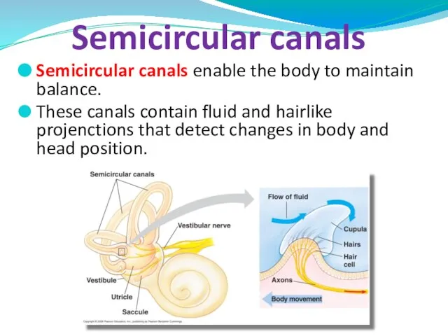
- 32. Semicircular canals enable the body to maintain balance. These canals contain fluid and hairlike projenctions that
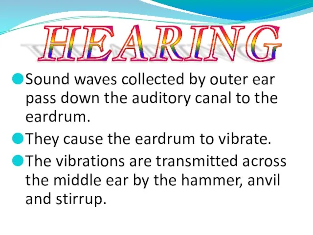
- 34. Sound waves collected by outer ear pass down the auditory canal to the eardrum. They cause
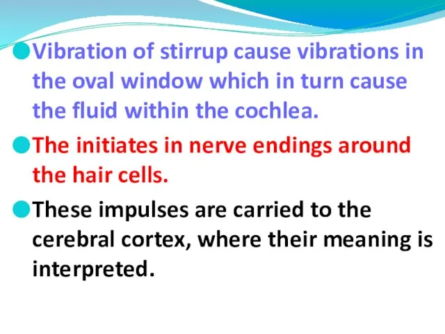
- 35. Vibration of stirrup cause vibrations in the oval window which in turn cause the fluid within
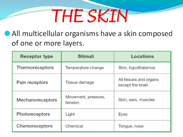
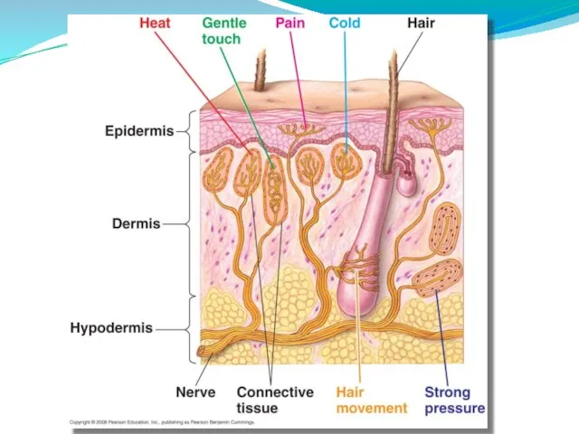
- 37. All multicellular organisms have a skin composed of one or more layers. THE SKIN
- 39. Functions of Skin It protects the inner layers of the body from physical and chemical effects.
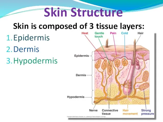
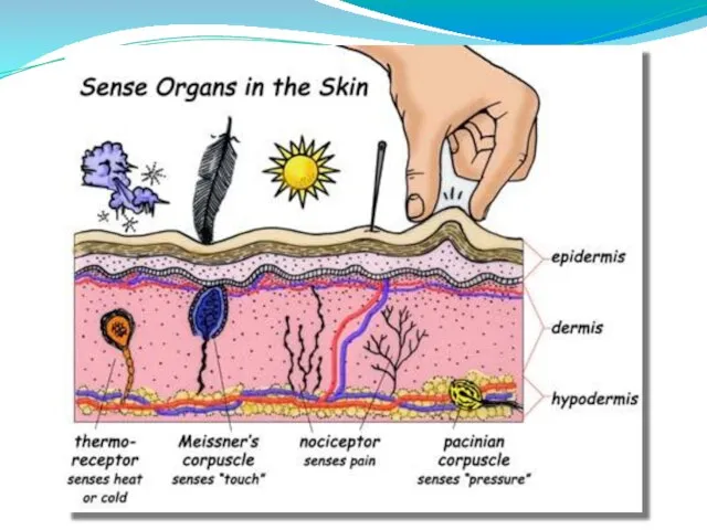
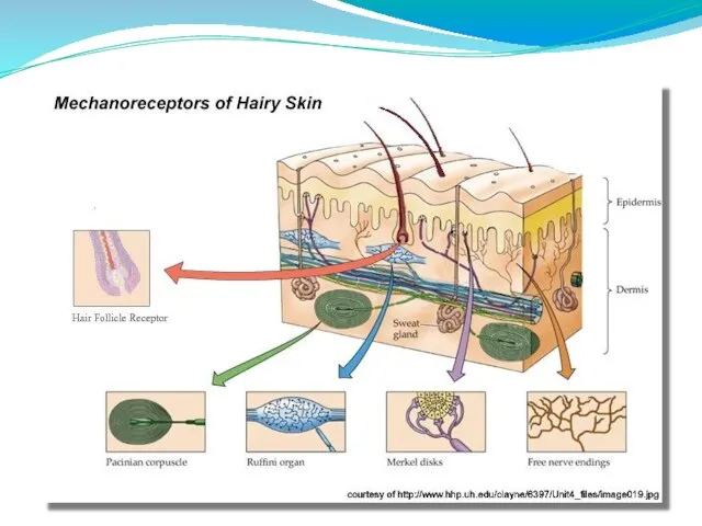
- 40. Skin is composed of 3 tissue layers: Epidermis Dermis Hypodermis Skin Structure
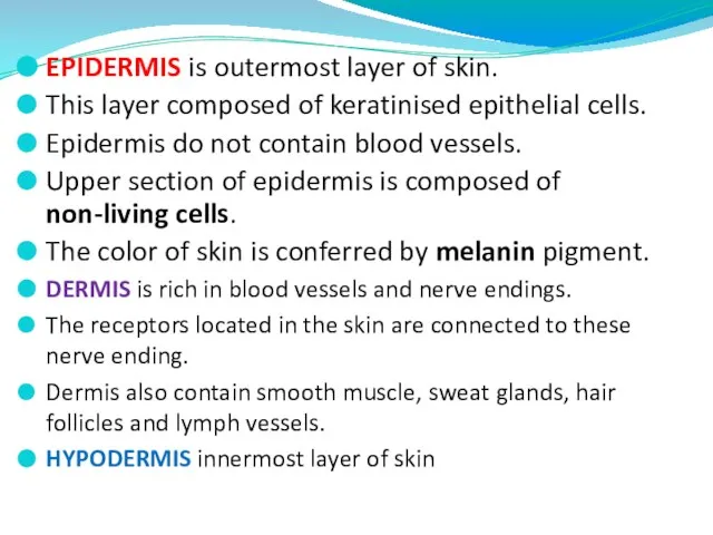
- 41. EPIDERMIS is outermost layer of skin. This layer composed of keratinised epithelial cells. Epidermis do not
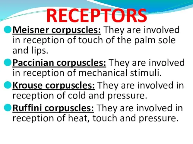
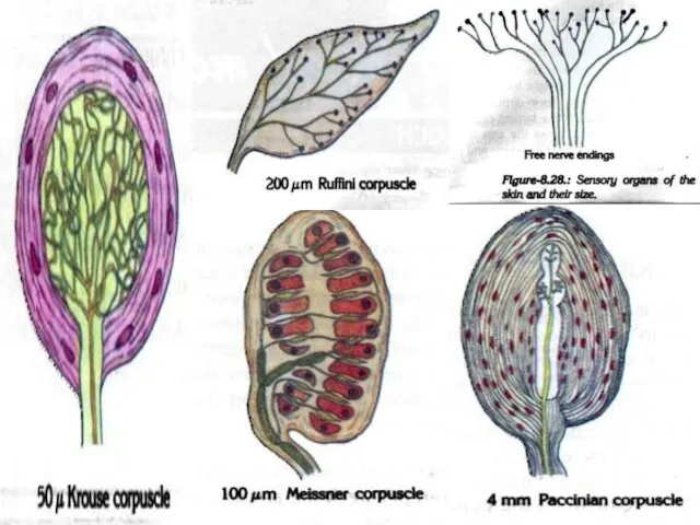
- 42. RECEPTORS Meisner corpuscles: They are involved in reception of touch of the palm sole and lips.
- 46. Sweat glands: They are present in all regions of the skin. They open onto the surface
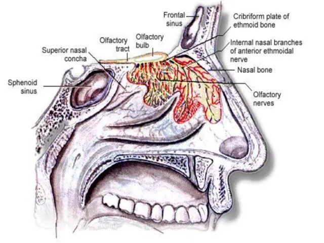
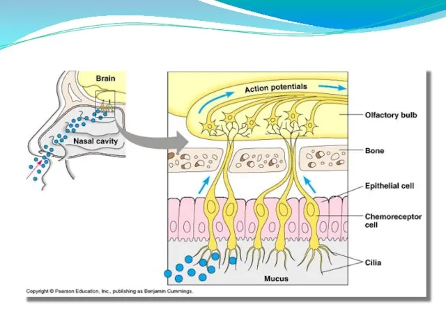
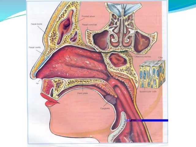
- 47. Nose is the organ of the body involved in both respiration and smell. The reception of
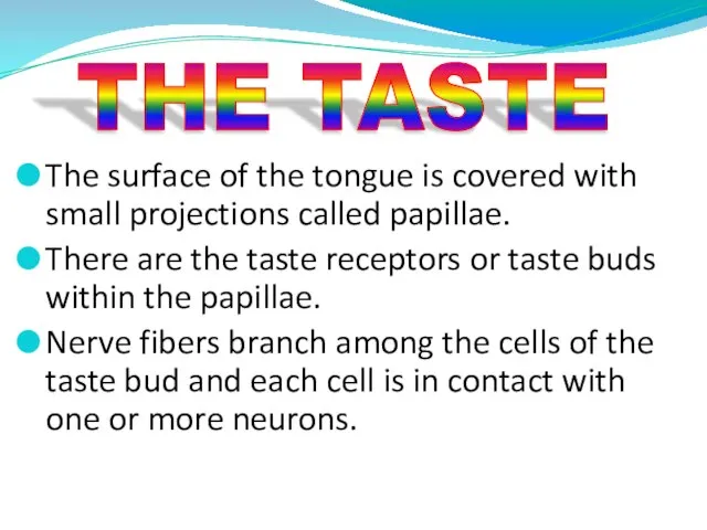
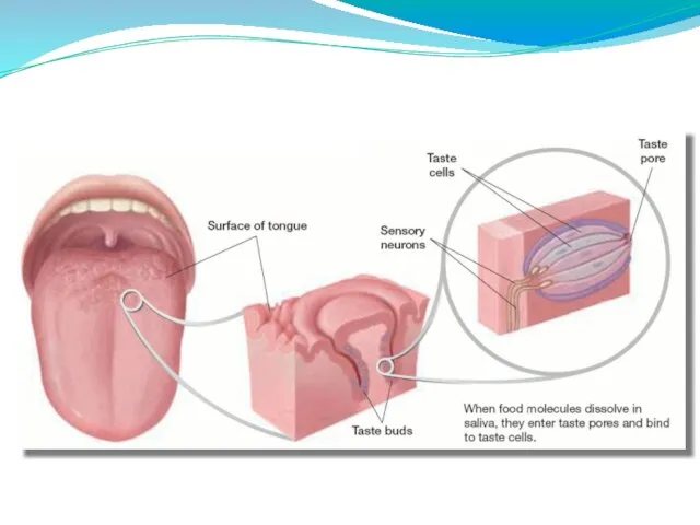
- 51. The surface of the tongue is covered with small projections called papillae. There are the taste
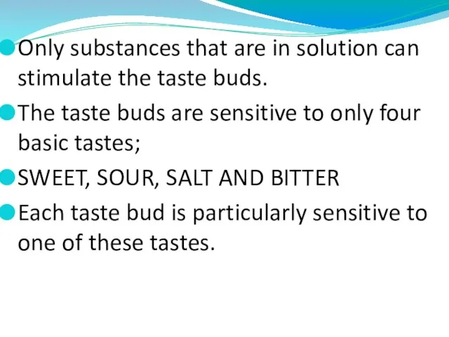
- 53. Only substances that are in solution can stimulate the taste buds. The taste buds are sensitive
- 54. When taste buds are stimulated, impulses are initiated by the sensory cells of the structure and
- 56. Скачать презентацию
Слайд 2Receptors provide information about both external and internal environments.
The receptors of the
Receptors provide information about both external and internal environments.
The receptors of the
Слайд 3Types of sensory receptors
Types of sensory receptors
Слайд 4Processing Sites and Lobes of the Cerebral Cortex
Processing Sites and Lobes of the Cerebral Cortex
Слайд 5It provides more than 80% of the information received about the external
It provides more than 80% of the information received about the external

Слайд 7
STRUCTURE OF EYE
Eyes contain 3 main parts;
Schlerenchyma
Choroid
Retina
STRUCTURE OF EYE
Eyes contain 3 main parts;
Schlerenchyma
Choroid
Retina
Слайд 8
Schlerenchyma
It is supportive structure of eye that protects inner structures of the
Schlerenchyma
It is supportive structure of eye that protects inner structures of the
Слайд 9CHOROID
Just inside the sclera is the choroid.
This layer contains many blood vessels.
CHOROID
Just inside the sclera is the choroid.
This layer contains many blood vessels.
Слайд 10The diameter of iris is related to the amount of light.
It narrows
The diameter of iris is related to the amount of light.
It narrows
Слайд 11THE RETINA
Behind the iris is the lens. It focuses the light on
THE RETINA
Behind the iris is the lens. It focuses the light on
Слайд 14Light entering the eye passes through the cornea, pupil, lens, and forms
Light entering the eye passes through the cornea, pupil, lens, and forms
Слайд 15Cones are sensitive to color and bright light.
Rods are sensitive to
Cones are sensitive to color and bright light.
Rods are sensitive to
Слайд 17A severe dificiency of vitamin A leads to a condition called night
A severe dificiency of vitamin A leads to a condition called night

Слайд 19While at rest, instead of focusing on the retina, the light rays
While at rest, instead of focusing on the retina, the light rays
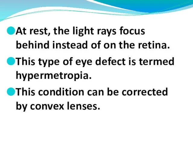
Слайд 20At rest, the light rays focus behind instead of on the retina.
At rest, the light rays focus behind instead of on the retina.
Слайд 21The human ear has 2 sensory functions.
One of them is hearing.
Other
The human ear has 2 sensory functions.
One of them is hearing.
Other
Слайд 22Structure of ears
Ears contains 3 main parts;
Outer ear
The middle ear
Inner ear
Structure of ears
Ears contains 3 main parts;
Outer ear
The middle ear
Inner ear
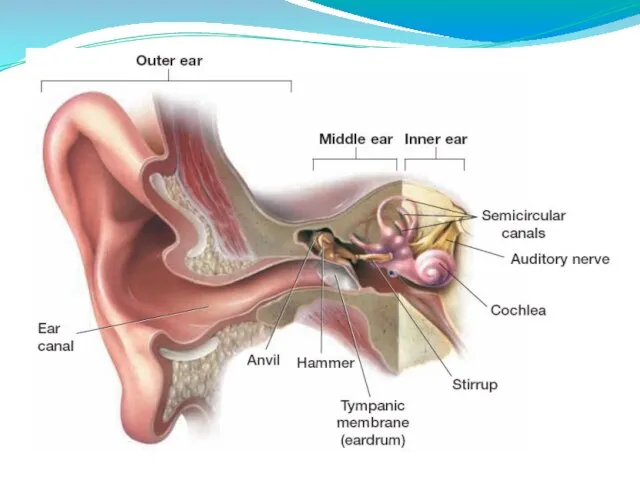
Слайд 241. OUTER EAR
Outer ear is composed of 3 parts.
These are pinna, auditory
1. OUTER EAR
Outer ear is composed of 3 parts.
These are pinna, auditory
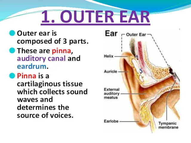
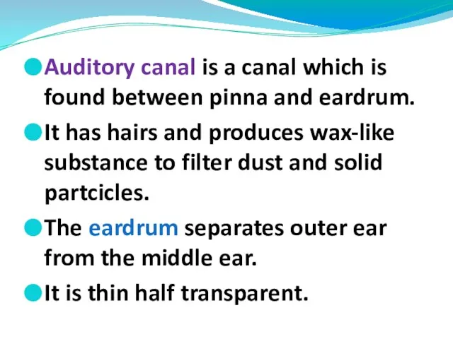
Слайд 25Auditory canal is a canal which is found between pinna and eardrum.
It
Auditory canal is a canal which is found between pinna and eardrum.
It
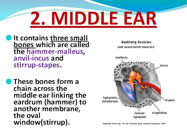
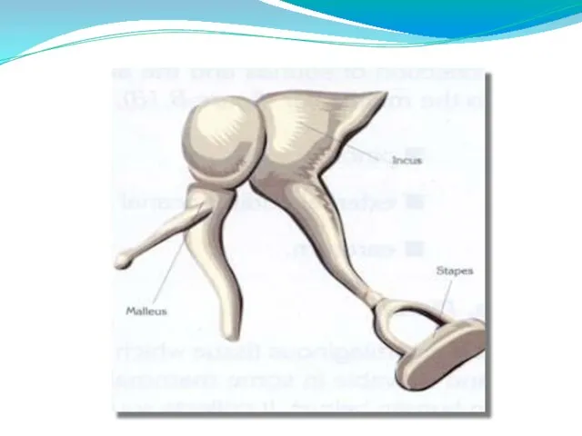
Слайд 262. MIDDLE EAR
It contains three small bones which are called the hammer-malleus,
2. MIDDLE EAR
It contains three small bones which are called the hammer-malleus,
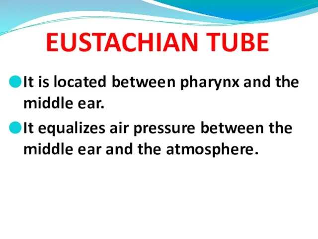
Слайд 28EUSTACHIAN TUBE
It is located between pharynx and the middle ear.
It equalizes air
EUSTACHIAN TUBE
It is located between pharynx and the middle ear.
It equalizes air
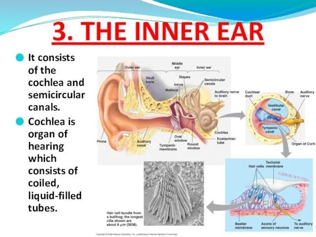
Слайд 293. THE INNER EAR
It consists of the cochlea and semicircular canals.
Cochlea is
3. THE INNER EAR
It consists of the cochlea and semicircular canals.
Cochlea is
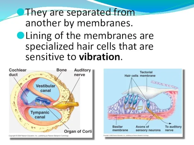
Слайд 30They are separated from another by membranes.
Lining of the membranes are specialized
They are separated from another by membranes.
Lining of the membranes are specialized
Слайд 32Semicircular canals enable the body to maintain balance.
These canals contain fluid and
Semicircular canals enable the body to maintain balance.
These canals contain fluid and

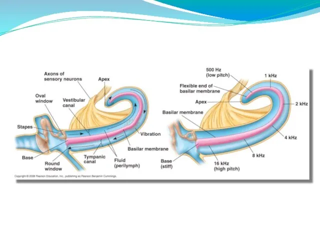
Слайд 34Sound waves collected by outer ear pass down the auditory canal to
Sound waves collected by outer ear pass down the auditory canal to
Слайд 35Vibration of stirrup cause vibrations in the oval window which in turn
Vibration of stirrup cause vibrations in the oval window which in turn

Слайд 37All multicellular organisms have a skin composed of one or more layers.
THE
All multicellular organisms have a skin composed of one or more layers.
THE
Слайд 39Functions of Skin
It protects the inner layers of the body from physical
Functions of Skin
It protects the inner layers of the body from physical
Слайд 40 Skin is composed of 3 tissue layers:
Epidermis
Dermis
Hypodermis
Skin Structure
Skin is composed of 3 tissue layers:
Epidermis
Dermis
Hypodermis
Skin Structure
Слайд 41EPIDERMIS is outermost layer of skin.
This layer composed of keratinised epithelial cells.
Epidermis
EPIDERMIS is outermost layer of skin.
This layer composed of keratinised epithelial cells.
Epidermis
Слайд 42RECEPTORS
Meisner corpuscles: They are involved in reception of touch of the palm
RECEPTORS
Meisner corpuscles: They are involved in reception of touch of the palm
Слайд 46 Sweat glands:
They are present in all regions of the skin. They
Sweat glands:
They are present in all regions of the skin. They
Слайд 47Nose is the organ of the body involved in both respiration and
Nose is the organ of the body involved in both respiration and
Слайд 51The surface of the tongue is covered with small projections called papillae.
There
The surface of the tongue is covered with small projections called papillae.
There
Слайд 53Only substances that are in solution can stimulate the taste buds.
The taste
Only substances that are in solution can stimulate the taste buds.
The taste
Слайд 54When taste buds are stimulated, impulses are initiated by the sensory cells
When taste buds are stimulated, impulses are initiated by the sensory cells
 Вов
Вов Бальні танці
 Состояние и перспективы развития системы аккредитации в сфере добровольного и обязательного подтверждения соответствия в Герман
 С любовью к животным
 Ооо Невская химия
 Психология человека
 Практические аспекты диагностики и лечения тромбозов у детей
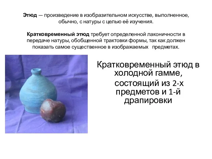 Кратковременный этюд в холодной гамме
 ПРОБЛЕМА КОРРУПЦИЯ В СОВРЕМЕННОЙ РОССИИ
 ПРОБЛЕМА ДЕЛОКАЛИЗАЦИИИСОХРАНЕНИЯ ЗНАНИЯ
 Практическая работа - тест по теме: "Симметрия"
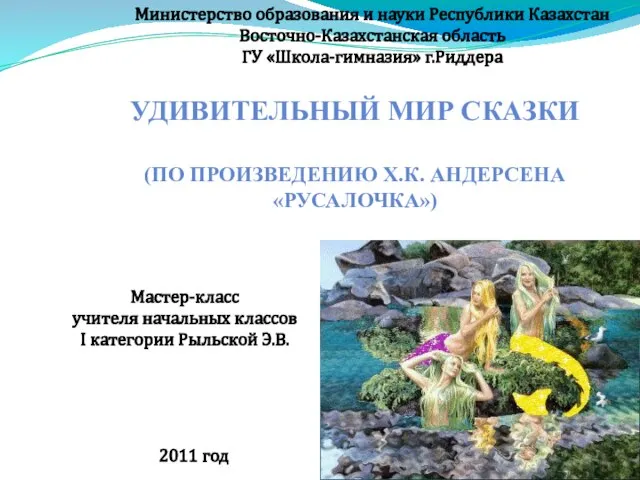 Презентация на тему Русалочка
  Вторая встреча. Миллион с Аязом 12-13 января
 Ubuntu + Python + Selenium=Легкий Старт
 Автоматизация звука Р в словах и предложениях
  Теоретическое планирование макроциклов, мезоциклов и микроциклов
 Подъёмы, спуск, торможения на лыжах
 Задачи на ноябрь, Почта России
 Презентация на тему Мутации (11 класс)
 20141109_1a._uznay_gornuyu_porodu
 Декодер Delta DMX 4-512
 Презентация на тему Цыгане - нация мира
 A Day to Remember. The Past Simple Tense
 Команда Борцы Экологического Движения
 День защиты детей
 Пункт поиска воевавших родственников
 Инструктаж
 Информационная безопасность: основные понятия и определения