- Transthoracic Echocardiography

Содержание
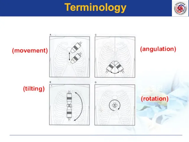
- 2. A. 이동 (movement) 기울임 (tilting) C. 경사 (angulation) 회전 (rotation) Terminology
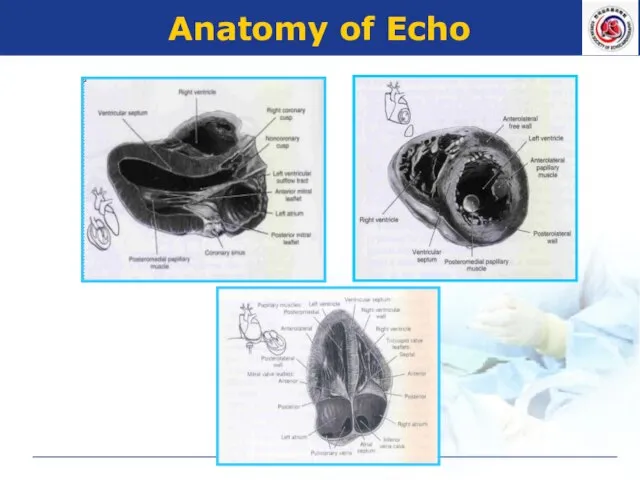
- 3. Anatomy of Echo
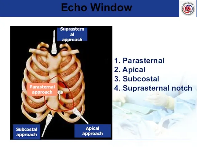
- 4. Echo Window 1. Parasternal 2. Apical 3. Subcostal 4. Suprasternal notch
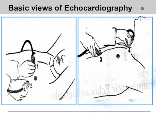
- 5. Basic views of Echocardiography Apical view Subcostal view Suprasternal view
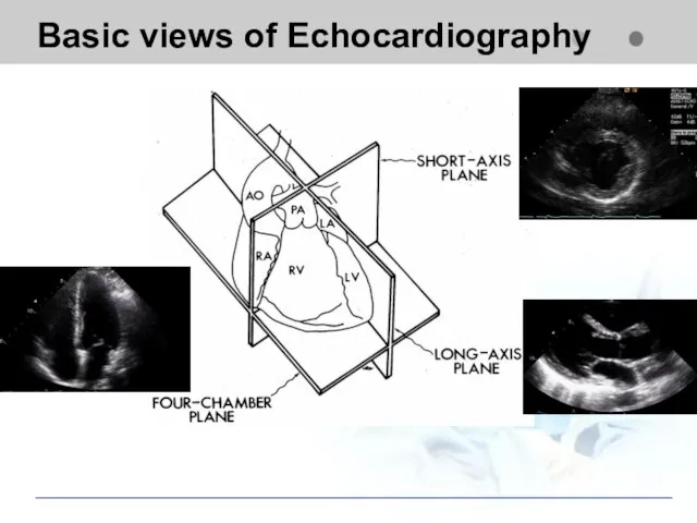
- 6. Basic views of Echocardiography
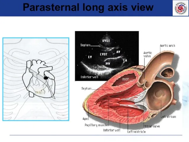
- 7. Parasternal long axis view
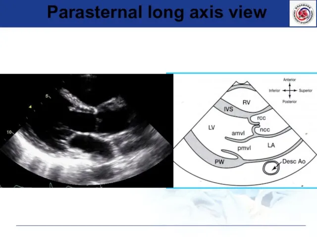
- 8. Parasternal long axis view
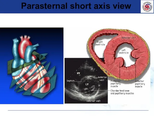
- 9. Parasternal short axis view
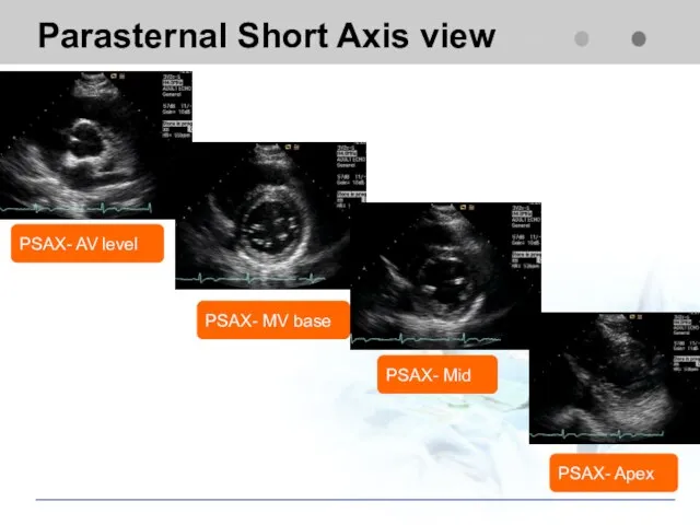
- 10. Parasternal Short Axis view PSAX- AV level PSAX- Mid PSAX- MV base PSAX- Apex
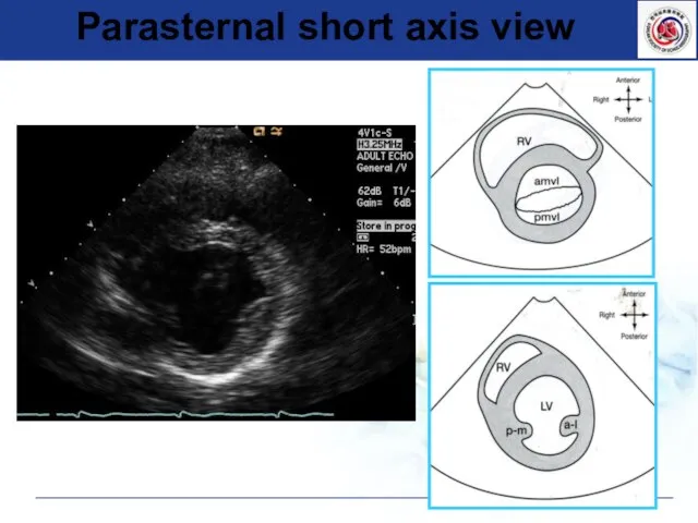
- 11. Parasternal short axis view
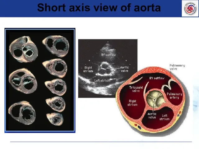
- 12. Short axis view of aorta
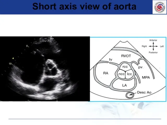
- 13. Short axis view of aorta
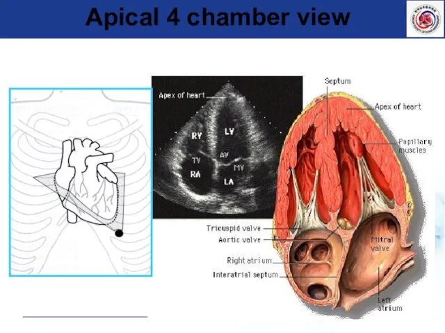
- 14. Apical 4 chamber view
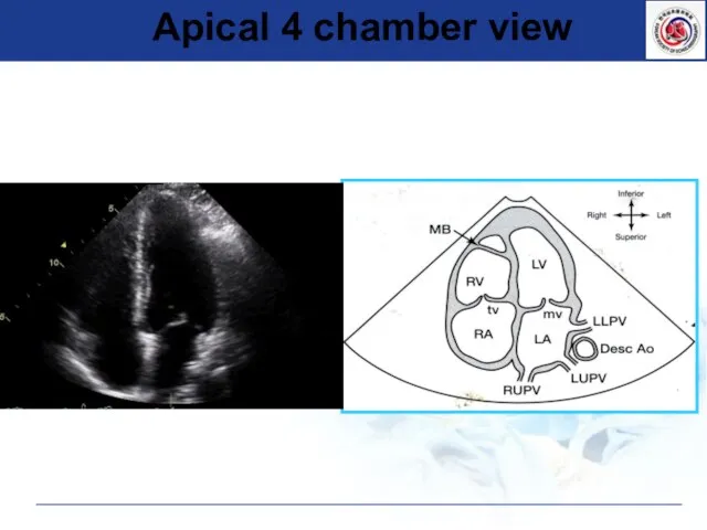
- 15. Apical 4 chamber view
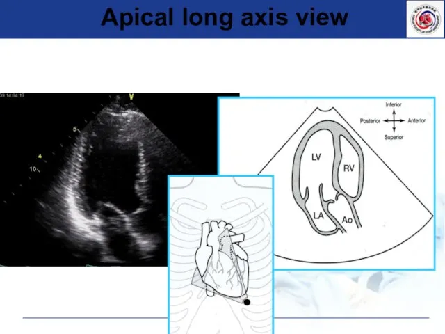
- 16. Apical long axis view
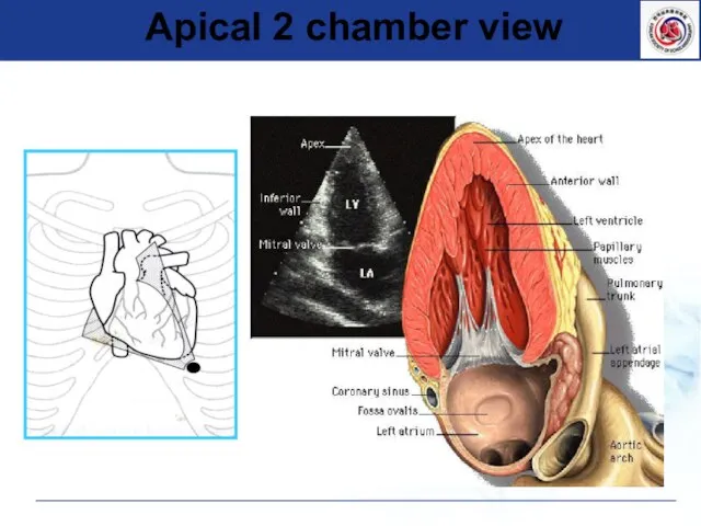
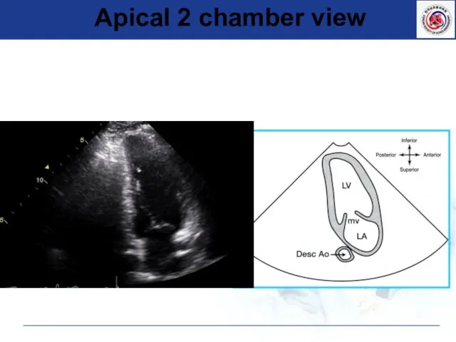
- 17. Apical 2 chamber view
- 18. Apical 2 chamber view
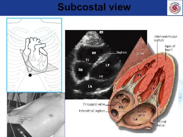
- 19. Subcostal view
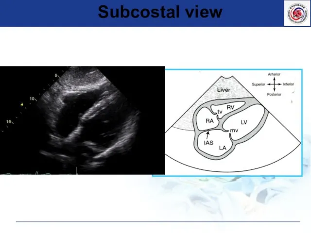
- 20. Subcostal view
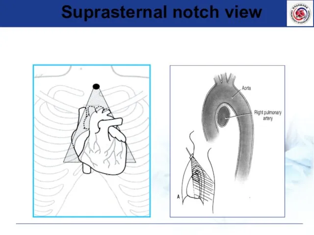
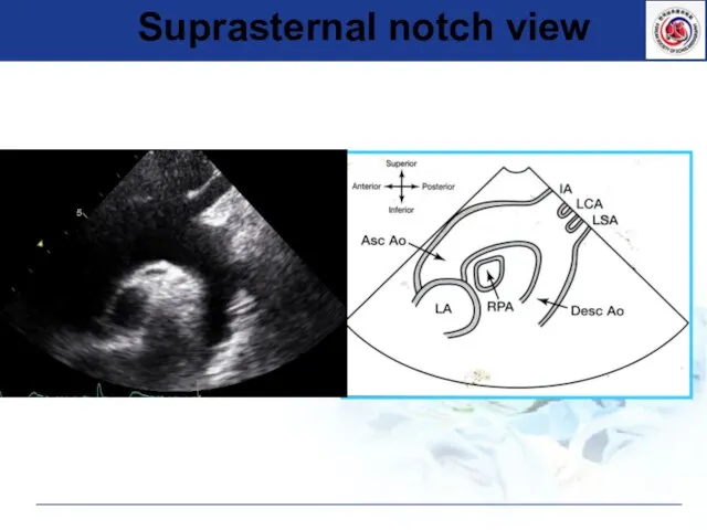
- 21. Suprasternal notch view
- 22. Suprasternal notch view
- 23. Measurement of Cardiac Chambers
- 24. ▶ Considering cardiac cycle : sinus rhythm : Multiple beats should be used in AF :
- 25. Respiration (at end-expiration) Image at minimum depth necessary Highest possible transducer frequency Adjust gains, dynamic range,
- 26. Factors affecting image quality Tester factors technique knowledge experience Machine factor Depth Gain Frame rate Resolution
- 27. 2D Image Optimization
- 28. 2D Image Optimization
- 29. Measure LV dimension
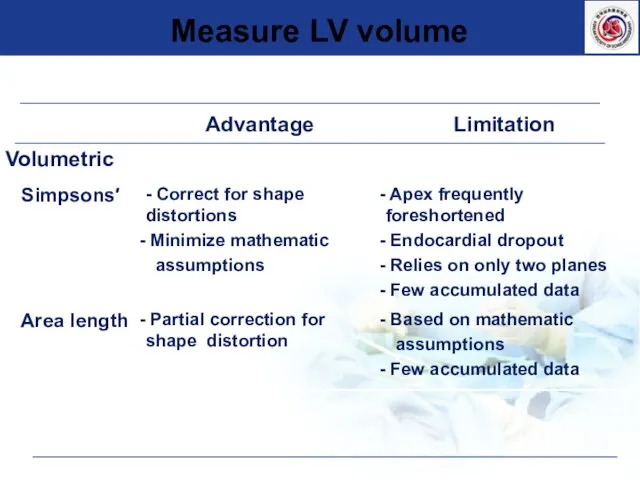
- 30. Measure LV volume
- 31. Measure LV mass
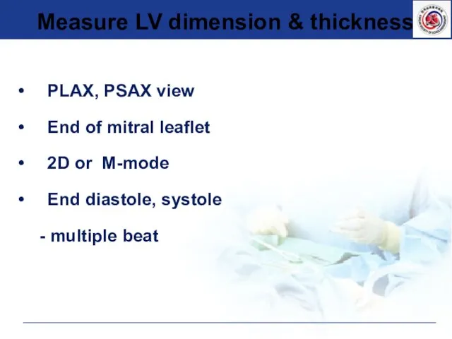
- 32. PLAX, PSAX view End of mitral leaflet 2D or M-mode End diastole, systole - multiple beat
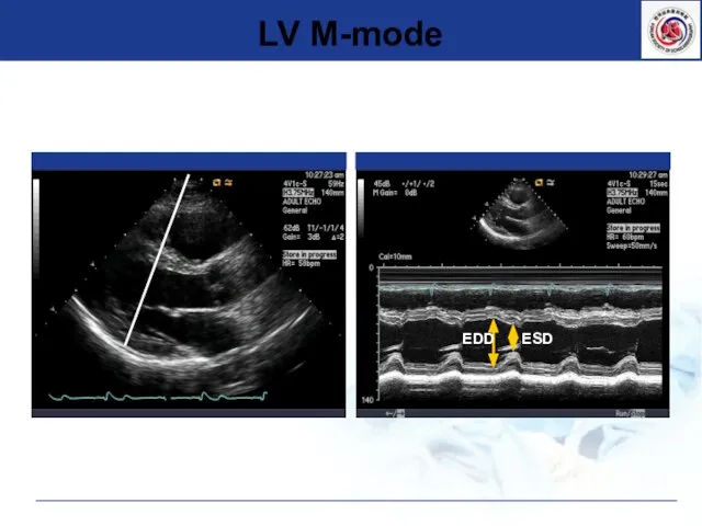
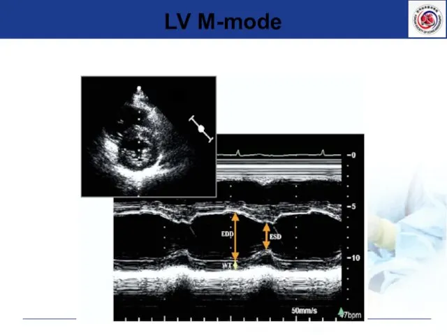
- 33. LV M-mode EDD ESD
- 34. LV M-mode
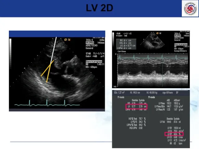
- 35. Oblique parasternal images를 피한다. LV 2D
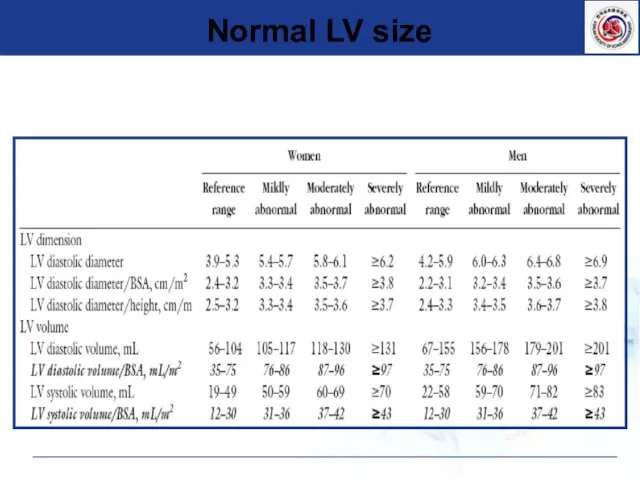
- 36. Normal LV size
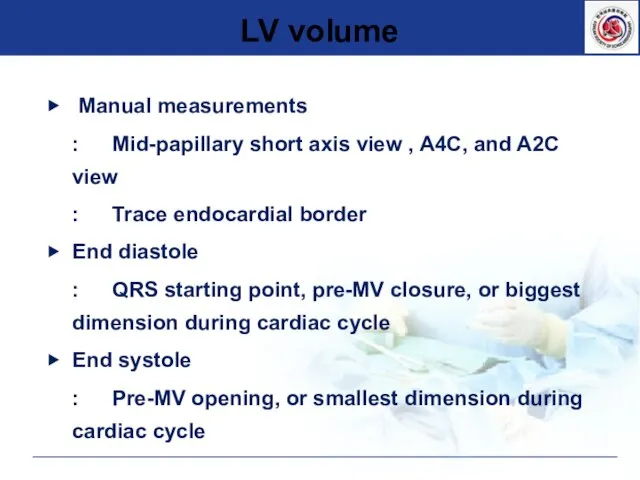
- 37. LV volume ▶ Manual measurements : Mid-papillary short axis view , A4C, and A2C view :
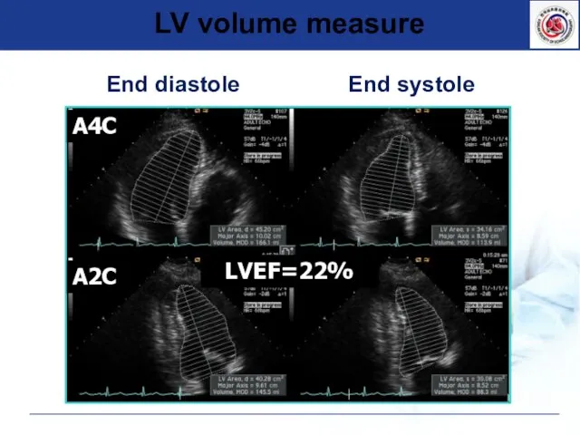
- 38. LV volume measure End diastole End systole A2C A4C LVEF=22%
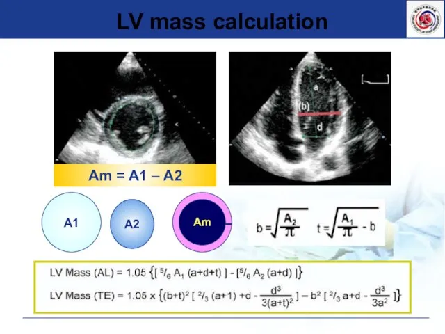
- 39. LV mass calculation A2 A1 Am Am = A1 – A2
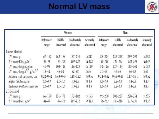
- 40. Normal LV mass
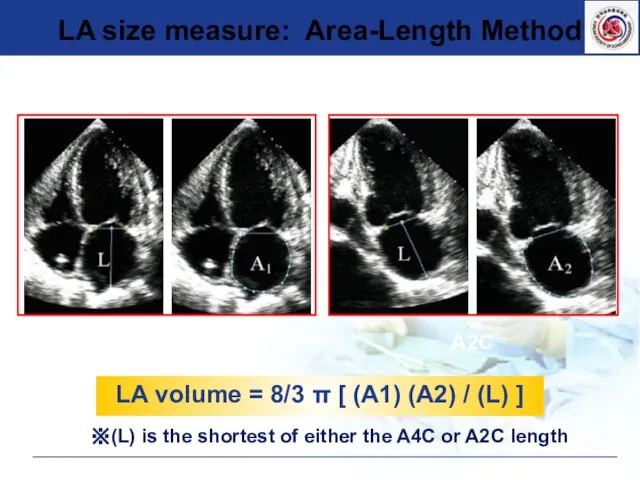
- 41. Measure LA size ▶ LV end systole, maximal LA size ▶ Avoid foreshortening of LA ▶
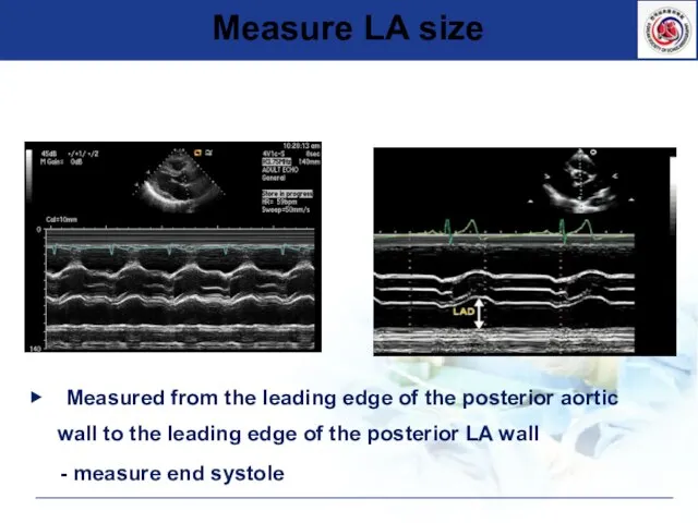
- 42. ▶ Measured from the leading edge of the posterior aortic wall to the leading edge of
- 43. A4C A2C LA volume = 8/3 π [ (A1) (A2) / (L) ] ※(L) is the
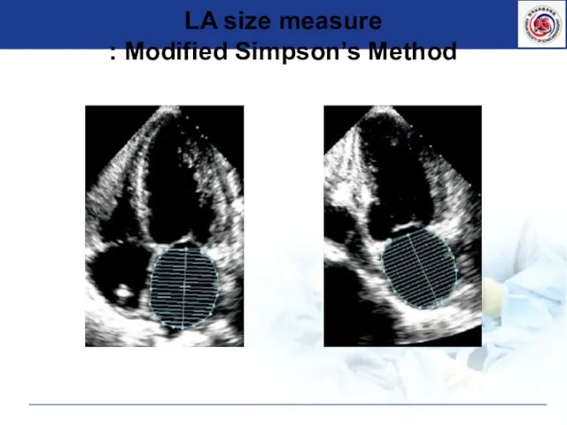
- 44. A4C A2C LA size measure : Modified Simpson’s Method
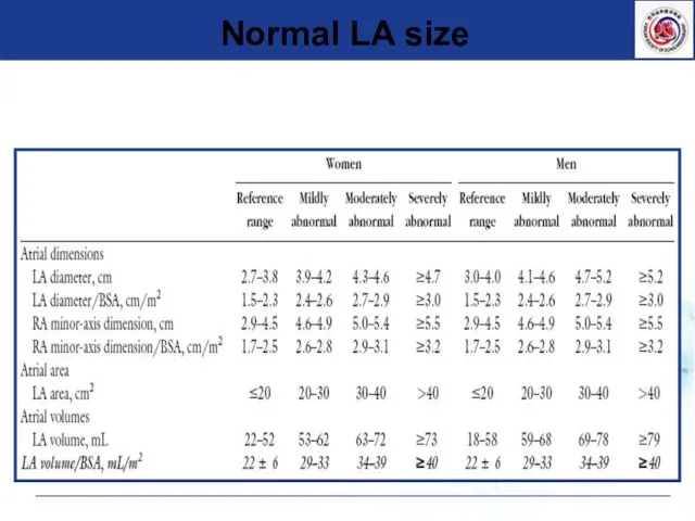
- 45. Normal LA size
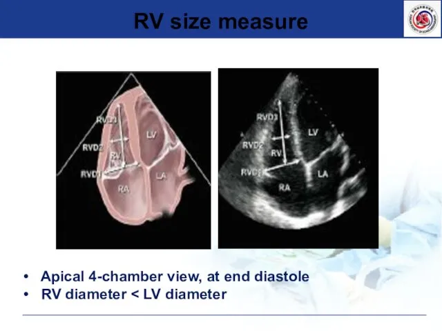
- 46. RV size measure Apical 4-chamber view, at end diastole RV diameter
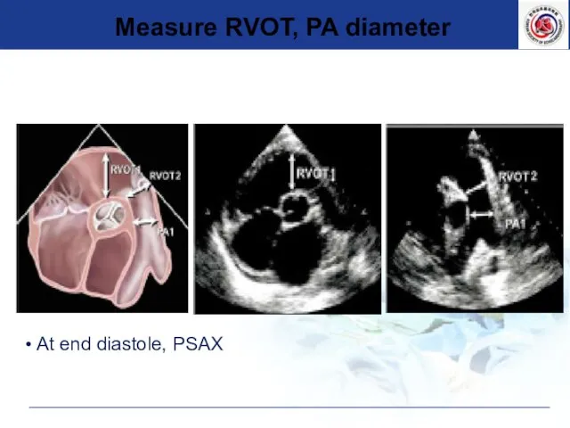
- 47. At end diastole, PSAX Measure RVOT, PA diameter
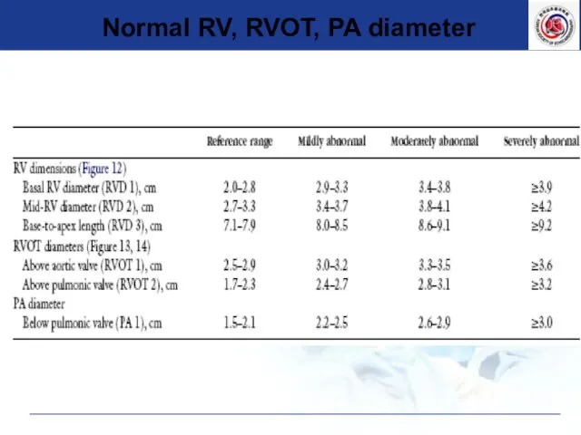
- 48. Normal RV, RVOT, PA diameter
- 50. Скачать презентацию
Слайд 3Anatomy of Echo
Anatomy of Echo
Слайд 4 Echo Window
1. Parasternal
2. Apical
3. Subcostal
4. Suprasternal notch
Echo Window
1. Parasternal
2. Apical
3. Subcostal
4. Suprasternal notch
Слайд 5Basic views of Echocardiography
Apical view
Subcostal view
Suprasternal view
Basic views of Echocardiography
Apical view
Subcostal view
Suprasternal view
Слайд 6Basic views of Echocardiography
Basic views of Echocardiography
Слайд 7 Parasternal long axis view
Parasternal long axis view
Слайд 8 Parasternal long axis view
Parasternal long axis view
Слайд 9 Parasternal short axis view
Parasternal short axis view
Слайд 10Parasternal Short Axis view
PSAX- AV level
PSAX- Mid
PSAX- MV base
PSAX- Apex
Parasternal Short Axis view
PSAX- AV level
PSAX- Mid
PSAX- MV base
PSAX- Apex
Слайд 11 Parasternal short axis view
Parasternal short axis view
Слайд 12 Short axis view of aorta
Short axis view of aorta
Слайд 13 Short axis view of aorta
Short axis view of aorta
Слайд 14 Apical 4 chamber view
Apical 4 chamber view
Слайд 15 Apical 4 chamber view
Apical 4 chamber view
Слайд 16 Apical long axis view
Apical long axis view
Слайд 17 Apical 2 chamber view
Apical 2 chamber view
Слайд 18 Apical 2 chamber view
Apical 2 chamber view
Слайд 19 Subcostal view
Subcostal view
Слайд 20 Subcostal view
Subcostal view
Слайд 21 Suprasternal notch view
Suprasternal notch view
Слайд 22 Suprasternal notch view
Suprasternal notch view
Слайд 23Measurement of Cardiac Chambers
Measurement of Cardiac Chambers
Слайд 24▶ Considering cardiac cycle
: sinus rhythm
: Multiple beats should be used in AF
: Avoid PVC or
▶ Considering cardiac cycle
: sinus rhythm
: Multiple beats should be used in AF
: Avoid PVC or
Слайд 25Respiration (at end-expiration)
Image at minimum depth necessary
Highest possible transducer frequency
Adjust gains, dynamic
Respiration (at end-expiration)
Image at minimum depth necessary
Highest possible transducer frequency
Adjust gains, dynamic
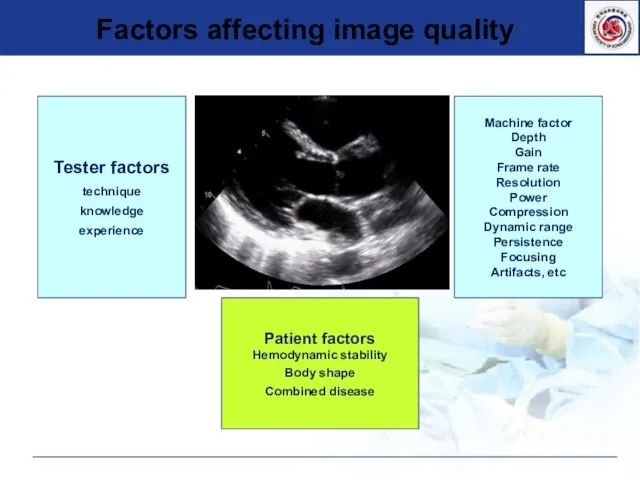
Слайд 26Factors affecting image quality
Tester factors
technique
knowledge
experience
Machine factor
Depth
Gain
Frame rate
Resolution
Power
Compression
Dynamic range
Persistence
Focusing
Artifacts, etc
Patient factors
Hemodynamic stability
Body shape
Combined
Factors affecting image quality
Tester factors
technique
knowledge
experience
Machine factor
Depth
Gain
Frame rate
Resolution
Power
Compression
Dynamic range
Persistence
Focusing
Artifacts, etc
Patient factors
Hemodynamic stability
Body shape
Combined
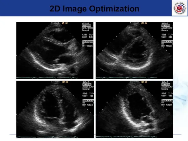
Слайд 272D Image Optimization
2D Image Optimization
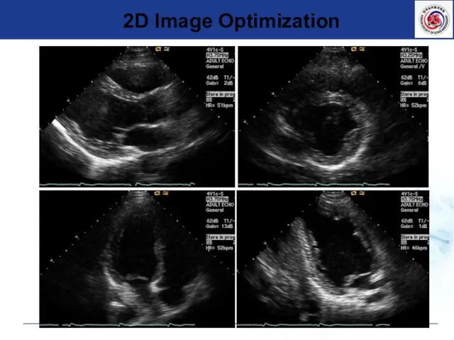
Слайд 282D Image Optimization
2D Image Optimization
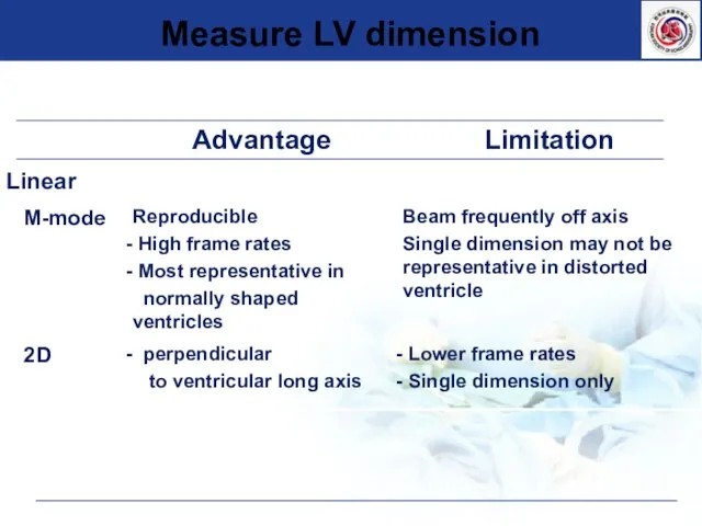
Слайд 29Measure LV dimension
Measure LV dimension
Слайд 30Measure LV volume
Measure LV volume
Слайд 31Measure LV mass
Measure LV mass

Слайд 32 PLAX, PSAX view
End of mitral leaflet
2D or M-mode
End diastole, systole
- multiple
PLAX, PSAX view
End of mitral leaflet
2D or M-mode
End diastole, systole
- multiple
Слайд 33LV M-mode
EDD
ESD
LV M-mode
EDD
ESD
Слайд 34LV M-mode
LV M-mode
Слайд 35Oblique parasternal images를 피한다.
LV 2D
Oblique parasternal images를 피한다.
LV 2D
Слайд 36Normal LV size
Normal LV size
Слайд 37LV volume
▶ Manual measurements
: Mid-papillary short axis view , A4C, and A2C view
: Trace
LV volume
▶ Manual measurements
: Mid-papillary short axis view , A4C, and A2C view
: Trace
Слайд 38LV volume measure
End diastole
End systole
A2C
A4C
LVEF=22%
LV volume measure
End diastole
End systole
A2C
A4C
LVEF=22%
Слайд 39LV mass calculation
A2
A1
Am
Am = A1 – A2
LV mass calculation
A2
A1
Am
Am = A1 – A2
Слайд 40Normal LV mass
Normal LV mass
Слайд 41Measure LA size
▶ LV end systole, maximal LA size
▶ Avoid foreshortening of LA
▶ LA length
Measure LA size
▶ LV end systole, maximal LA size
▶ Avoid foreshortening of LA
▶ LA length
Слайд 42▶ Measured from the leading edge of the posterior aortic wall to the
▶ Measured from the leading edge of the posterior aortic wall to the
Слайд 43A4C
A2C
LA volume = 8/3 π [ (A1) (A2) / (L) ]
※(L) is
A4C
A2C
LA volume = 8/3 π [ (A1) (A2) / (L) ]
※(L) is
Слайд 44A4C
A2C
LA size measure
: Modified Simpson’s Method
A4C
A2C
LA size measure
: Modified Simpson’s Method
Слайд 45Normal LA size
Normal LA size
Слайд 46RV size measure
Apical 4-chamber view, at end diastole
RV diameter <
RV size measure
Apical 4-chamber view, at end diastole
RV diameter <
Слайд 47 At end diastole, PSAX
Measure RVOT, PA diameter
At end diastole, PSAX
Measure RVOT, PA diameter
Слайд 48Normal RV, RVOT, PA diameter
Normal RV, RVOT, PA diameter
 «ЛИФБА БАЙРАМЫ
 .
. Международные валютно-кредитные отношения
 Основные понятия контекстной рекламы
 8 класс. Тема 1. События и вероятности
 Презентация на тему Сказки Пушкина
 Теория потока М. Чиксентмихайи
 Соревнование AGI
 Покров Пресвятой Богородицы
 Выпускной аттестационный проект. Дополнительная общеразвивающая программа Добро пожаловать в Мастерскую Светелка
 Специфика деятельности управляющего гостиничным комплексом
 Академия новостей Academ.info
 Публичный доклад директора МОУДОД «ДТДиМ» г.Воркуты.
 Дима 50
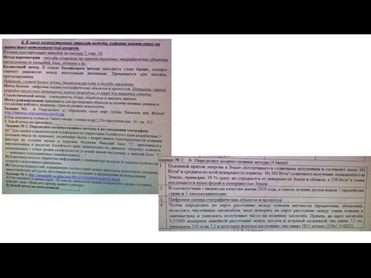 картогр. методы
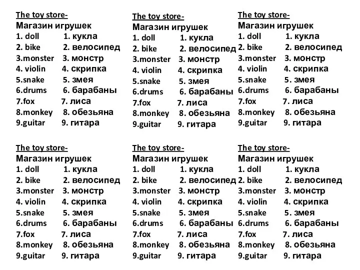 The toy store. Магазин игрушек
 ЖИЗНЬ ЕЖЕЙ
 Главное управление проектами комстэк монтаж и эксплуатация слаботочных систем
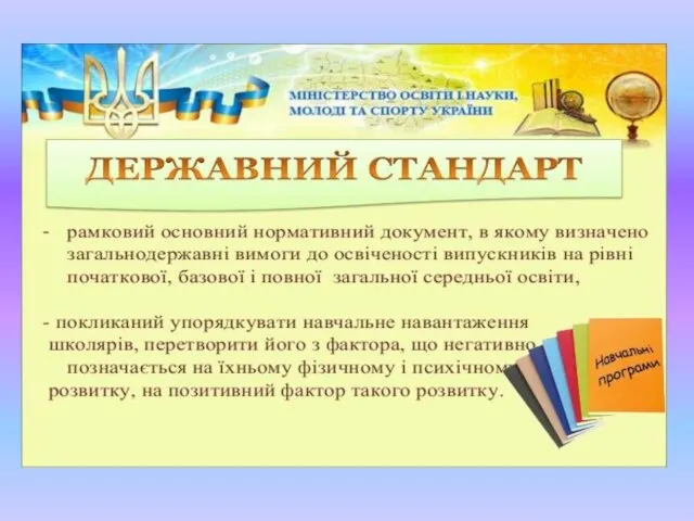 Державний стандарт
 Преобразование рисунка
 ИТОГИ 2008 – 2009 учебного года
 Девушка Рита
 Педагогический совет «Личность современного учителя в Новой школе» Муниципальный конкурс «Учитель года – 2012»
 Большие и маленькие дела социального педагога Ступиной Л.Н. и воспитанников нашего центра
 The Most Absurd Inventions of All Time
 Нанотехнологии - инвестиционный ресурс развития Удмуртской Республики
 Презентация на тему Опыление у цветковых растений
  Развитие речи