- Tuberculosis Disease

Содержание
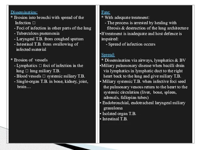
- 2. Tuberculosis – Clinical Features Localized type may be asymptomatic Low grade remittent fever, night sweats, malaise,
- 3. Tuberculosis – Diagnosis History, physical examination, radiological findings “consolidation & cavitation” Identification of the acid-fast bacilli
- 4. Tuberculosis – Prognosis Depends on: The extent of the disease and the patient immune status Secondary
- 5. Tuberculosis – Chronic Consequences Pulmonary fibrosis The lung lesions may heal with fibrosis at any stage,
- 6. Infection in Immunocompromised Individuals Mycobacterial infection of all types are increased in immunocompromised individuals and is
- 7. Atypical Mycobacterial Infection These infections are caused by a group of non-tuberculous mycobacteria of which the
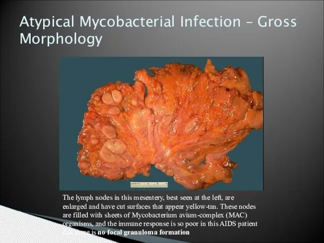
- 8. The lymph nodes in this mesentery, best seen at the left, are enlarged and have cut
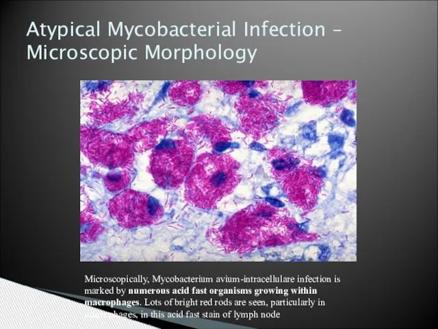
- 9. Microscopically, Mycobacterium avium-intracellulare infection is marked by numerous acid fast organisms growing within macrophages. Lots of
- 14. Скачать презентацию
Слайд 3Tuberculosis – Diagnosis
History, physical examination, radiological findings “consolidation & cavitation”
Identification of
Tuberculosis – Diagnosis
History, physical examination, radiological findings “consolidation & cavitation”
Identification of
Слайд 4Tuberculosis – Prognosis
Depends on:
The extent of the disease and the
Tuberculosis – Prognosis
Depends on:
The extent of the disease and the

Слайд 5Tuberculosis – Chronic Consequences
Pulmonary fibrosis
The lung lesions may heal with fibrosis at
Tuberculosis – Chronic Consequences
Pulmonary fibrosis
The lung lesions may heal with fibrosis at
Слайд 6Infection in Immunocompromised Individuals
Mycobacterial infection of all types are increased in
Infection in Immunocompromised Individuals
Mycobacterial infection of all types are increased in

Слайд 7Atypical Mycobacterial Infection
These infections are caused by a group of non-tuberculous mycobacteria
Atypical Mycobacterial Infection
These infections are caused by a group of non-tuberculous mycobacteria
Слайд 8The lymph nodes in this mesentery, best seen at the left, are
The lymph nodes in this mesentery, best seen at the left, are
Слайд 9Microscopically, Mycobacterium avium-intracellulare infection is marked by numerous acid fast organisms growing
Microscopically, Mycobacterium avium-intracellulare infection is marked by numerous acid fast organisms growing
 Positive traits of character - Persistence
 слайд-шоу фокусника-иллюзиониста Леонида Зангиева
 Ножницы вашей мечты
 Металлы. Общая характеристика металлов (нахождение в природе и физические свойства)
 Оказание первой помощи. Современные требования
 ГАРАНТИРОВАННЫЙ ОБЪЕМ БЕСПЛАТНОЙ МЕДИЦИНСКОЙ ПОМОЩИ(ГОБМП)
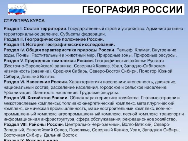 ГЕОГРАФИЯ РОССИИ СТРУКТУРА КУРСА
 Геологичекое строение и рельеф
 История происхождения фамилий жителей села Батурино
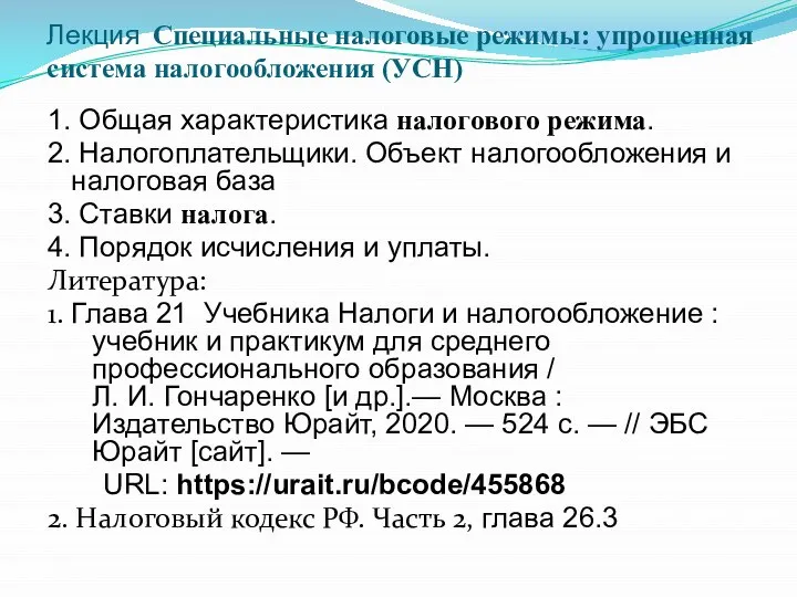 Специальные налоговые режимы_Упрощенная система налогобложения
 «Компьютерная зависимость детей»
 Реклама в фильме Римские каникулы (1953)
 Описание памятника архитектуры
 Coca-Cola
 Унитазы Culto
 Графическй дизайн в школьной тетраде
 От Сиднея вдоль Большого Водораздельного хребта
 Matematika_19_09_1
 Портфолио Агентства маркетинговых коммуникаций Чистяковой Ирины PR сопровождение проекта "ГлавМаркет"
 Критерии и показатели оценки деятельности таможенных органов России
  Развитие культуры
 Иркутск, 2011
 Дорога на родину И.С.Тургенева
 Теория привязанностей
 Рабочая группаРоссийской академии медицинских наук
 Стоимость нефти и авиаГСМ
 Мучные изделия
 Понятие вектора