Biosynthesized selenium nanoparticles, quantum dots, complex drug based on viburnum juice on selenium nanoparticles
- Biosynthesized selenium nanoparticles, quantum dots, complex drug based on viburnum juice on selenium nanoparticles

Содержание
- 2. Research objectives: 1. To get acquainted with some species of plants, bacteria, fungi, protozoa, features of
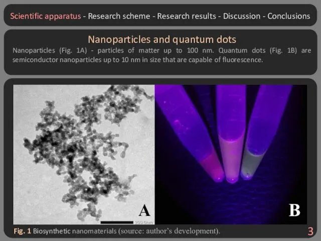
- 3. Fig. 1 Biosynthetic nanomaterials (source: author’s development). Nanoparticles and quantum dots Nanoparticles (Fig. 1A) - particles
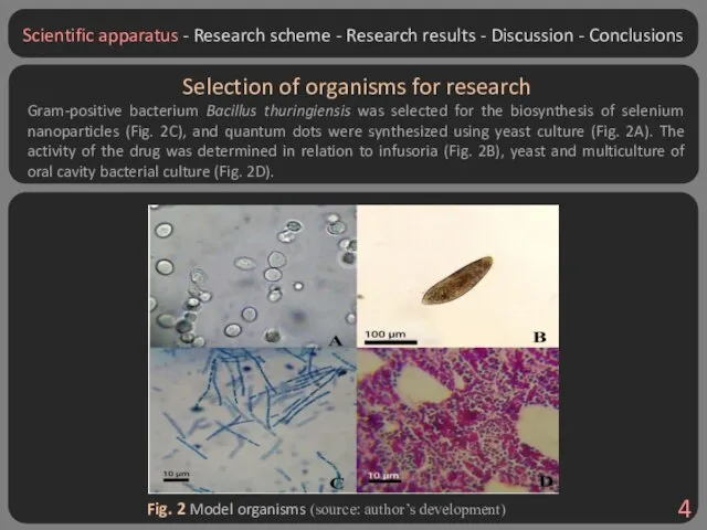
- 4. Fig. 2 Model organisms (source: author’s development) Selection of organisms for research Gram-positive bacterium Bacillus thuringiensis
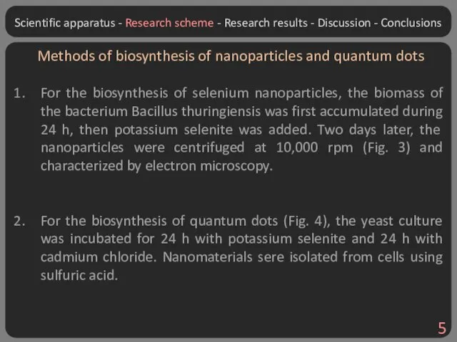
- 5. Methods of biosynthesis of nanoparticles and quantum dots For the biosynthesis of selenium nanoparticles, the biomass
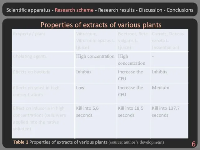
- 6. Table 1 Properties of extracts of various plants (source: author’s development) Properties of extracts of various
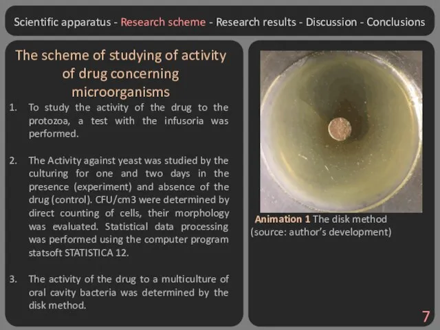
- 7. Animation 1 The disk method (source: author’s development) The scheme of studying of activity of drug
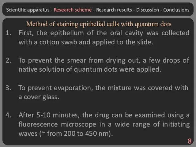
- 8. Method of staining epithelial cells with quantum dots First, the epithelium of the oral cavity was
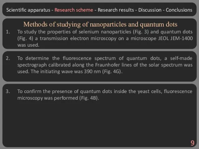
- 9. Methods of studying of nanoparticles and quantum dots To study the properties of selenium nanoparticles (Fig.
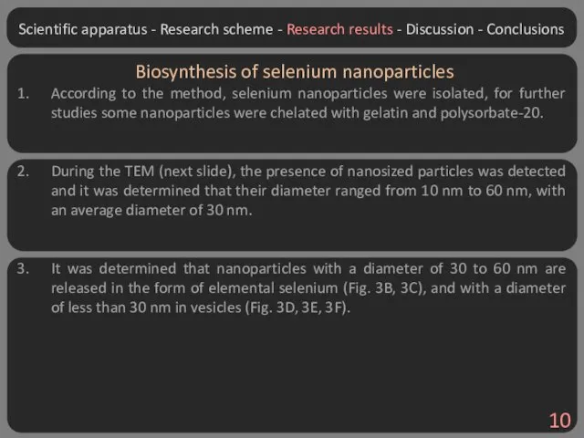
- 10. Biosynthesis of selenium nanoparticles According to the method, selenium nanoparticles were isolated, for further studies some
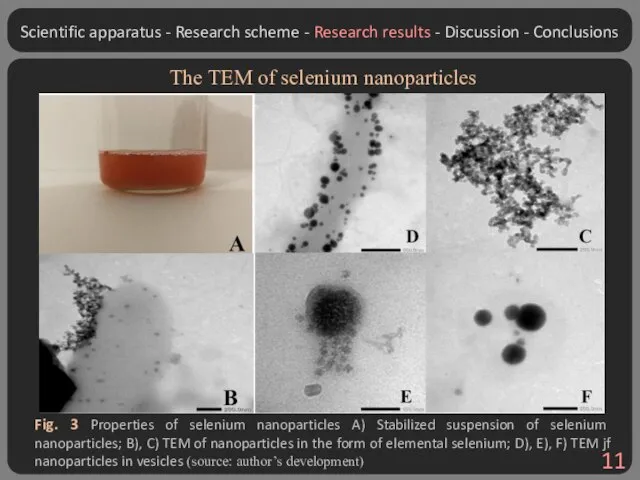
- 11. Fig. 3 Properties of selenium nanoparticles A) Stabilized suspension of selenium nanoparticles; B), C) TEM of
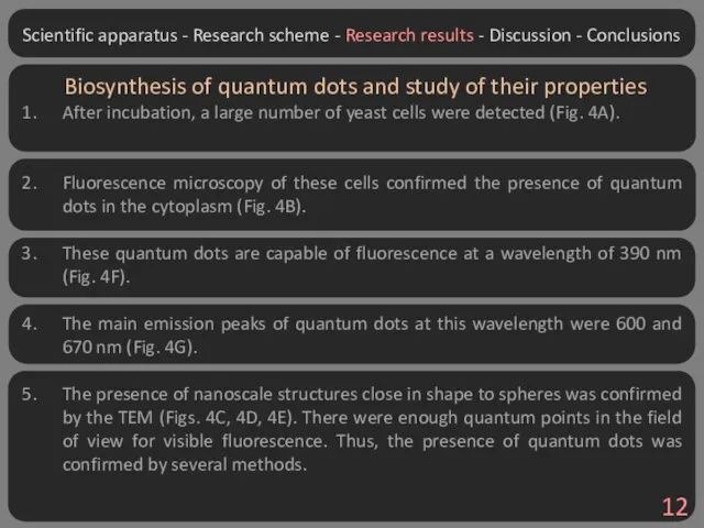
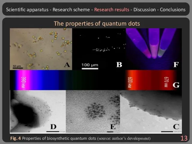
- 12. Biosynthesis of quantum dots and study of their properties After incubation, a large number of yeast
- 13. Fig. 4 Properties of biosynthetic quantum dots (source: author’s development) The properties of quantum dots Scientific
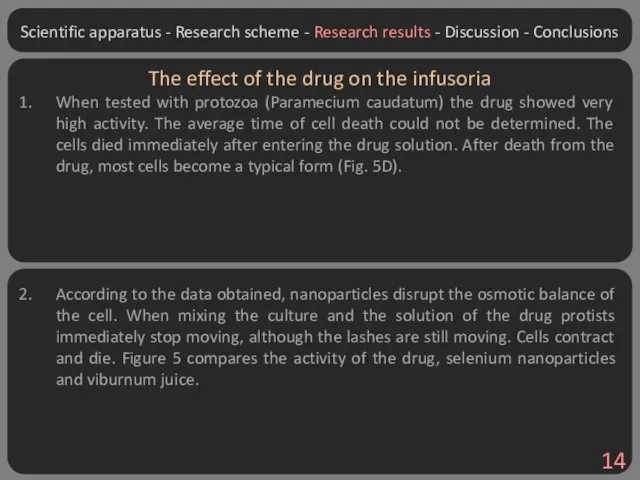
- 14. The effect of the drug on the infusoria When tested with protozoa (Paramecium caudatum) the drug
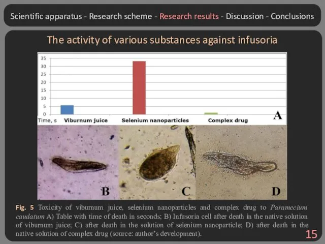
- 15. The activity of various substances against infusoria Fig. 5 Toxicity of viburnum juice, selenium nanoparticles and
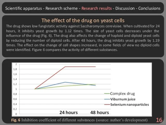
- 16. The effect of the drug on yeast cells The drug shows low fungistatic activity against Saccharomyces
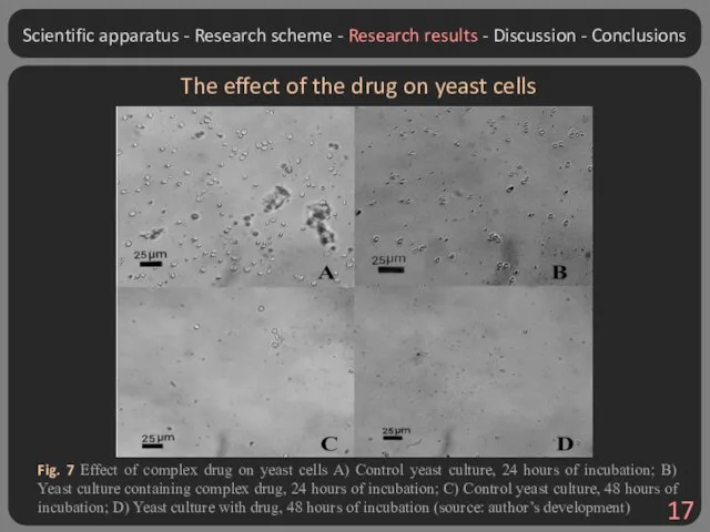
- 17. The effect of the drug on yeast cells Fig. 7 Effect of complex drug on yeast
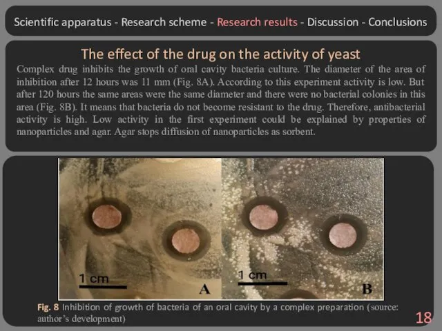
- 18. The effect of the drug on the activity of yeast Complex drug inhibits the growth of
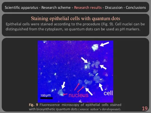
- 19. Staining epithelial cells with quantum dots Epithelial cells were stained according to the procedure (Fig. 9).
- 20. Significance Most hypotheses were confirmed during the study. For the first time a complex antibacterial drug
- 21. Research prospects: The prospect of the study is to conduct X-ray diffraction analysis to more accurately
- 22. Personal contribution Defining the topic and scheme of the study. Conducting most of the experiments (except
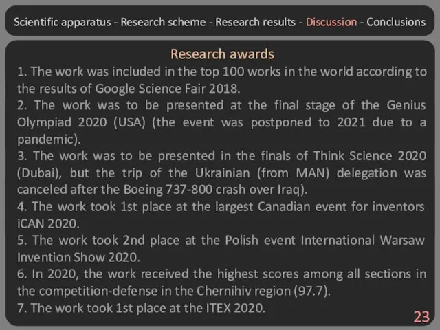
- 23. Scientific apparatus - Research scheme - Research results - Discussion - Conclusions Research awards 1. The
- 25. Скачать презентацию
Слайд 3Fig. 1 Biosynthetic nanomaterials (source: author’s development).
Nanoparticles and quantum dots
Nanoparticles (Fig. 1A)
Fig. 1 Biosynthetic nanomaterials (source: author’s development).
Nanoparticles and quantum dots
Nanoparticles (Fig. 1A)
Слайд 4Fig. 2 Model organisms (source: author’s development)
Selection of organisms for research
Gram-positive bacterium
Fig. 2 Model organisms (source: author’s development)
Selection of organisms for research
Gram-positive bacterium
Слайд 5Methods of biosynthesis of nanoparticles and quantum dots
For the biosynthesis of selenium
Methods of biosynthesis of nanoparticles and quantum dots
For the biosynthesis of selenium
Слайд 6Table 1 Properties of extracts of various plants (source: author’s development)
Properties of
Table 1 Properties of extracts of various plants (source: author’s development)
Properties of
Слайд 7Animation 1 The disk method (source: author’s development)
The scheme of studying of
Animation 1 The disk method (source: author’s development)
The scheme of studying of
Слайд 8Method of staining epithelial cells with quantum dots
First, the epithelium of the
Method of staining epithelial cells with quantum dots
First, the epithelium of the
Слайд 9Methods of studying of nanoparticles and quantum dots
To study the properties of
Methods of studying of nanoparticles and quantum dots
To study the properties of
Слайд 10Biosynthesis of selenium nanoparticles
According to the method, selenium nanoparticles were isolated, for
Biosynthesis of selenium nanoparticles
According to the method, selenium nanoparticles were isolated, for
Слайд 11Fig. 3 Properties of selenium nanoparticles A) Stabilized suspension of selenium nanoparticles;
Fig. 3 Properties of selenium nanoparticles A) Stabilized suspension of selenium nanoparticles;
Слайд 12Biosynthesis of quantum dots and study of their properties
After incubation, a large
Biosynthesis of quantum dots and study of their properties
After incubation, a large
Слайд 13Fig. 4 Properties of biosynthetic quantum dots (source: author’s development)
The properties of
Fig. 4 Properties of biosynthetic quantum dots (source: author’s development)
The properties of
Слайд 14The effect of the drug on the infusoria
When tested with protozoa (Paramecium
The effect of the drug on the infusoria
When tested with protozoa (Paramecium
Слайд 15The activity of various substances against infusoria
Fig. 5 Toxicity of viburnum juice,
The activity of various substances against infusoria
Fig. 5 Toxicity of viburnum juice,
Слайд 16The effect of the drug on yeast cells
The drug shows low fungistatic
The effect of the drug on yeast cells
The drug shows low fungistatic
Слайд 17The effect of the drug on yeast cells
Fig. 7 Effect of complex
The effect of the drug on yeast cells
Fig. 7 Effect of complex
Слайд 18The effect of the drug on the activity of yeast
Complex drug inhibits
The effect of the drug on the activity of yeast
Complex drug inhibits
Слайд 19Staining epithelial cells with quantum dots
Epithelial cells were stained according to the
Staining epithelial cells with quantum dots
Epithelial cells were stained according to the
Слайд 20Significance
Most hypotheses were confirmed during the study. For the first time a
Significance
Most hypotheses were confirmed during the study. For the first time a
Слайд 21Research prospects:
The prospect of the study is to conduct X-ray diffraction analysis
Research prospects:
The prospect of the study is to conduct X-ray diffraction analysis
Слайд 22Personal contribution
Defining the topic and scheme of the study.
Conducting most of the
Personal contribution
Defining the topic and scheme of the study.
Conducting most of the
Слайд 23Scientific apparatus - Research scheme - Research results - Discussion - Conclusions
Research
Scientific apparatus - Research scheme - Research results - Discussion - Conclusions
Research
 Бионика, как наука
 Пути и направления эволюции
 Пол и гендер
 ПОЛЕЗНАЯ АЗБУКА ПИТАНИЯ Проблема: не всегда умеем правильно выбирать продукты для здорового питания (не знаем о пользе ов
 Гигиена кормов и кормления животных
 Чудесные цветники весной
 Холодолюбивая фауна в антропогенном периоде
 Строение и функции клетки. Цитоплазма и её органоиды
 Презентация на тему Первоцветы Особенности раннецветущих растений
  Рассматривание клеток и тканей в оптический микроскоп
 Отдел голосеменные
 Аноклазе: кудонь жуватат
 Общая характеристика обмена веществ и преобразование энергии
 Обмен углеводов
 Філогенія Excavata
 Дыхательная система. Полость носа, гортань, трахея
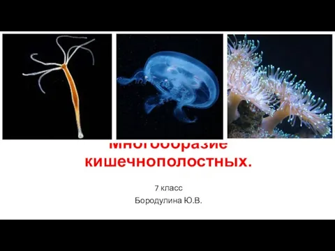 Многообразие кишечнополостных (7 класс)
 Слайды по теме Царство Грибы
 Соматический и автономный отделы нервной системы
 Филин
Филин Вирусология и открытие вирусов
 Свертывание крови. Группы крови. Переливание. 8 класс
 Витаминдер және олардың маңызы
 Медицинская арахноэнтомология
 Вирусы грибов
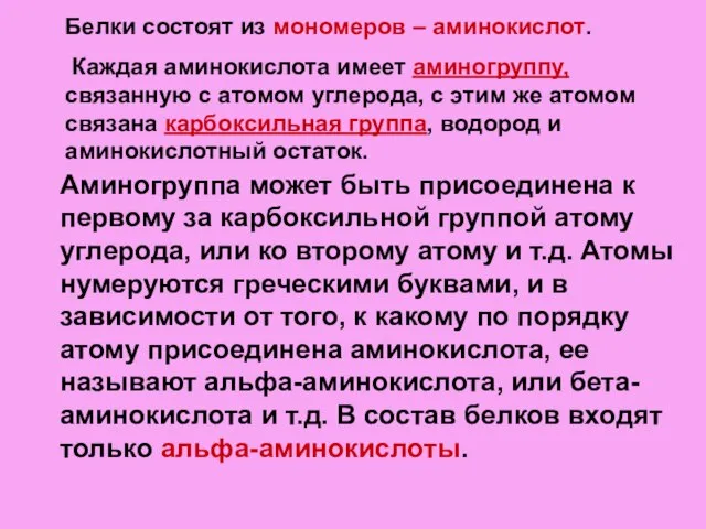 Белки. Функции белков
 Развитие жизни в криптозое
 Les kaktas