- An introduction to medical parasitology

Содержание
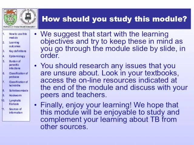
- 2. How should you study this module? We suggest that start with the learning objectives and try
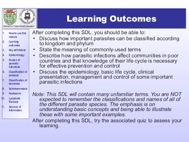
- 3. Learning Outcomes After completing this SDL, you should be able to: Discuss how important parasites can
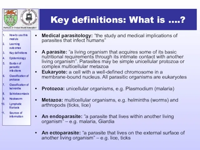
- 4. Key definitions: What is ….? Medical parasitology: “the study and medical implications of parasites that infect
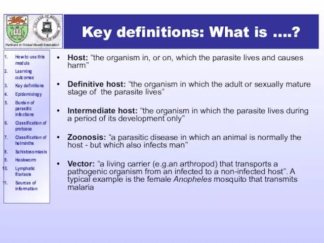
- 5. Key definitions: What is ….? Host: “the organism in, or on, which the parasite lives and
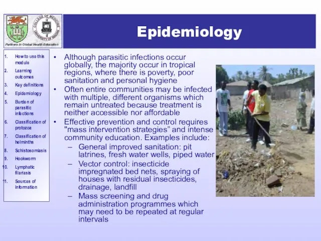
- 6. Epidemiology Although parasitic infections occur globally, the majority occur in tropical regions, where there is poverty,
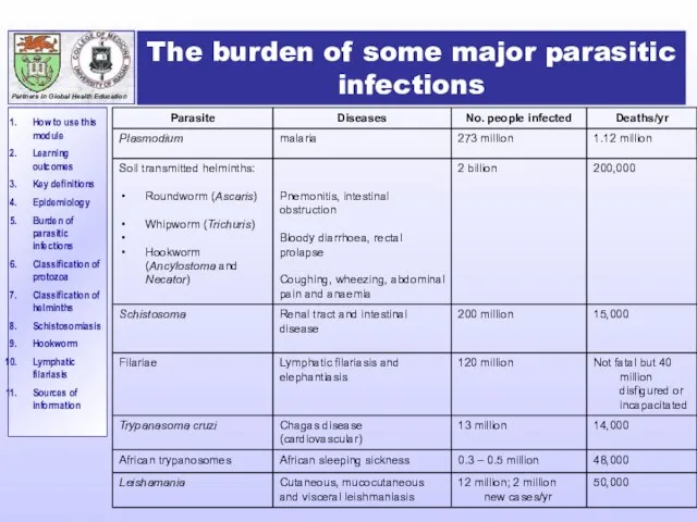
- 7. The burden of some major parasitic infections
- 8. Taxonomic classification of parasitic organisms The classification of parasites is controversial - there is no universally
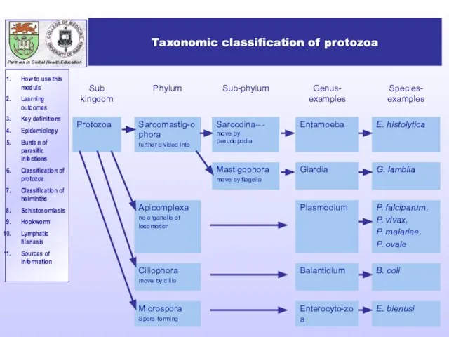
- 9. Taxonomic classification of protozoa
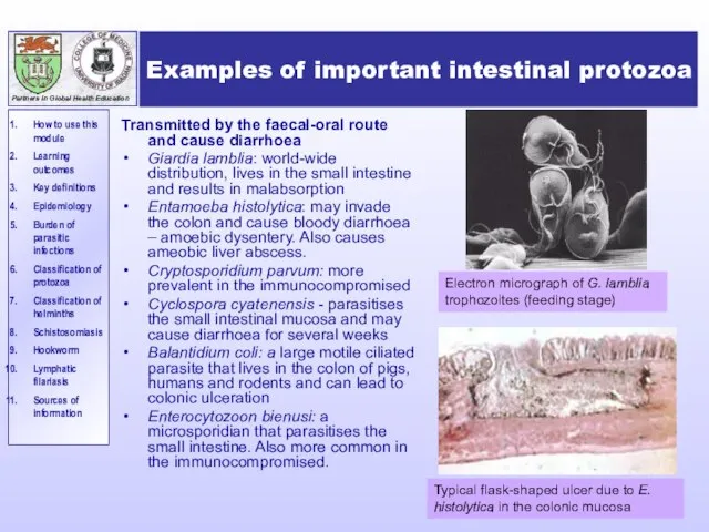
- 10. Examples of important intestinal protozoa Transmitted by the faecal-oral route and cause diarrhoea Giardia lamblia: world-wide
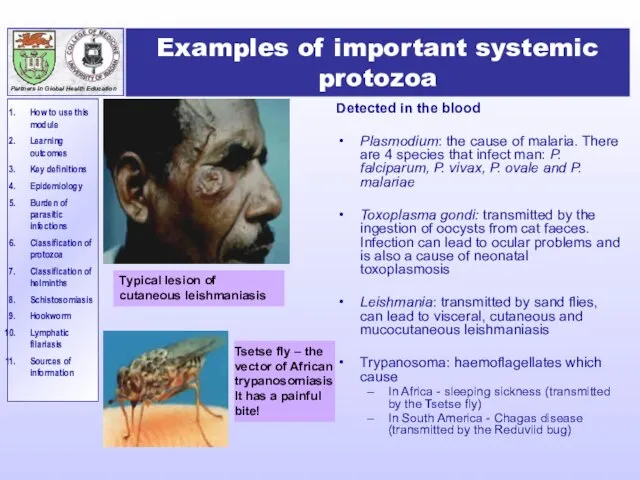
- 11. Examples of important systemic protozoa Detected in the blood Plasmodium: the cause of malaria. There are
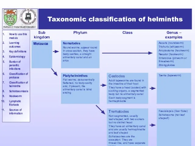
- 12. Taxonomic classification of helminths
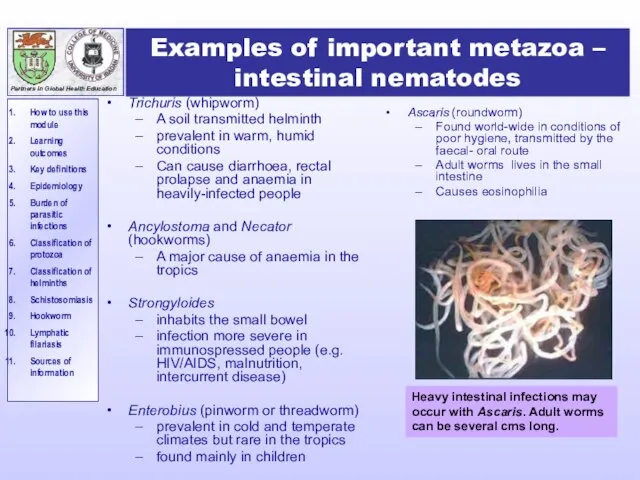
- 13. Examples of important metazoa – intestinal nematodes Trichuris (whipworm) A soil transmitted helminth prevalent in warm,
- 14. Examples of important metazoa –systemic nematodes Filaria including: Onchocerca volvulus – Transmitted by the simulium black
- 15. Examples of important flatworms - cestodes Intestinal - (“tapeworms”) Taenia saginata worldwide acquired by ingestion of
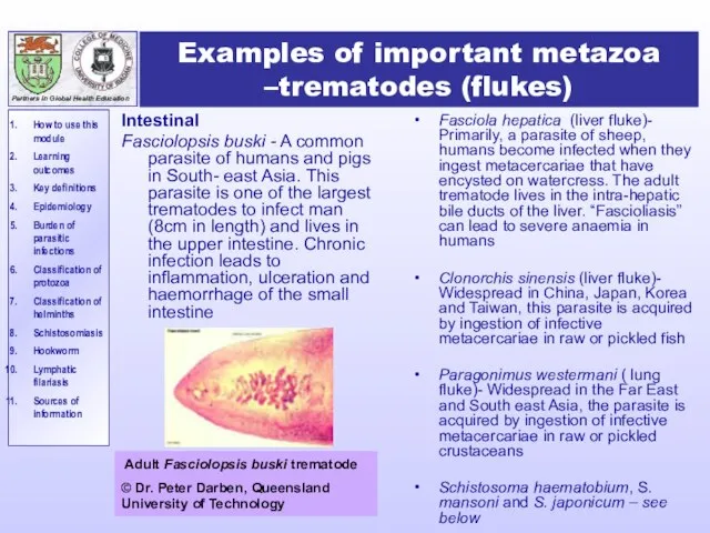
- 16. Examples of important metazoa –trematodes (flukes) Intestinal Fasciolopsis buski - A common parasite of humans and
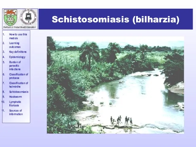
- 17. Schistosomiasis (bilharzia)
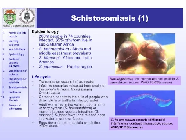
- 18. Schistosomiasis (1) Epidemiology 200m people in 74 countries infected, 85% of whom live in sub-Saharan Africa
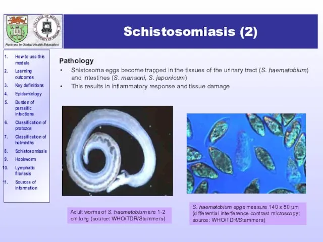
- 19. Schistosomiasis (2) Pathology Shistosoma eggs become trapped in the tissues of the urinary tract (S. haematobium)
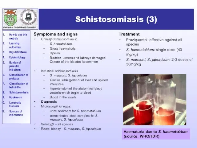
- 20. Schistosomiasis (3) Symptoms and signs Urinary Schistosomiasis: S. hamatobium Gross haematuria Dysuria Bladder, ureters and kidneys
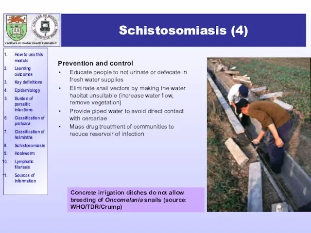
- 21. Schistosomiasis (4) Prevention and control Educate people to not urinate or defecate in fresh water supplies
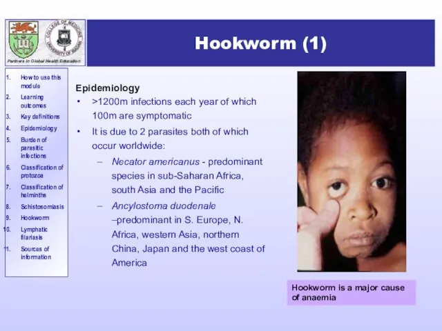
- 22. Hookworm (1) Epidemiology >1200m infections each year of which 100m are symptomatic It is due to
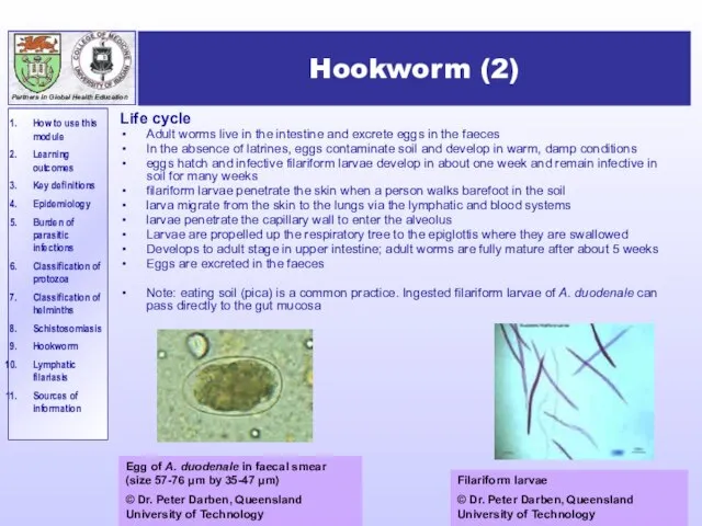
- 23. Hookworm (2) Life cycle Adult worms live in the intestine and excrete eggs in the faeces
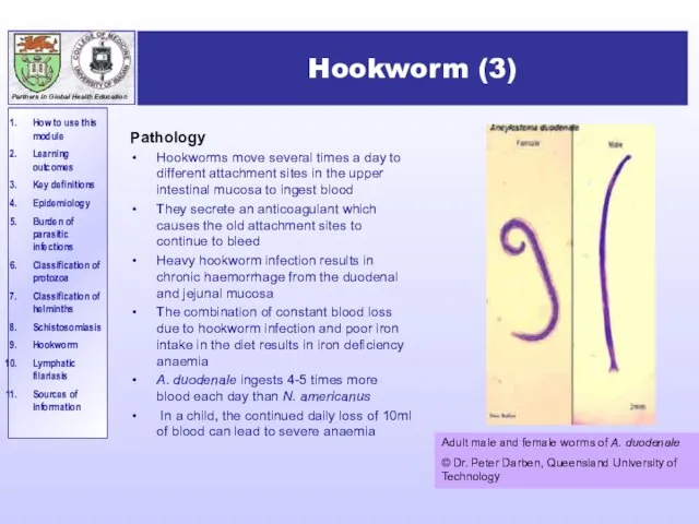
- 24. Hookworm (3) Pathology Hookworms move several times a day to different attachment sites in the upper
- 25. Hookworm (4) Symptoms and signs Minor Often itchy papules are found at the site where the
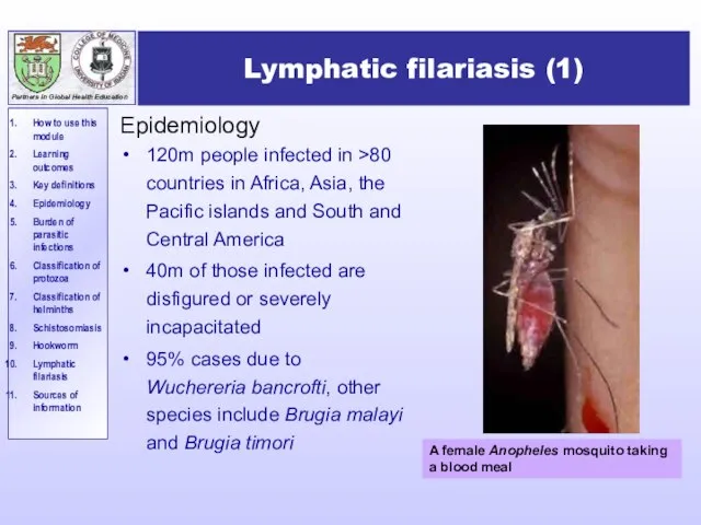
- 26. Lymphatic filariasis (1) Epidemiology 120m people infected in >80 countries in Africa, Asia, the Pacific islands
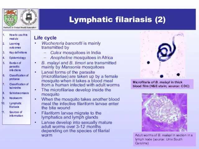
- 27. Lymphatic filariasis (2) Life cycle Wuchereria bancrofti is mainly transmitted by Culex mosquitoes in India Anopheline
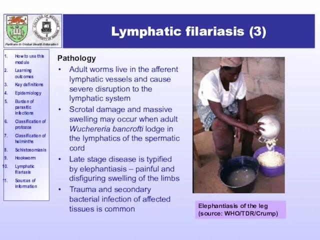
- 28. Lymphatic filariasis (3) Pathology Adult worms live in the afferent lymphatic vessels and cause severe disruption
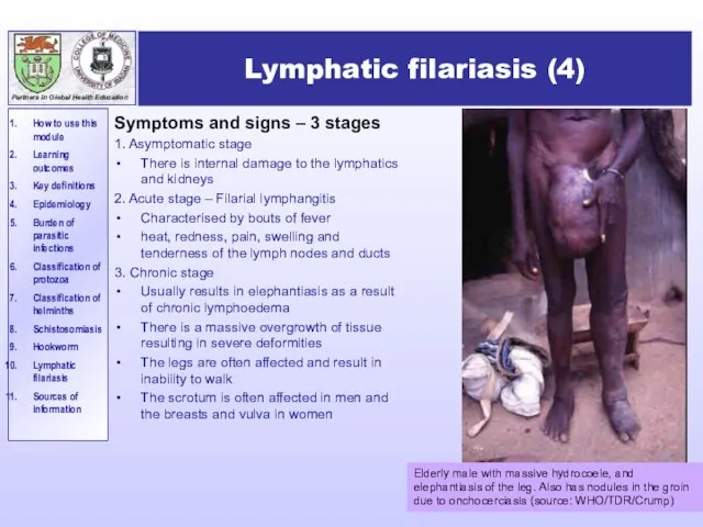
- 29. Lymphatic filariasis (4) Symptoms and signs – 3 stages 1. Asymptomatic stage There is internal damage
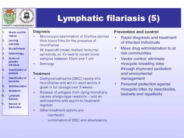
- 30. Lymphatic filariasis (5) Prevention and control Rapid diagnosis and treatment of infected individuals Mass drug administration
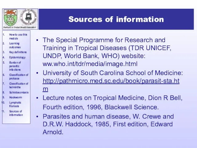
- 31. Sources of information The Special Programme for Research and Training in Tropical Diseases (TDR UNICEF, UNDP,
- 33. Скачать презентацию
Слайд 3Learning Outcomes
After completing this SDL, you should be able to:
Discuss how important
Learning Outcomes
After completing this SDL, you should be able to:
Discuss how important
Слайд 4Key definitions: What is ….?
Medical parasitology: “the study and medical implications of
Key definitions: What is ….?
Medical parasitology: “the study and medical implications of
Слайд 5Key definitions: What is ….?
Host: “the organism in, or on, which the
Key definitions: What is ….?
Host: “the organism in, or on, which the
Слайд 6Epidemiology
Although parasitic infections occur globally, the majority occur in tropical regions, where
Epidemiology
Although parasitic infections occur globally, the majority occur in tropical regions, where
Слайд 7The burden of some major parasitic infections
The burden of some major parasitic infections
Слайд 8Taxonomic classification of parasitic organisms
The classification of parasites is controversial - there
Taxonomic classification of parasitic organisms
The classification of parasites is controversial - there
Слайд 9Taxonomic classification of protozoa
Taxonomic classification of protozoa
Слайд 10Examples of important intestinal protozoa
Transmitted by the faecal-oral route and cause diarrhoea
Giardia
Examples of important intestinal protozoa
Transmitted by the faecal-oral route and cause diarrhoea
Giardia
Слайд 11Examples of important systemic protozoa
Detected in the blood
Plasmodium: the cause of malaria.
Examples of important systemic protozoa
Detected in the blood
Plasmodium: the cause of malaria.
Слайд 12Taxonomic classification of helminths
Taxonomic classification of helminths
Слайд 13Examples of important metazoa – intestinal nematodes
Trichuris (whipworm)
A soil transmitted helminth
prevalent in
Examples of important metazoa – intestinal nematodes
Trichuris (whipworm)
A soil transmitted helminth
prevalent in
Слайд 14Examples of important metazoa –systemic nematodes
Filaria including:
Onchocerca volvulus – Transmitted by the
Examples of important metazoa –systemic nematodes
Filaria including:
Onchocerca volvulus – Transmitted by the
Слайд 15Examples of important flatworms - cestodes
Intestinal - (“tapeworms”)
Taenia saginata
worldwide
acquired by ingestion
Examples of important flatworms - cestodes
Intestinal - (“tapeworms”)
Taenia saginata
worldwide
acquired by ingestion
Слайд 16Examples of important metazoa –trematodes (flukes)
Intestinal
Fasciolopsis buski - A common parasite of
Examples of important metazoa –trematodes (flukes)
Intestinal
Fasciolopsis buski - A common parasite of
Слайд 17Schistosomiasis (bilharzia)
Schistosomiasis (bilharzia)
Слайд 18Schistosomiasis (1)
Epidemiology
200m people in 74 countries infected, 85% of whom live in
Schistosomiasis (1)
Epidemiology
200m people in 74 countries infected, 85% of whom live in
Слайд 19Schistosomiasis (2)
Pathology
Shistosoma eggs become trapped in the tissues of the urinary tract
Schistosomiasis (2)
Pathology
Shistosoma eggs become trapped in the tissues of the urinary tract
Слайд 20Schistosomiasis (3)
Symptoms and signs
Urinary Schistosomiasis:
S. hamatobium
Gross haematuria
Dysuria
Bladder, ureters and kidneys damaged Cancer
Schistosomiasis (3)
Symptoms and signs
Urinary Schistosomiasis:
S. hamatobium
Gross haematuria
Dysuria
Bladder, ureters and kidneys damaged Cancer
Слайд 21Schistosomiasis (4)
Prevention and control
Educate people to not urinate or defecate in fresh
Schistosomiasis (4)
Prevention and control
Educate people to not urinate or defecate in fresh
Слайд 22Hookworm (1)
Epidemiology
>1200m infections each year of which 100m are symptomatic
It is due
Hookworm (1)
Epidemiology
>1200m infections each year of which 100m are symptomatic
It is due
Слайд 23Hookworm (2)
Life cycle
Adult worms live in the intestine and excrete eggs in
Hookworm (2)
Life cycle
Adult worms live in the intestine and excrete eggs in
Слайд 24Hookworm (3)
Pathology
Hookworms move several times a day to different attachment sites in
Hookworm (3)
Pathology
Hookworms move several times a day to different attachment sites in
Слайд 25Hookworm (4)
Symptoms and signs
Minor
Often itchy papules are found at the site where
Hookworm (4)
Symptoms and signs
Minor
Often itchy papules are found at the site where
Слайд 26Lymphatic filariasis (1)
Epidemiology
120m people infected in >80 countries in Africa, Asia, the
Lymphatic filariasis (1)
Epidemiology
120m people infected in >80 countries in Africa, Asia, the
Слайд 27Lymphatic filariasis (2)
Life cycle
Wuchereria bancrofti is mainly transmitted by
Culex mosquitoes in
Lymphatic filariasis (2)
Life cycle
Wuchereria bancrofti is mainly transmitted by
Culex mosquitoes in
Слайд 28Lymphatic filariasis (3)
Pathology
Adult worms live in the afferent lymphatic vessels and cause
Lymphatic filariasis (3)
Pathology
Adult worms live in the afferent lymphatic vessels and cause
Слайд 29Lymphatic filariasis (4)
Symptoms and signs – 3 stages
1. Asymptomatic stage
There is internal
Lymphatic filariasis (4)
Symptoms and signs – 3 stages
1. Asymptomatic stage
There is internal
Слайд 30Lymphatic filariasis (5)
Prevention and control
Rapid diagnosis and treatment of infected individuals
Mass drug
Lymphatic filariasis (5)
Prevention and control
Rapid diagnosis and treatment of infected individuals
Mass drug
Слайд 31Sources of information
The Special Programme for Research and Training in Tropical Diseases
Sources of information
The Special Programme for Research and Training in Tropical Diseases
 Производственная практика. ООО Сибирская ярмарка
 Узники концлагерей
 Повышение эффективности разработки нефтяных месторождений
 Презентация на тему Висячие Сады Семирамиды
 Be First
 МОУ Малыкайская средняя общеобразовательная школа им. М.В. МегежекскогоМетодический семинар: Диагностика уровня обученности и
  Презентация на тему Тепловые двигатели ДВС
  Число PHI
 Кто такие астрономы и что такое астрономия
 Банки: от древности до современности
 Правописание безударных окончаний существительных
 Агентство маркетинговых исследований
 Техника движения ног при выполнении нападающего удара
 Крылья. Конструкции и виды крыльев. Варианты для фотосессий и фотозон
 20141005_yaponiya_11_klass_1_chast
 Азия (2 класс)
 Презентация ко дню учителя
 Где с 31 декабря на 1 января не празднуют новый год
 s1665326323
 Источники муниципального права
 Руководство родителей и учащихсяГородская Школьная Информационная Система
 8 июля – день семьи , любви и верности!
 The Numeral
 Зейне Тұрсынкж слаид
 Правовые последствия. Брекзит и реформирование права
 Презентация
 Биография и творчество А.Г.Алексина
 С Л О В А Р И