- IDENTIFICATION OF PATHOGENIC BACTERIA IN CLINICAL MICROBIOLOGY

Содержание
- 2. Identification of Gram Positive Cocci(I) Staphylococcus species Gram stain – Gram positive cocci in clusters Motility
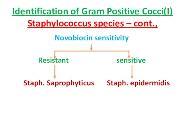
- 3. Identification of Gram Positive Cocci(I) Staphylococcus species – cont., Novobiocin sensitivity Resistant sensitive Staph. Saprophyticus Staph.
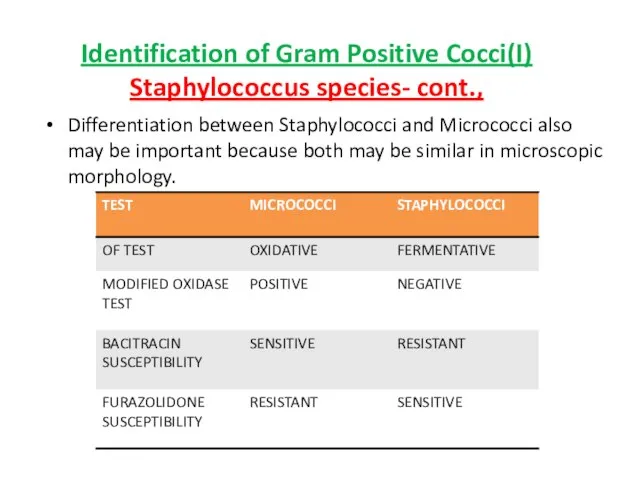
- 4. Differentiation between Staphylococci and Micrococci also may be important because both may be similar in microscopic
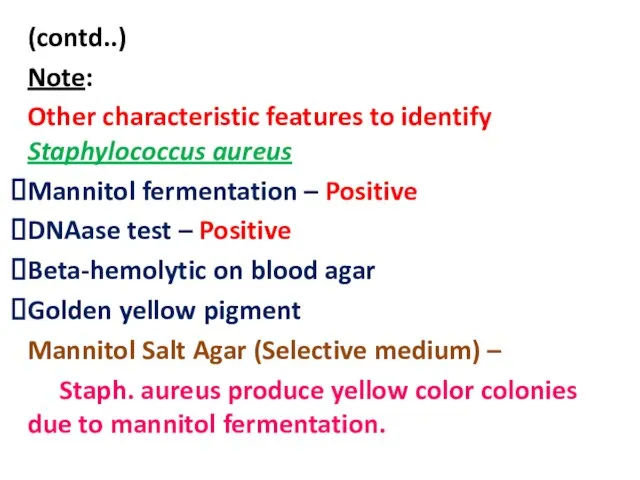
- 5. (contd..) Note: Other characteristic features to identify Staphylococcus aureus Mannitol fermentation – Positive DNAase test –
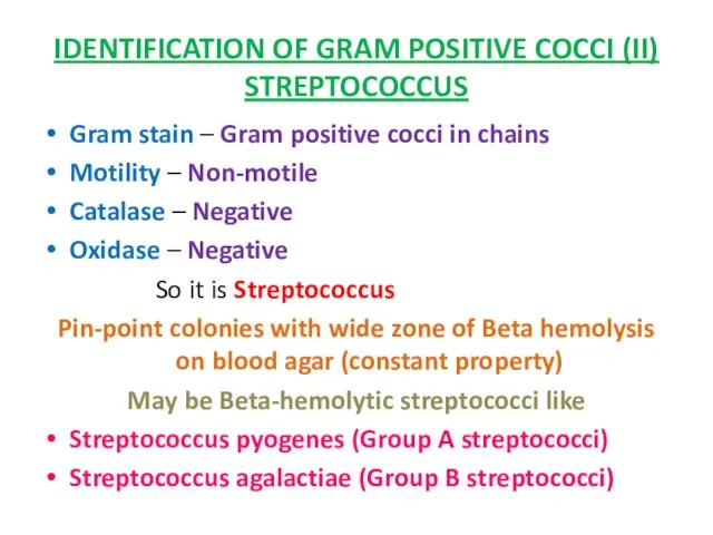
- 6. IDENTIFICATION OF GRAM POSITIVE COCCI (II) STREPTOCOCCUS Gram stain – Gram positive cocci in chains Motility
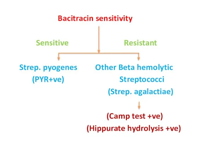
- 7. Bacitracin sensitivity Sensitive Resistant Strep. pyogenes Other Beta hemolytic (PYR+ve) Streptococci (Strep. agalactiae) (Camp test +ve)
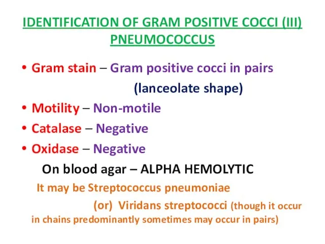
- 8. IDENTIFICATION OF GRAM POSITIVE COCCI (III) PNEUMOCOCCUS Gram stain – Gram positive cocci in pairs (lanceolate
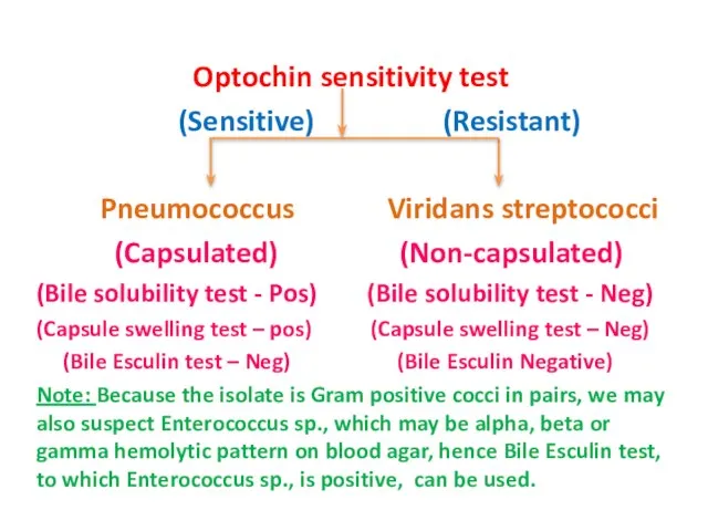
- 9. Optochin sensitivity test (Sensitive) (Resistant) Pneumococcus Viridans streptococci (Capsulated) (Non-capsulated) (Bile solubility test - Pos) (Bile
- 10. Note: Pneumococcus – cause of Lobar pneumonia so it is most likely to be present in
- 11. IDENTIFICATION OF GRAM POSITIVE COCCI- IV ENTEROCOCCUS Gram stain – Gram positive cocci in pairs Motility
- 12. BILE ESCULIN HYDROLYSIS TEST Positive Negative Group D streptococcus Pneumococcus Enterococcus Viridans streptococci (Grow in MacConkey’s
- 13. Growth in 6.5% salt (Salt tolerance test) Positive (Growth) Negative (No growth) Enterococcus Group D streptococcus
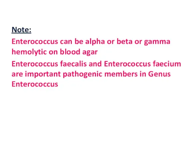
- 14. Note: Enterococcus can be alpha or beta or gamma hemolytic on blood agar Enterococcus faecalis and
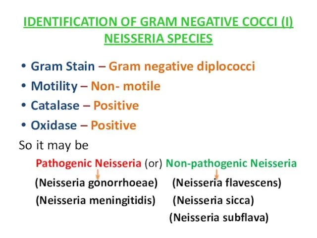
- 15. IDENTIFICATION OF GRAM NEGATIVE COCCI (I) NEISSERIA SPECIES Gram Stain – Gram negative diplococci Motility –
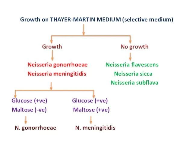
- 16. Growth on THAYER-MARTIN MEDIUM (selective medium) Growth No growth Neisseria gonorrhoeae Neisseria flavescens Neisseria meningitidis Neisseria
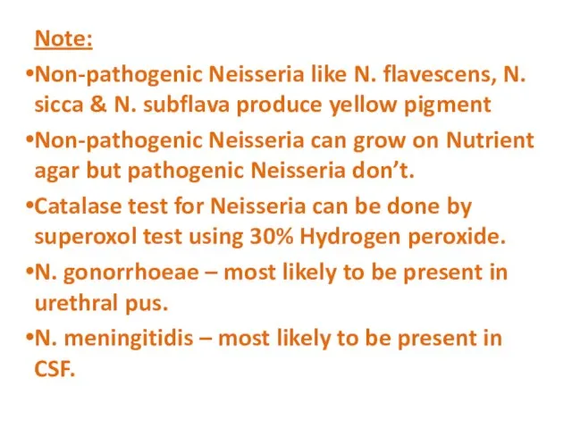
- 17. Note: Non-pathogenic Neisseria like N. flavescens, N. sicca & N. subflava produce yellow pigment Non-pathogenic Neisseria
- 18. IDENTIFICATION OF GRAM POSITIVE BACILLI List of Gram positive Bacilli Corynebacterium sp. Listeria sp. Erysipelothrix rhusiopathiae
- 19. Spore (Present) (Absent) Bacillus sp. Corynebacterium sp. Clostridium sp. Listeria sp. Ersipelothrix sp. Lactobacillus sp. Bacillus
- 20. Bacillus species Motile Non-motile Other Bacillus sp. Bacillus anthracis (McFadyean’s reaction +ve)
- 21. Catalase (+) (-) Corynebacterium Erysipelothrix Listeria Lactobacillus kurthia Actinomyces Beta hemolytic on BA H2S Production (+)
- 22. Listeria Corynebacterium (Esculin Hydrolysis) (+) (-) Listeria Corynebacterium (Motile at 250C) (Non-motile) (Non-Motile at 370C)
- 23. Lactobacillus Actinomyces (Branching filaments) (+) (-) Actinomyces Lactobacillus
- 24. Note: Other examples of anaerobic Gram positive bacilli – Eubacterium, Propionibacterium, Bifidobacterium, Mobilincus. Remember, Actinomyces and
- 25. IDENTIFICATION OF GRAM NEGATIVE BACILLI (I) ESCHERICHIA COLI /E.COLI Gram stain – Gram negative bacilli Motility
- 26. MacConkey’s agar – Dry, flat LF colonies Motility – Motile So it may be E.coli, Citrobacter,
- 27. Citrate test (+) (-) Citrobacter E.coli Serratia Citrobacter Serratia (No red pigment) (Red pigment)
- 28. IMVIC REACTIONS FOR E.COLI Indole – (+) Methyl red – (+) Voges-Proskauer – (-) Citrate -
- 29. IDENTIFICATION OF GNB (II) KLEBSIELLA SPECIES Gram stain – Gram Negative bacilli Motility – Non-motile Catalase
- 30. On MacConkey’s agar – Mucoid LF Colonies Motility – Non-motile So it may be Klebsiella species
- 31. Indole test (+) (-) Klebsiella oxytoca Klebsiella rhinoscleromatis Klebsiella pneumoniae Klebsiella ozanae (Urease +ve) (Urease –ve)
- 32. Malonate (+) (-) Klebsiella rhinoscleromatis Klebsiella ozanae (VP POSITIVE) (VP POSITIVE)
- 33. IMVIC REACTIONS FOR KLEBSIELLA PNEUMONIAE Indole – (-) Methyl red – (-) Voges-Proskauer – (+) Citrate
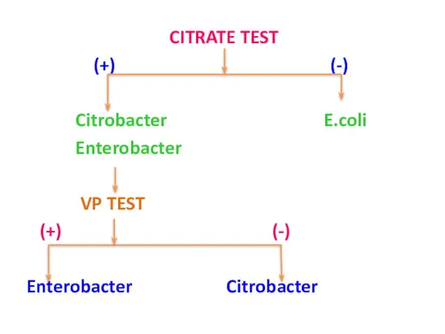
- 34. IDENTIFICATION OF GNB (III) CITROBACTER Gram stain – Gram negative bacilli Motility – Motile Catalase –
- 35. E.coli Enterobacter Citrobacter Citrate test (+) (-) Citrobacter E. coli Enterobacter
- 36. VP TEST (+) (-) Enterobacter Citrobacter H2S Production (-) (+) C. amalonauticus C. freundii C. koseri/diversus
- 37. C. amalonauticus C. koseri/diversus MALONATE ADONITOL (+) (-) C. koseri/diversus C. amalonauticus
- 38. Other reactions of Citrobacter Indole – (+/-) MR – (+) VP – (-) Urease – (weakly
- 39. IDENTIFICATION OF GNB (IV) ENTEROBACTER Gram stain – Gram Negative Bacilli Motility – Motile Catalase –
- 40. CITRATE TEST (+) (-) Citrobacter E.coli Enterobacter VP TEST (+) (-) Enterobacter Citrobacter
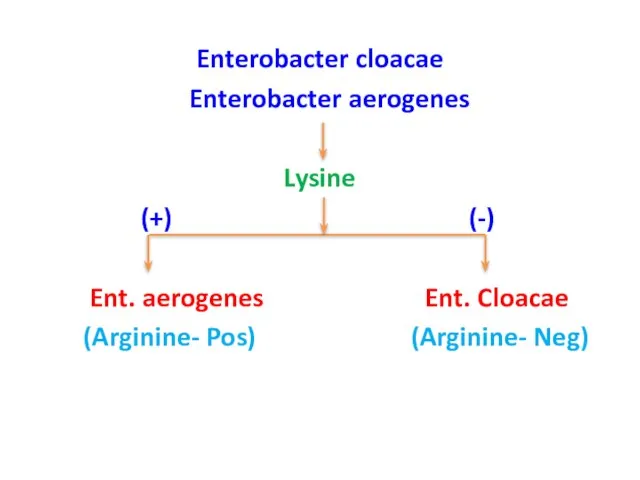
- 41. Enterobacter cloacae Enterobacter aerogenes Lysine (+) (-) Ent. aerogenes Ent. Cloacae (Arginine- Pos) (Arginine- Neg)
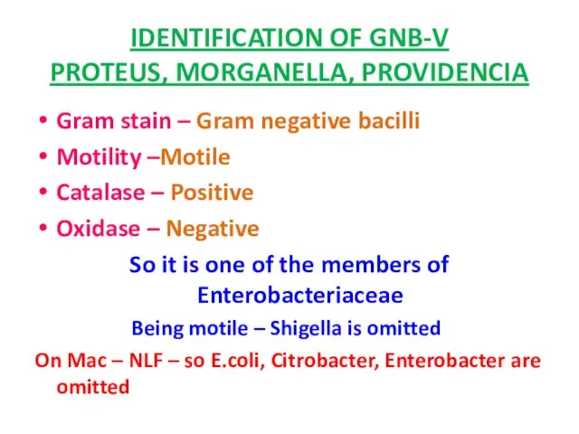
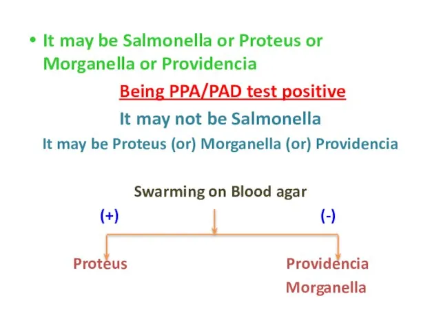
- 42. IDENTIFICATION OF GNB-V PROTEUS, MORGANELLA, PROVIDENCIA Gram stain – Gram negative bacilli Motility –Motile Catalase –
- 43. It may be Salmonella or Proteus or Morganella or Providencia Being PPA/PAD test positive It may
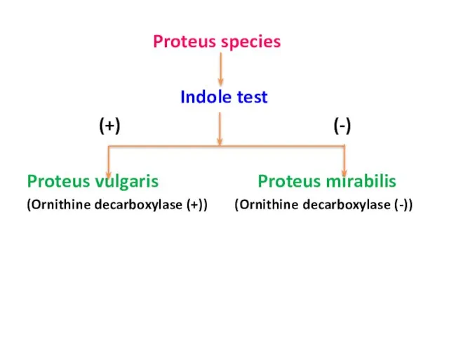
- 44. Proteus species Indole test (+) (-) Proteus vulgaris Proteus mirabilis (Ornithine decarboxylase (+)) (Ornithine decarboxylase (-))
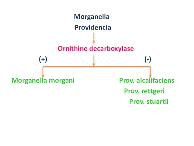
- 45. Morganella Providencia Ornithine decarboxylase (+) (-) Morganella morgani Prov. alcalifaciens Prov. rettgeri Prov. stuartii
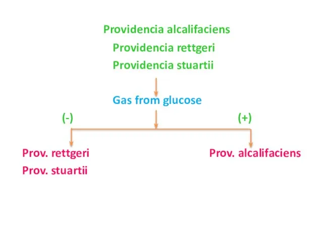
- 46. Providencia alcalifaciens Providencia rettgeri Providencia stuartii Gas from glucose (-) (+) Prov. rettgeri Prov. alcalifaciens Prov.
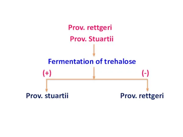
- 47. Prov. rettgeri Prov. Stuartii Fermentation of trehalose (+) (-) Prov. stuartii Prov. rettgeri
- 48. Other biochemicals for Proteus species MR – (+) VP – (-) Urease - (+) Citrate –
- 49. IDENTIFICATION OF GNB – VI SHIGELLA SPECIES Gram stain – Gram negative bacilli Motility – Non-motile
- 50. Mannitol fermentation (+) (-) Shigella dysenteriae Shigella flexneri Shigella boydii Shigella sonnei
- 51. Shigella flexneri Shigella boydii Shigella sonnei ONGP (-) (+) Shigella flexneri Shigella sonnei Shigella boydii
- 52. Note: Shigella dysenteriae type 1 is always catalase negative. Differentiation between Sh. Flexneri and Sh. Boydii
- 53. Other biochemicals for Shigella species Indole – (+/-) MR – (+) VP – (-) Citrate –
- 54. IDENTIFICATION OF GNB – VII SALMONELLA SPECIES Gram stain – Gram negative bacilli Motility – Motile
- 55. PPA/PDA (+) (-) Proteus sp. Salmonella sp. (Swarming on BA (+)) (Swarming on BA (-)) S.
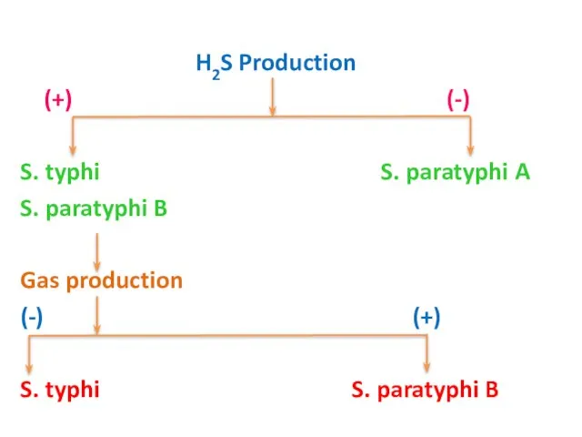
- 56. H2S Production (+) (-) S. typhi S. paratyphi A S. paratyphi B Gas production (-) (+)
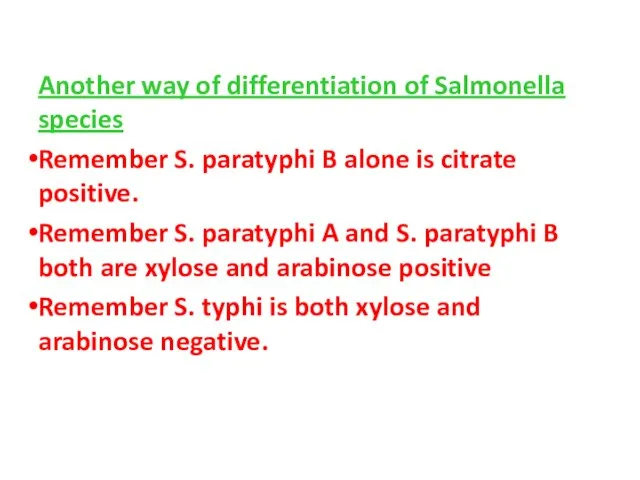
- 57. Another way of differentiation of Salmonella species Remember S. paratyphi B alone is citrate positive. Remember
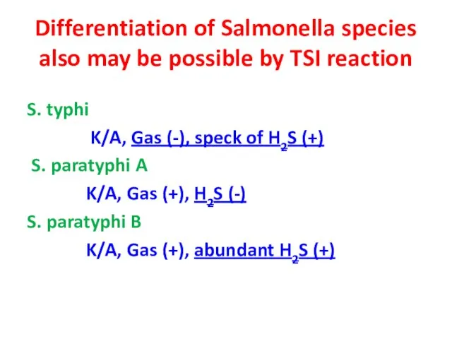
- 58. Differentiation of Salmonella species also may be possible by TSI reaction S. typhi K/A, Gas (-),
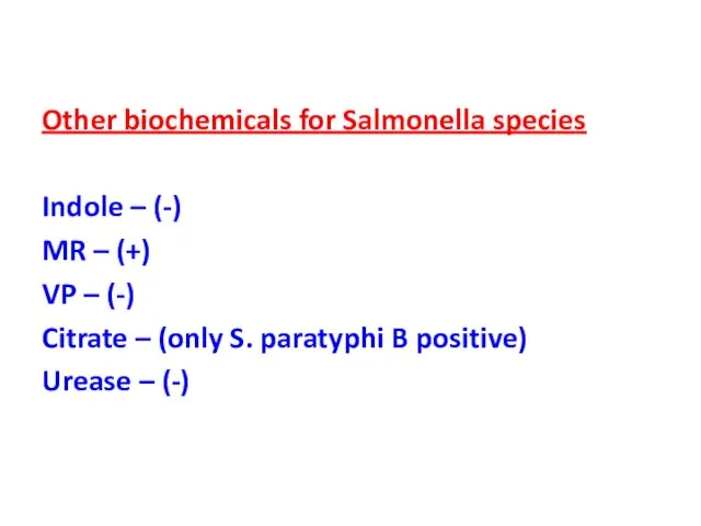
- 59. Other biochemicals for Salmonella species Indole – (-) MR – (+) VP – (-) Citrate –
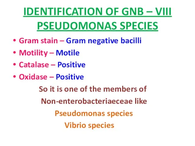
- 60. IDENTIFICATION OF GNB – VIII PSEUDOMONAS SPECIES Gram stain – Gram negative bacilli Motility – Motile
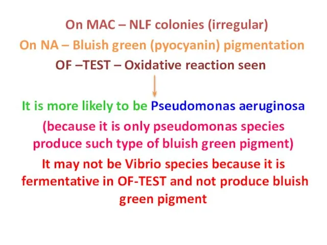
- 61. On MAC – NLF colonies (irregular) On NA – Bluish green (pyocyanin) pigmentation OF –TEST –
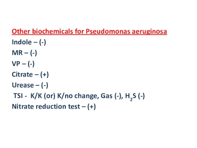
- 62. Other biochemicals for Pseudomonas aeruginosa Indole – (-) MR – (-) VP – (-) Citrate –
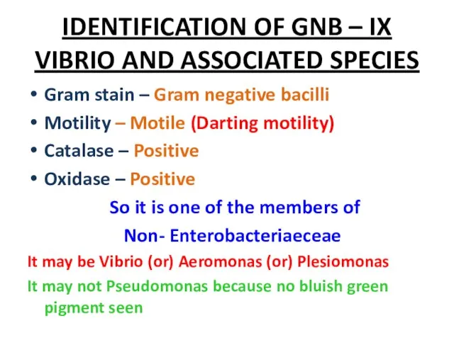
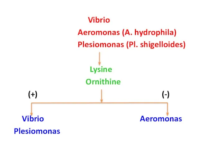
- 63. IDENTIFICATION OF GNB – IX VIBRIO AND ASSOCIATED SPECIES Gram stain – Gram negative bacilli Motility
- 64. Vibrio Aeromonas (A. hydrophila) Plesiomonas (Pl. shigelloides) Lysine Ornithine (+) (-) Vibrio Aeromonas Plesiomonas
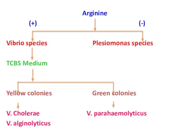
- 65. Arginine (+) (-) Vibrio species Plesiomonas species TCBS Medium Yellow colonies Green colonies V. Cholerae V.
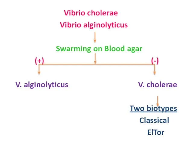
- 66. Vibrio cholerae Vibrio alginolyticus Swarming on Blood agar (+) (-) V. alginolyticus V. cholerae Two biotypes
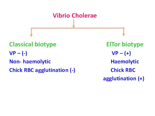
- 67. Vibrio Cholerae Classical biotype ElTor biotype VP – (-) VP – (+) Non- haemolytic Haemolytic Chick
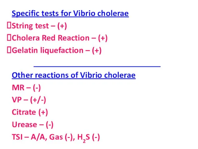
- 68. Specific tests for Vibrio cholerae String test – (+) Cholera Red Reaction – (+) Gelatin liquefaction
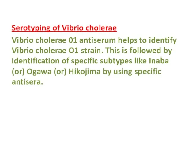
- 69. Serotyping of Vibrio cholerae Vibrio cholerae 01 antiserum helps to identify Vibrio cholerae O1 strain. This
- 71. Скачать презентацию
Слайд 2Identification of Gram Positive Cocci(I)
Staphylococcus species
Gram stain – Gram positive cocci in
Identification of Gram Positive Cocci(I)
Staphylococcus species
Gram stain – Gram positive cocci in
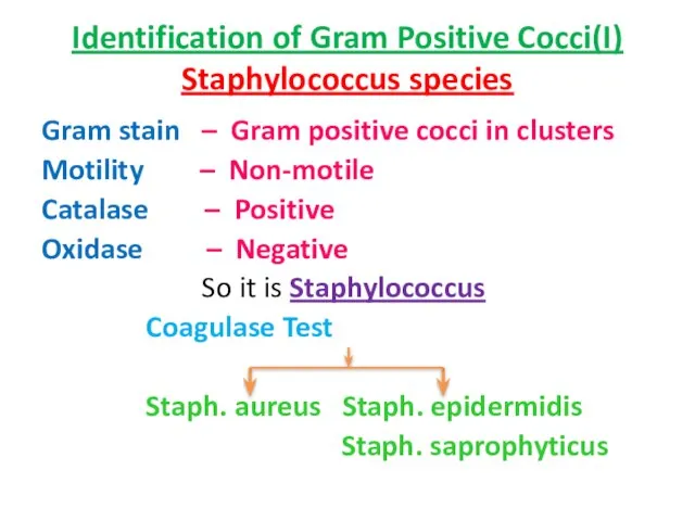
Слайд 3Identification of Gram Positive Cocci(I)
Staphylococcus species – cont.,
Novobiocin sensitivity
Resistant sensitive
Identification of Gram Positive Cocci(I)
Staphylococcus species – cont.,
Novobiocin sensitivity
Resistant sensitive
Слайд 4Differentiation between Staphylococci and Micrococci also may be important because both may
Differentiation between Staphylococci and Micrococci also may be important because both may
Слайд 5(contd..)
Note:
Other characteristic features to identify Staphylococcus aureus
Mannitol fermentation – Positive
DNAase
(contd..)
Note:
Other characteristic features to identify Staphylococcus aureus
Mannitol fermentation – Positive
DNAase
Слайд 6IDENTIFICATION OF GRAM POSITIVE COCCI (II)
STREPTOCOCCUS
Gram stain – Gram positive
IDENTIFICATION OF GRAM POSITIVE COCCI (II)
STREPTOCOCCUS
Gram stain – Gram positive
Слайд 7Bacitracin sensitivity
Sensitive Resistant
Strep. pyogenes Other Beta hemolytic
(PYR+ve)
Bacitracin sensitivity
Sensitive Resistant
Strep. pyogenes Other Beta hemolytic
(PYR+ve)
Слайд 8IDENTIFICATION OF GRAM POSITIVE COCCI (III)
PNEUMOCOCCUS
Gram stain – Gram positive cocci in
IDENTIFICATION OF GRAM POSITIVE COCCI (III)
PNEUMOCOCCUS
Gram stain – Gram positive cocci in
Слайд 9Optochin sensitivity test
(Sensitive) (Resistant)
Pneumococcus Viridans streptococci
(Capsulated) (Non-capsulated)
(Bile solubility
Optochin sensitivity test
(Sensitive) (Resistant)
Pneumococcus Viridans streptococci
(Capsulated) (Non-capsulated)
(Bile solubility
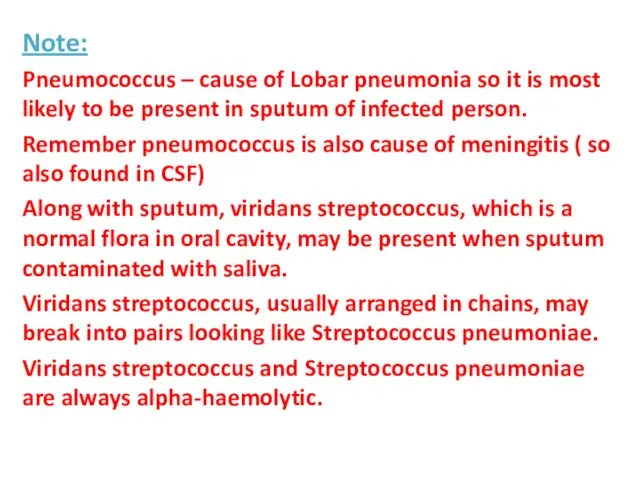
Слайд 10Note:
Pneumococcus – cause of Lobar pneumonia so it is most likely to
Note:
Pneumococcus – cause of Lobar pneumonia so it is most likely to
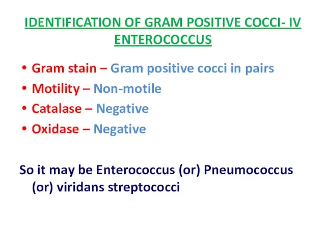
Слайд 11IDENTIFICATION OF GRAM POSITIVE COCCI- IV
ENTEROCOCCUS
Gram stain – Gram positive cocci in
IDENTIFICATION OF GRAM POSITIVE COCCI- IV
ENTEROCOCCUS
Gram stain – Gram positive cocci in
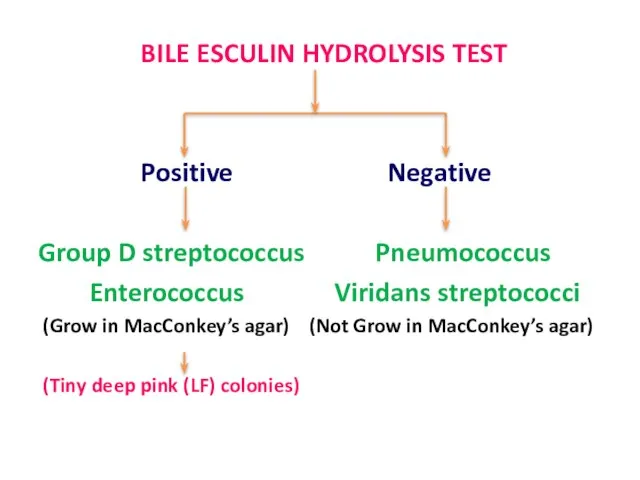
Слайд 12 BILE ESCULIN HYDROLYSIS TEST
Positive Negative
Group D streptococcus Pneumococcus
Enterococcus Viridans
BILE ESCULIN HYDROLYSIS TEST
Positive Negative
Group D streptococcus Pneumococcus
Enterococcus Viridans
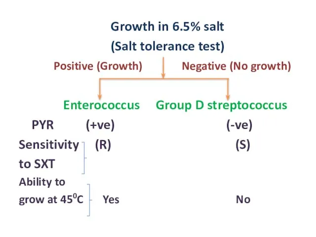
Слайд 13 Growth in 6.5% salt
(Salt tolerance test)
Positive (Growth) Negative (No
Growth in 6.5% salt
(Salt tolerance test)
Positive (Growth) Negative (No
Слайд 14Note:
Enterococcus can be alpha or beta or gamma hemolytic on blood agar
Enterococcus can be alpha or beta or gamma hemolytic on blood agar
Слайд 15IDENTIFICATION OF GRAM NEGATIVE COCCI (I)
NEISSERIA SPECIES
Gram Stain – Gram negative
IDENTIFICATION OF GRAM NEGATIVE COCCI (I)
NEISSERIA SPECIES
Gram Stain – Gram negative
Слайд 16 Growth on THAYER-MARTIN MEDIUM (selective medium)
Growth No growth
Neisseria gonorrhoeae Neisseria
Growth on THAYER-MARTIN MEDIUM (selective medium)
Growth No growth
Neisseria gonorrhoeae Neisseria
Слайд 17Note:
Non-pathogenic Neisseria like N. flavescens, N. sicca & N. subflava produce yellow
Note:
Non-pathogenic Neisseria like N. flavescens, N. sicca & N. subflava produce yellow
Слайд 18IDENTIFICATION OF GRAM POSITIVE BACILLI
List of Gram positive Bacilli
Corynebacterium sp.
Listeria
IDENTIFICATION OF GRAM POSITIVE BACILLI
List of Gram positive Bacilli
Corynebacterium sp.
Listeria

Слайд 19 Spore
(Present) (Absent)
Bacillus sp. Corynebacterium sp.
Clostridium sp. Listeria
Spore
(Present) (Absent)
Bacillus sp. Corynebacterium sp.
Clostridium sp. Listeria
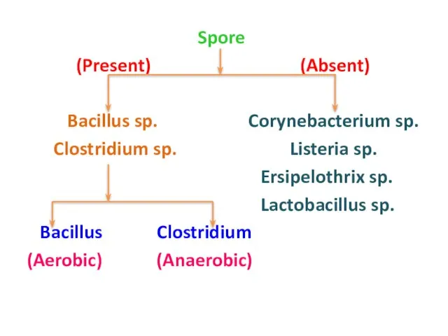
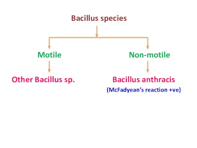
Слайд 20 Bacillus species
Motile Non-motile
Other Bacillus sp. Bacillus anthracis
(McFadyean’s reaction +ve)
Bacillus species
Motile Non-motile
Other Bacillus sp. Bacillus anthracis
(McFadyean’s reaction +ve)
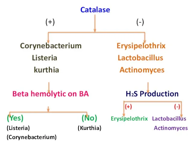
Слайд 21 Catalase
(+) (-)
Corynebacterium Erysipelothrix
Listeria Lactobacillus
kurthia Actinomyces
Beta
Catalase
(+) (-)
Corynebacterium Erysipelothrix
Listeria Lactobacillus
kurthia Actinomyces
Beta
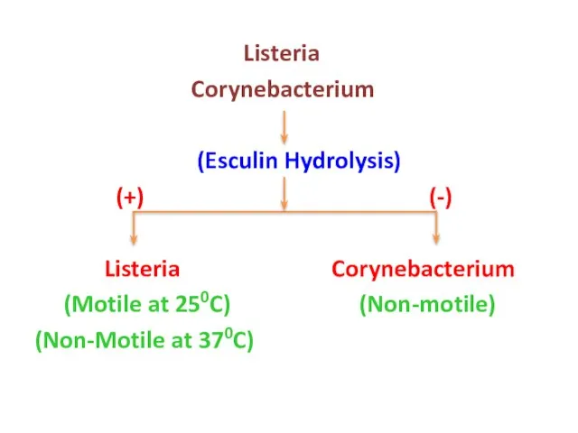
Слайд 22 Listeria
Corynebacterium
(Esculin Hydrolysis)
(+) (-)
Listeria Corynebacterium
(Motile at 250C)
Listeria
Corynebacterium
(Esculin Hydrolysis)
(+) (-)
Listeria Corynebacterium
(Motile at 250C)
Слайд 23 Lactobacillus
Actinomyces
(Branching filaments)
(+) (-)
Actinomyces Lactobacillus
Lactobacillus
Actinomyces
(Branching filaments)
(+) (-)
Actinomyces Lactobacillus

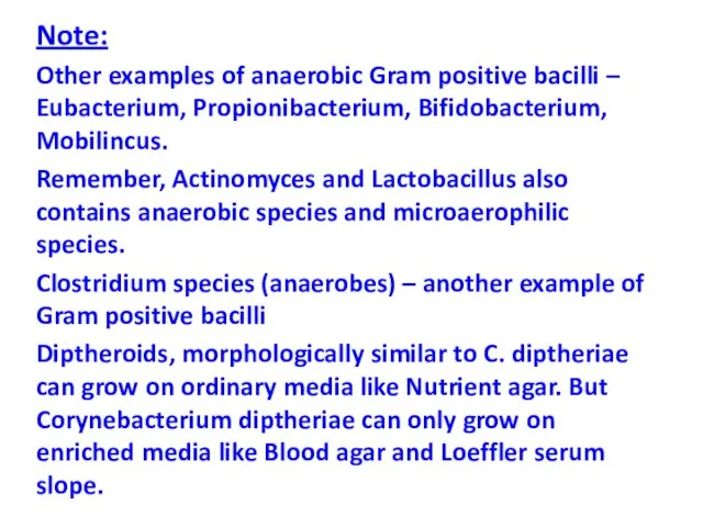
Слайд 24Note:
Other examples of anaerobic Gram positive bacilli – Eubacterium, Propionibacterium, Bifidobacterium, Mobilincus.
Remember,
Note:
Other examples of anaerobic Gram positive bacilli – Eubacterium, Propionibacterium, Bifidobacterium, Mobilincus.
Remember,
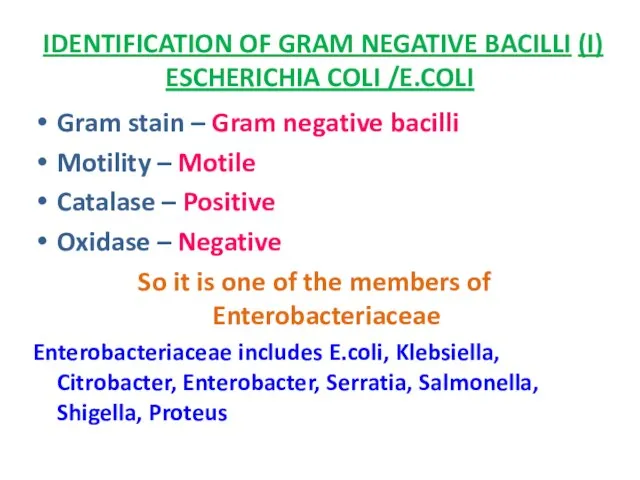
Слайд 25IDENTIFICATION OF GRAM NEGATIVE BACILLI (I)
ESCHERICHIA COLI /E.COLI
Gram stain – Gram negative
IDENTIFICATION OF GRAM NEGATIVE BACILLI (I)
ESCHERICHIA COLI /E.COLI
Gram stain – Gram negative
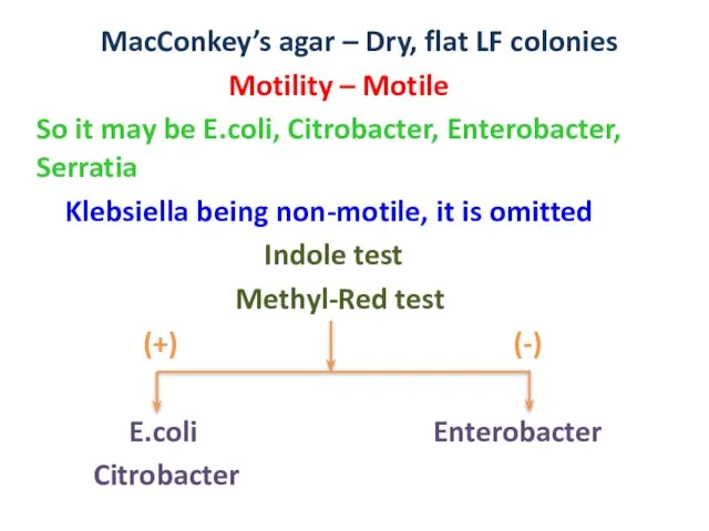
Слайд 26 MacConkey’s agar – Dry, flat LF colonies
Motility – Motile
So it
MacConkey’s agar – Dry, flat LF colonies
Motility – Motile
So it
Слайд 27 Citrate test
(+) (-)
Citrobacter E.coli
Serratia
Citrobacter Serratia
(No red
Citrate test
(+) (-)
Citrobacter E.coli
Serratia
Citrobacter Serratia
(No red
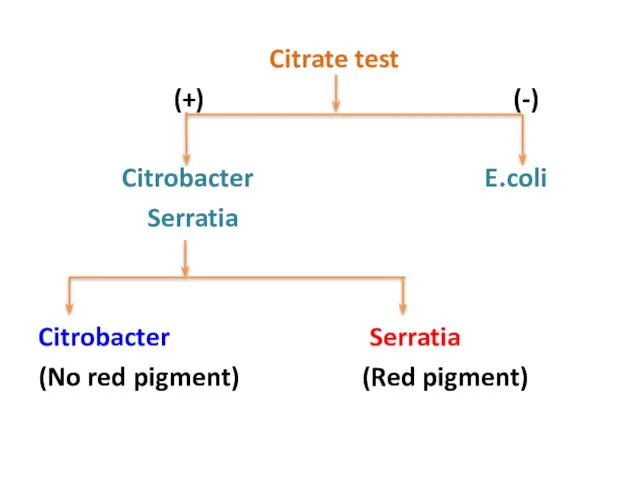
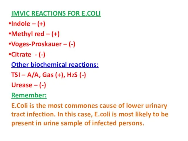
Слайд 28IMVIC REACTIONS FOR E.COLI
Indole – (+)
Methyl red – (+)
Voges-Proskauer – (-)
Citrate -
IMVIC REACTIONS FOR E.COLI
Indole – (+)
Methyl red – (+)
Voges-Proskauer – (-)
Citrate -
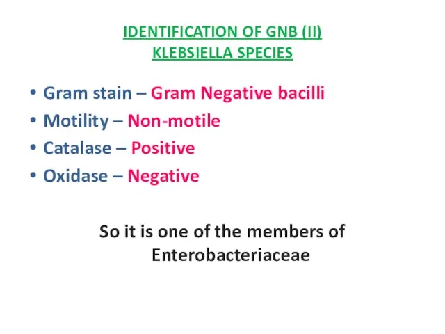
Слайд 29IDENTIFICATION OF GNB (II)
KLEBSIELLA SPECIES
Gram stain – Gram Negative bacilli
Motility –
IDENTIFICATION OF GNB (II)
KLEBSIELLA SPECIES
Gram stain – Gram Negative bacilli
Motility –
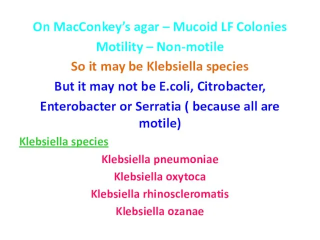
Слайд 30On MacConkey’s agar – Mucoid LF Colonies
Motility – Non-motile
So it may be
On MacConkey’s agar – Mucoid LF Colonies
Motility – Non-motile
So it may be
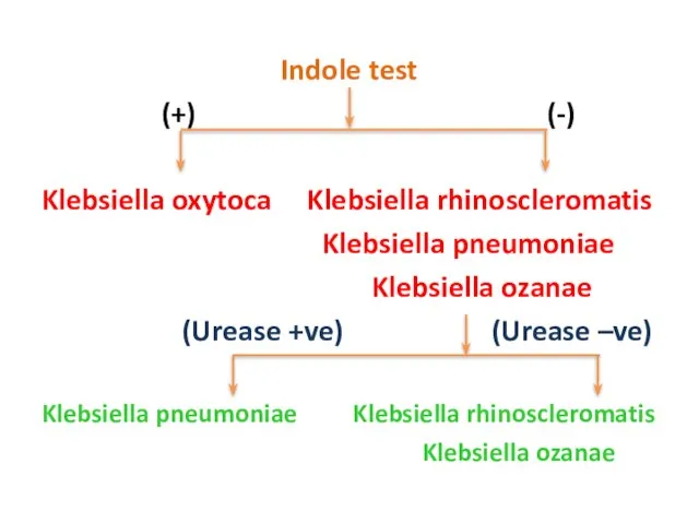
Слайд 31 Indole test
(+) (-)
Klebsiella oxytoca Klebsiella rhinoscleromatis
Klebsiella pneumoniae
Klebsiella
Indole test
(+) (-)
Klebsiella oxytoca Klebsiella rhinoscleromatis
Klebsiella pneumoniae
Klebsiella
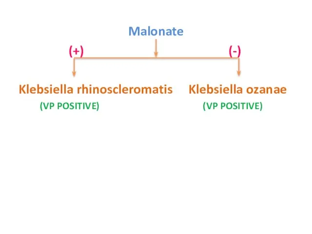
Слайд 32 Malonate
(+) (-)
Klebsiella rhinoscleromatis Klebsiella ozanae
(VP POSITIVE)
Malonate
(+) (-)
Klebsiella rhinoscleromatis Klebsiella ozanae
(VP POSITIVE)
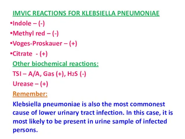
Слайд 33IMVIC REACTIONS FOR KLEBSIELLA PNEUMONIAE
Indole – (-)
Methyl red – (-)
Voges-Proskauer – (+)
Citrate
IMVIC REACTIONS FOR KLEBSIELLA PNEUMONIAE
Indole – (-)
Methyl red – (-)
Voges-Proskauer – (+)
Citrate
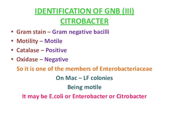
Слайд 34IDENTIFICATION OF GNB (III)
CITROBACTER
Gram stain – Gram negative bacilli
Motility – Motile
IDENTIFICATION OF GNB (III)
CITROBACTER
Gram stain – Gram negative bacilli
Motility – Motile
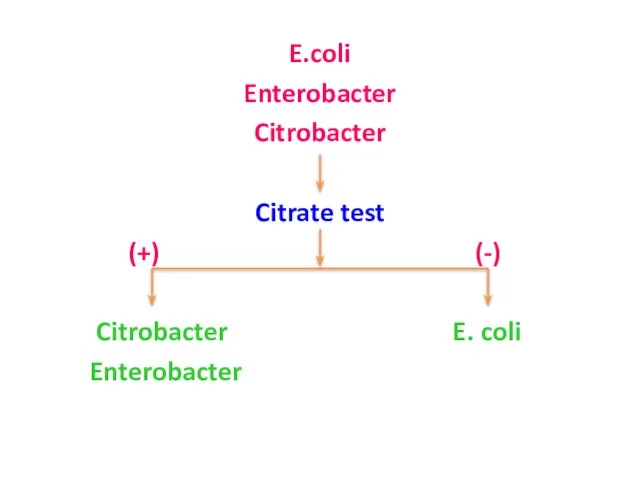
Слайд 35E.coli
Enterobacter
Citrobacter
Citrate test
(+) (-)
Citrobacter E. coli
Enterobacter
E.coli
Enterobacter
Citrobacter
Citrate test
(+) (-)
Citrobacter E. coli
Enterobacter
Слайд 36VP TEST
(+) (-)
Enterobacter Citrobacter
H2S Production
(-) (+)
VP TEST
(+) (-)
Enterobacter Citrobacter
H2S Production
(-) (+)
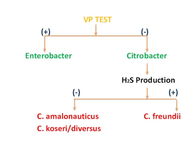
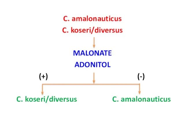
Слайд 37C. amalonauticus
C. koseri/diversus
MALONATE
ADONITOL
(+) (-)
C. koseri/diversus C. amalonauticus
C. amalonauticus
C. koseri/diversus
MALONATE
ADONITOL
(+) (-)
C. koseri/diversus C. amalonauticus
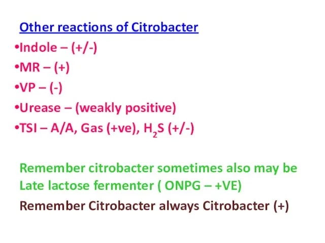
Слайд 38Other reactions of Citrobacter
Indole – (+/-)
MR – (+)
VP – (-)
Urease –
Other reactions of Citrobacter
Indole – (+/-)
MR – (+)
VP – (-)
Urease –
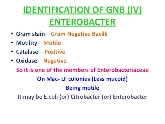
Слайд 39IDENTIFICATION OF GNB (IV)
ENTEROBACTER
Gram stain – Gram Negative Bacilli
Motility – Motile
IDENTIFICATION OF GNB (IV)
ENTEROBACTER
Gram stain – Gram Negative Bacilli
Motility – Motile
Слайд 40 CITRATE TEST
(+) (-)
Citrobacter E.coli
Enterobacter
VP TEST
CITRATE TEST
(+) (-)
Citrobacter E.coli
Enterobacter
VP TEST
Слайд 41Enterobacter cloacae
Enterobacter aerogenes
Lysine
(+) (-)
Ent. aerogenes Ent.
Enterobacter cloacae
Enterobacter aerogenes
Lysine
(+) (-)
Ent. aerogenes Ent.
Слайд 42IDENTIFICATION OF GNB-V
PROTEUS, MORGANELLA, PROVIDENCIA
Gram stain – Gram negative bacilli
Motility –Motile
IDENTIFICATION OF GNB-V
PROTEUS, MORGANELLA, PROVIDENCIA
Gram stain – Gram negative bacilli
Motility –Motile
Слайд 43It may be Salmonella or Proteus or Morganella or Providencia
Being
It may be Salmonella or Proteus or Morganella or Providencia
Being
Слайд 44 Proteus species
Indole test
(+) (-)
Proteus vulgaris Proteus mirabilis
(Ornithine
Proteus species
Indole test
(+) (-)
Proteus vulgaris Proteus mirabilis
(Ornithine
Слайд 45 Morganella
Providencia
Ornithine decarboxylase
(+) (-)
Morganella morgani Prov. alcalifaciens
Morganella
Providencia
Ornithine decarboxylase
(+) (-)
Morganella morgani Prov. alcalifaciens
Слайд 46 Providencia alcalifaciens
Providencia rettgeri
Providencia stuartii
Gas from glucose
Providencia alcalifaciens
Providencia rettgeri
Providencia stuartii
Gas from glucose
Слайд 47 Prov. rettgeri
Prov. Stuartii
Fermentation of trehalose
(+) (-)
Prov. rettgeri
Prov. Stuartii
Fermentation of trehalose
(+) (-)
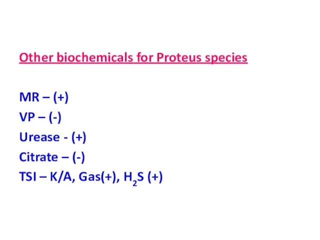
Слайд 48Other biochemicals for Proteus species
MR – (+)
VP – (-)
Urease - (+)
Citrate
MR – (+)
VP – (-)
Urease - (+)
Citrate
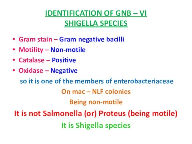
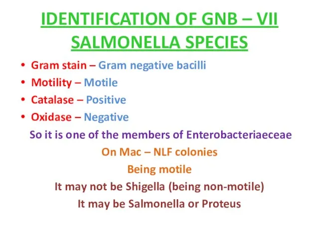
Слайд 49IDENTIFICATION OF GNB – VI
SHIGELLA SPECIES
Gram stain – Gram negative bacilli
Motility
IDENTIFICATION OF GNB – VI
SHIGELLA SPECIES
Gram stain – Gram negative bacilli
Motility
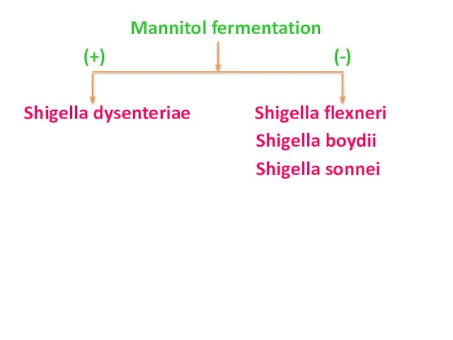
Слайд 50 Mannitol fermentation
(+) (-)
Shigella dysenteriae Shigella flexneri
Shigella boydii
Shigella
Mannitol fermentation
(+) (-)
Shigella dysenteriae Shigella flexneri
Shigella boydii
Shigella
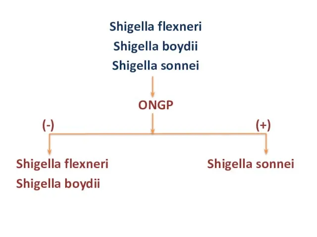
Слайд 51Shigella flexneri
Shigella boydii
Shigella sonnei
ONGP
(-) (+)
Shigella flexneri Shigella sonnei
Shigella boydii
Shigella flexneri
Shigella boydii
Shigella sonnei
ONGP
(-) (+)
Shigella flexneri Shigella sonnei
Shigella boydii
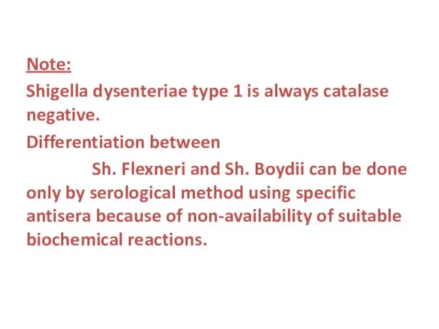
Слайд 52Note:
Shigella dysenteriae type 1 is always catalase negative.
Differentiation between
Sh. Flexneri
Shigella dysenteriae type 1 is always catalase negative.
Differentiation between
Sh. Flexneri
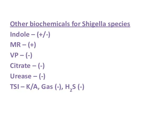
Слайд 53Other biochemicals for Shigella species
Indole – (+/-)
MR – (+)
VP – (-)
Citrate
Other biochemicals for Shigella species
Indole – (+/-)
MR – (+)
VP – (-)
Citrate
Слайд 54IDENTIFICATION OF GNB – VII
SALMONELLA SPECIES
Gram stain – Gram negative bacilli
Motility
IDENTIFICATION OF GNB – VII
SALMONELLA SPECIES
Gram stain – Gram negative bacilli
Motility
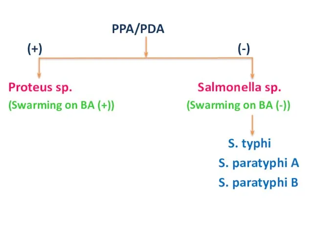
Слайд 55 PPA/PDA
(+) (-)
Proteus sp. Salmonella sp.
(Swarming on BA (+)) (Swarming
PPA/PDA
(+) (-)
Proteus sp. Salmonella sp.
(Swarming on BA (+)) (Swarming
Слайд 56 H2S Production
(+) (-)
S. typhi S. paratyphi A
S. paratyphi B
Gas
H2S Production
(+) (-)
S. typhi S. paratyphi A
S. paratyphi B
Gas
Слайд 57Another way of differentiation of Salmonella species
Remember S. paratyphi B alone is
Another way of differentiation of Salmonella species
Remember S. paratyphi B alone is
Слайд 58Differentiation of Salmonella species also may be possible by TSI reaction
S.
Differentiation of Salmonella species also may be possible by TSI reaction
S.
Слайд 59Other biochemicals for Salmonella species
Indole – (-)
MR – (+)
VP – (-)
Citrate –
Indole – (-)
MR – (+)
VP – (-)
Citrate –
Слайд 60IDENTIFICATION OF GNB – VIII
PSEUDOMONAS SPECIES
Gram stain – Gram negative bacilli
Motility
IDENTIFICATION OF GNB – VIII
PSEUDOMONAS SPECIES
Gram stain – Gram negative bacilli
Motility
Слайд 61 On MAC – NLF colonies (irregular)
On NA – Bluish green (pyocyanin)
On MAC – NLF colonies (irregular)
On NA – Bluish green (pyocyanin)
Слайд 62Other biochemicals for Pseudomonas aeruginosa
Indole – (-)
MR – (-)
VP – (-)
Citrate –
Indole – (-)
MR – (-)
VP – (-)
Citrate –
Слайд 63IDENTIFICATION OF GNB – IX
VIBRIO AND ASSOCIATED SPECIES
Gram stain – Gram negative
IDENTIFICATION OF GNB – IX
VIBRIO AND ASSOCIATED SPECIES
Gram stain – Gram negative
Слайд 64 Vibrio
Aeromonas (A. hydrophila)
Plesiomonas (Pl. shigelloides)
Lysine
Vibrio
Aeromonas (A. hydrophila)
Plesiomonas (Pl. shigelloides)
Lysine
Слайд 65 Arginine
(+) (-)
Vibrio species Plesiomonas species
TCBS Medium
Yellow colonies Green colonies
V. Cholerae
Arginine
(+) (-)
Vibrio species Plesiomonas species
TCBS Medium
Yellow colonies Green colonies
V. Cholerae
Слайд 66 Vibrio cholerae
Vibrio alginolyticus
Swarming on Blood agar
(+) (-)
Vibrio cholerae
Vibrio alginolyticus
Swarming on Blood agar
(+) (-)
Слайд 67 Vibrio Cholerae
Classical biotype ElTor biotype
VP – (-) VP – (+)
Non-
Vibrio Cholerae
Classical biotype ElTor biotype
VP – (-) VP – (+)
Non-
Слайд 68Specific tests for Vibrio cholerae
String test – (+)
Cholera Red Reaction – (+)
Gelatin
Specific tests for Vibrio cholerae
String test – (+)
Cholera Red Reaction – (+)
Gelatin
Слайд 69Serotyping of Vibrio cholerae
Vibrio cholerae 01 antiserum helps to identify Vibrio
Serotyping of Vibrio cholerae
Vibrio cholerae 01 antiserum helps to identify Vibrio
 Система дополнительного образования детей: прошлое и современность
 Краткосрочное и стратегическое финансовое планирование для поддержки пути модернизации государственной системы Решения Oracle EPM (H
 Таможенное декларирование товаров
 Локальная форма социального обеспечения
 Физическая культура и спорт в профилактике заболеваний и укрепления здоровья
 Процессуальные решения и документы, сроки и процессуальные издержки
 Методика подготовки учащихся к ЕГЭ по истории
 Параллельность в пространстве
 Презентация на тему Владимир Иванович Вернадский
 MBA Webinar. Harvard Business School On-line Узнать бизнес-школу лучше еще никогда не было так просто! Потому что нам важен ваш результат ® !
 Музеи Оренбурга
 Пельмени-ушки, русская традиция
 ООО «ВЛИ Восток»
 Музыкальный театр
 Гжель. Народные промыслы. Их истоки и современное развитие
 Человек в управлении организации
 Решение задач по химическим уравнениям
 «Жуткие» растения тропиков
 База данных и СУБД: основные понятия
 Гидрометеорологический мониторинг Юнтоловского заказника
 Правописание слов с сочетаниями ЧК - ЧН
 Школьный психолог
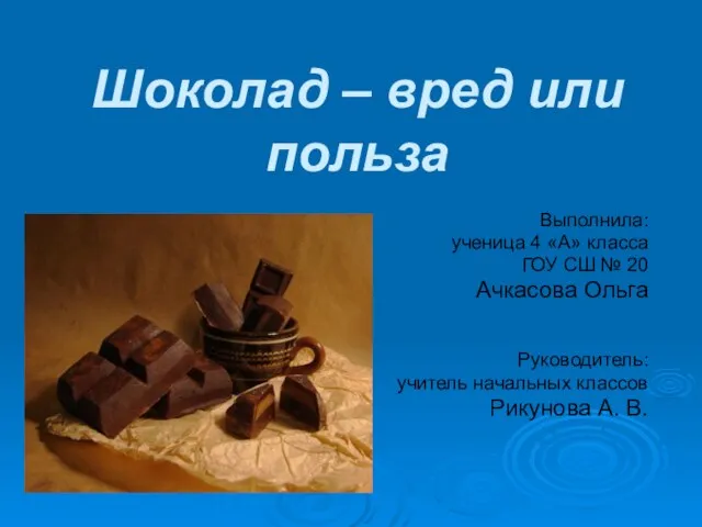 Шоколад – вред или польза
 Презентация на тему Остров Кипр
  Где зимуют птицы (1 класс)
 Презентация на тему Жизнь на селе в старину 3 класс
  Правоведение
 Can you swim?