- State Establishment “N.N. Alexandrov National Cancer Centre of Belarus”

Содержание
- 2. THE COUNCIL OF MINISTERS OF THE BYELORUSSIAN SSR Resolution On Intensifying Cancer from May 23, 1959
- 3. 1960 y. N.N. Alexandrov – the founder and first director near a model of the future
- 4. Administration building
- 5. Modernized building for oncological mammalogical department
- 6. Laboratory building
- 7. Recreational pavilions in pedestrian zones on the Centre territory
- 8. With the Decree of President of the Republic of Belarus Alexandr Grigoryevich Lukashenko from July 6,
- 9. SE N.N.Alexandrov NCCB SE COH NCCB 5 regional ODs 6 interdistrict ODs and MCOD Specialized Medical
- 10. Centre Structure At the base of the Centre, the BelMAPO Oncology Department (2 Doctors of Sciences
- 11. Research Trends organizing anticancer struggle, studying cancer epidemiology and prophylaxis developing new technologies for diagnosing malignant
- 12. Center Bedding 820 beds 12 beds— the Resuscitation Department
- 13. Hospitalized Patients 19343 Annually more than 200 foreign patients from the USA, France, Russia, India, Iraq,
- 14. Diagnostic Base of the Centre
- 15. Practically, the whole specter of biochemical, clinical, immunohistological, radioisotopic, molecular and genetic investigations is performed. Laboratory
- 16. Laboratory of Molecular Oncogenomics Specter of Performed Investigations Detecting mutations in genes of hemodialysis system Detecting
- 17. Translocation t(11,14) under lymphoma from mantle zone cells Fluorescent microscope Axioskop 40 FISH-Laboratory FISH ─ fluorescent
- 18. Laboratory of Molecular Cytogenetics
- 19. Morphological Methods of Investigation
- 20. Distribution of protein ER in tumour cell nuclei at BC, x40. Immunohistochemical staining MKAT (clone 1D5,
- 21. Distribution of protein c-erbB-2 in tumour cell nuclei at BC, x40. Immunohistochemical staining MKAT (dilution 1:300,
- 22. Telepathology system for giving on-line consultations on morphological preparations Modern equipment for biopsy material automated paraffin
- 23. Radiodiagnosis The department is equipped with modern, mainly, digital diagnostic instruments securing the use of an
- 24. Computer roentgen tomographs
- 25. Magnetic resonance tomograph
- 26. One-photon emission tomograph
- 27. Angiographic investigations
- 28. Ultrasound investigations USI scanners in the expert class
- 29. Intraoperative ultrasound investigation
- 30. Diagnosing Bladder Cancer on the Basis of Photodynamic Effect Bladder cancer manifestation after intravesicular introduction of
- 31. Efficacy of Clinical Use of 5-ALA (Alamin) of the Belarusian Production Additional malignant bladder tumours are
- 32. Highly Technological Methods of Treatment
- 33. Surgical Activities Totally, there were made 2007 — 9 323 operations 2008 — 10 628 operations.
- 34. Highly Technological Medical Interventions Combined operations with resection and prosthetics of arch and chest aorta, vertebrae
- 35. Pancreatectomy under total pancreas cancer, with pylorus constrictor being saved Gastropancreatoduodenal resections under locally spread stomach
- 36. Multicomponent management of patients with primary liver cancer and colorectal cancer with metastases in liver using
- 37. method of combined and radiation treatment of patients with supratentorial gliomas and a metastatic brain lesion
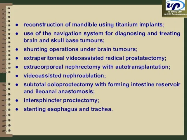
- 38. reconstruction of mandible using titanium implants; use of the navigation system for diagnosing and treating brain
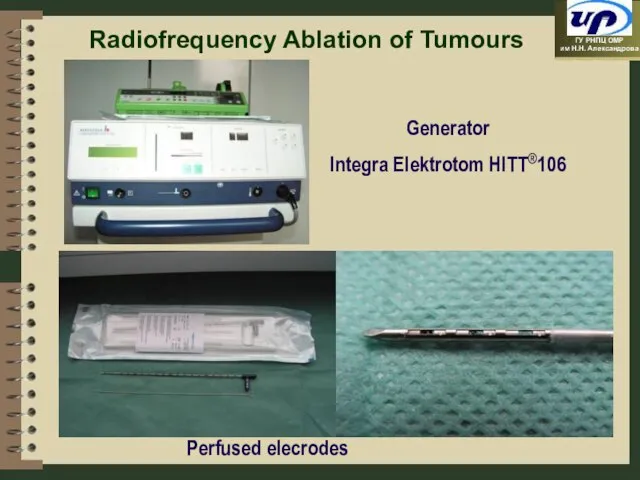
- 39. Radiofrequency Ablation of Tumours Perfused elecrodes Generator Integra Elektrotom HITT®106
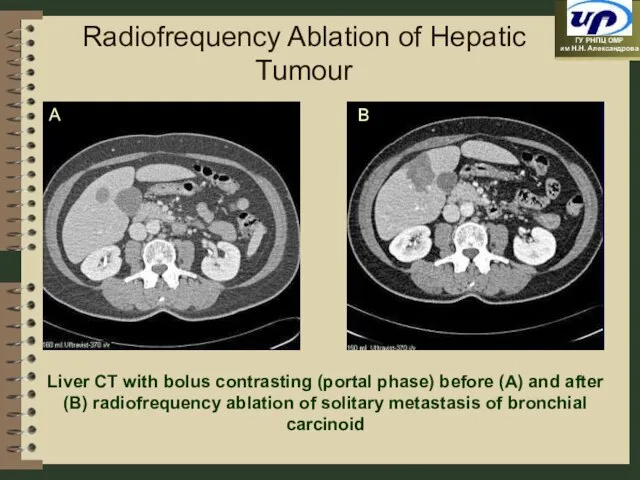
- 40. Radiofrequency Ablation of Hepatic Tumour Liver CT with bolus contrasting (portal phase) before (A) and after
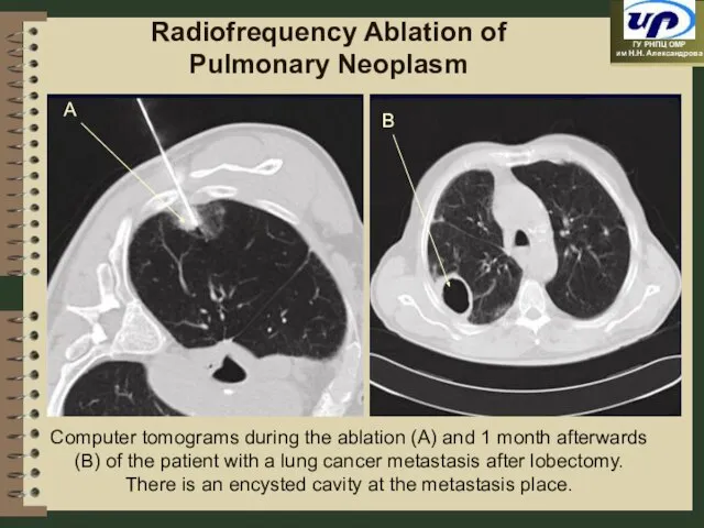
- 41. Radiofrequency Ablation of Pulmonary Neoplasm Computer tomograms during the ablation (A) and 1 month afterwards (B)
- 42. Radiofrequency Ablation of Kidney Neoplasm
- 43. Endoscopic Operations Laparascopic: splenectomy,adrenalectomy, nephrectomy, radical prostatectomy, obstructive resection of sigmoid colon and others. Thoracoscopic: lobectomy,
- 44. OPERATIONS UNDER HEAD AND NECK TUMOURS
- 45. Upper maxilla cancer with growing into the orbit and spreading into the anterior cranial fossa CRANIOFACIAL
- 46. CRANIOFACIAL RESECTION Cranial and facial stage View of the wound after removing preparation Macropreparation
- 47. TECHNOLOGY OF TRACHEOESOPHAGEAL SHUNTING WITH VOCAL PROSTHESIS INSERTING SET FOR INSERTING VOCAL PROSTHESES
- 48. ONE-STAGE REPARATION OF VOCAL AND ESOPHAGEAL FUNCTIONS PHARYNGOSTOMA TRACHEOSTOMA
- 49. TRACHEOESOPHAGEAL ANASTOMOSIS FORMING The trochar is inserted via pharynx
- 50. PROSTHESIS FIXATION IN CONDUCTOR The prosthesis is fixed in a conductor
- 51. PHARYNX ANTERIOR WALL FORMING WITH LOCAL TISSUES Vocal prosthesis
- 52. THE SKIN DEFECT IS REMOVED WITH SKIN AND MUSCULAR PECTORAL GRAFT
- 53. INSERTED VOCAL PROSTHESIS
- 54. Plastic Operations in Patients with Breast Cancer
- 55. Patient Z, 34 y.o. Diagnosis: left breast cancer T2N0M0G2. The condition after a complex treatment in
- 56. The same patient. The condition after a delayed mammaplasty with a free TRAM-graft in 1999.
- 57. The same patient. The condition after the reconstruction of mamillary and areolar complex in 2001.
- 58. Right breast cancer Т2N1М0. The condition after bilateral subcutaneous mastectomy and one-stage mammaplasty through a combined
- 59. Postmastectomy Syndrome
- 60. Plasty of Soft Tissues
- 63. Rhabdomyosarcoma of Left Forearm Soft Tissues
- 64. The same patient: plasty with a radial fixed vascular pedicle flap
- 65. Operations Preserving Organs under Bone Tumours
- 66. Modular Endoprostheses
- 69. Endoprosthetics with saddle-like prosthesis
- 71. Lung Cancer and Mediastrium Tumours
- 72. Lung Tumour Invading Left Atrium Lumen
- 73. Tumour in Left Atrium Lumen
- 74. The lung is removed, the left atrium wall defect is substituted with xenopericardium
- 75. MSCT before Operation
- 76. Detecting Site of Thoracic and Abdominal Aorta with Tumour Esophagus Celiac trunk SMA Left kidney vessels
- 77. Spine Left kidney vessels Aorta The site of aorta with tumour is resected. Celia trunk
- 78. Esophagus Left kidney vessels Aorta prosthesis The prosthesis is made to aorta. Celia trunk SMА
- 79. Macropreparation
- 80. Extracorporeal nephrectomy with autotransplantation
- 81. View of reconstructed kidney
- 82. Ileocystoplasty according to R. Hautmann (Modified) after Radical Cystectomy due to Bladder Cancer
- 83. Operating Block
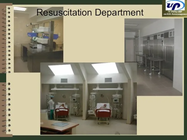
- 84. Resuscitation Department
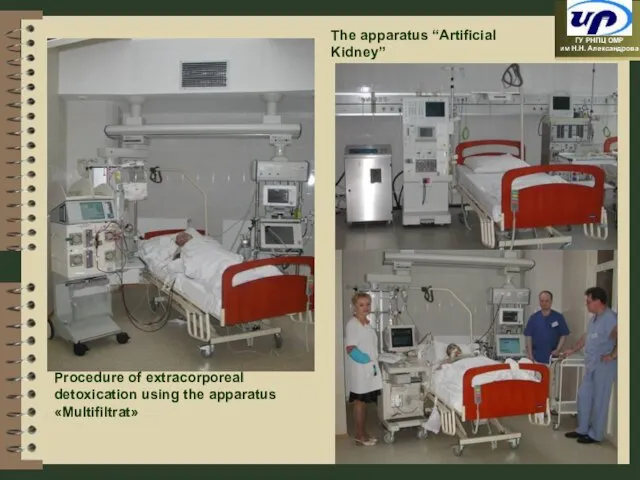
- 85. The apparatus “Artificial Kidney” Procedure of extracorporeal detoxication using the apparatus «Multifiltrat»
- 86. Radiotherapy
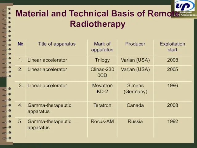
- 87. Material and Technical Basis of Remote Radiotherapy
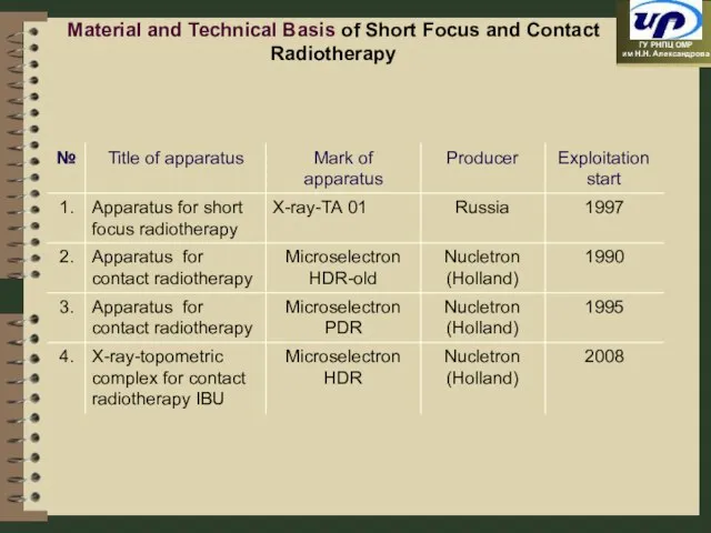
- 88. Material and Technical Basis of Short Focus and Contact Radiotherapy
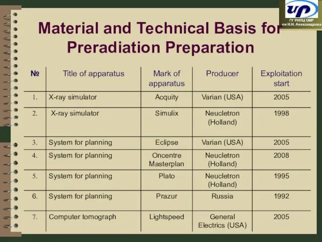
- 89. Material and Technical Basis for Preradiation Preparation
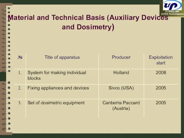
- 90. Material and Technical Basis (Auxiliary Devices and Dosimetry)
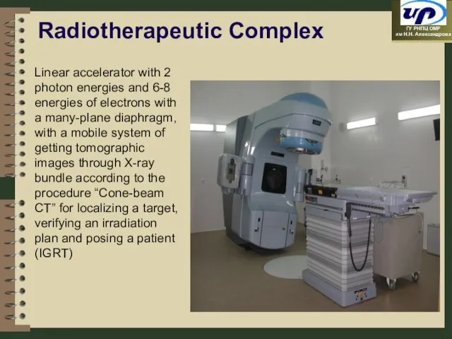
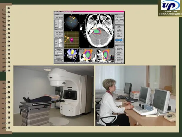
- 91. Linear accelerator with 2 photon energies and 6-8 energies of electrons with a many-plane diaphragm, with
- 93. High Technologies in Radiotherapy Three-dimensional conformal radiotherapy Radiotherapy with modulating dose intensity Stereotaxic radiotherapy / radiosurgery
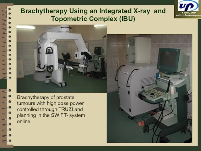
- 94. Brachytherapy Using an Integrated X-ray and Topometric Complex (IBU) Brachytherapy of prostate tumours with high dose
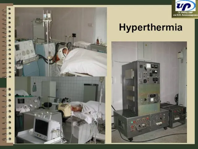
- 95. Hyperthermia
- 96. Equipment for Photodynamic Therapy and Diagnosis «Metalaz-M» «Kamin-Video» «Lesa-6» «LD-680»
- 97. 6 months afterwards Patient C., 34 y.o., CIN III. 26.10.07. – PDT Before treatment Photodynamic Therapy
- 98. Dynamics of Malignant Neoplasm Morbidity and Mortality in the Republic of Belarus 194,3 (-3,1%) 329,1 414,1
- 99. Europe Belarus European Union Annals of Oncology 16: 481-488, 2005 Belarusian Cancer-Register, 2008 Correlation of the
- 101. Скачать презентацию
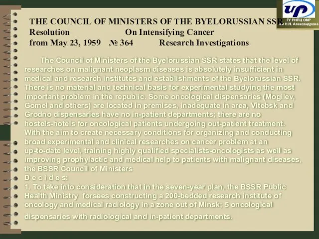
Слайд 2THE COUNCIL OF MINISTERS OF THE BYELORUSSIAN SSR
Resolution On Intensifying Cancer
from May 23,
THE COUNCIL OF MINISTERS OF THE BYELORUSSIAN SSR
Resolution On Intensifying Cancer
from May 23,
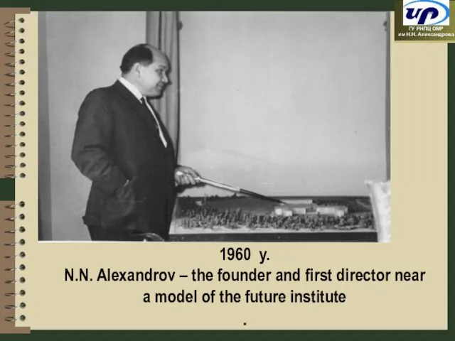
Слайд 31960 y.
N.N. Alexandrov – the founder and first director near a model of
1960 y.
N.N. Alexandrov – the founder and first director near a model of
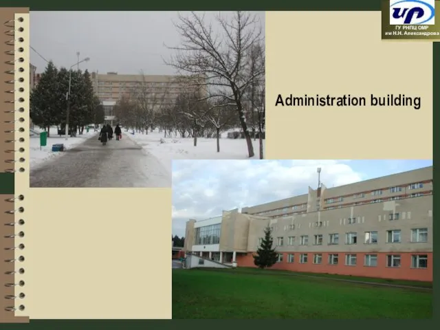
Слайд 4Administration building
Administration building
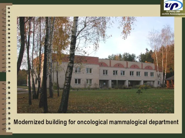
Слайд 5Modernized building for oncological mammalogical department
Modernized building for oncological mammalogical department
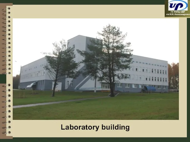
Слайд 6Laboratory building
Laboratory building
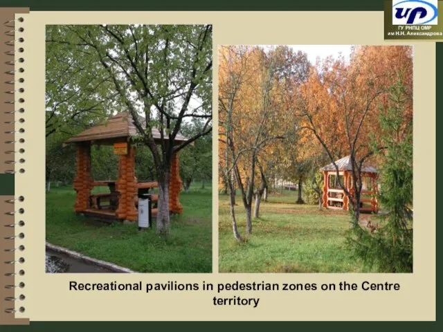
Слайд 7Recreational pavilions in pedestrian zones on the Centre
territory
Recreational pavilions in pedestrian zones on the Centre
territory
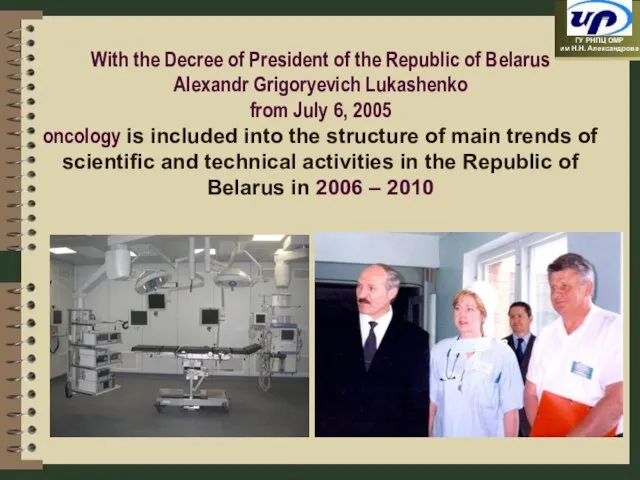
Слайд 8With the Decree of President of the Republic of Belarus
Alexandr Grigoryevich
With the Decree of President of the Republic of Belarus Alexandr Grigoryevich
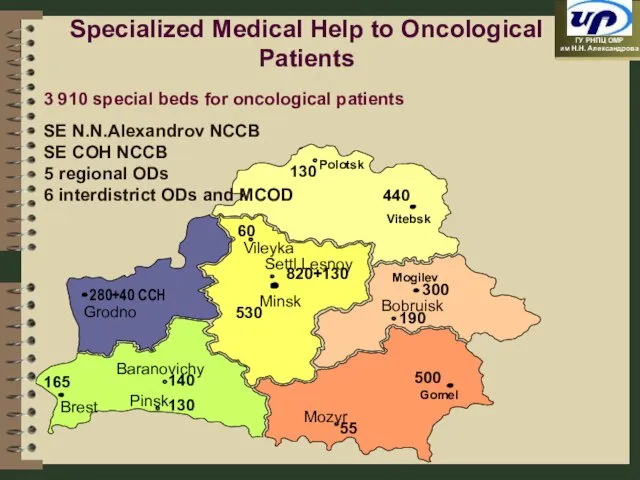
Слайд 9SE N.N.Alexandrov NCCB
SE COH NCCB
5 regional ODs
6 interdistrict ODs and MCOD
Specialized Medical
SE N.N.Alexandrov NCCB
SE COH NCCB
5 regional ODs
6 interdistrict ODs and MCOD
Specialized Medical
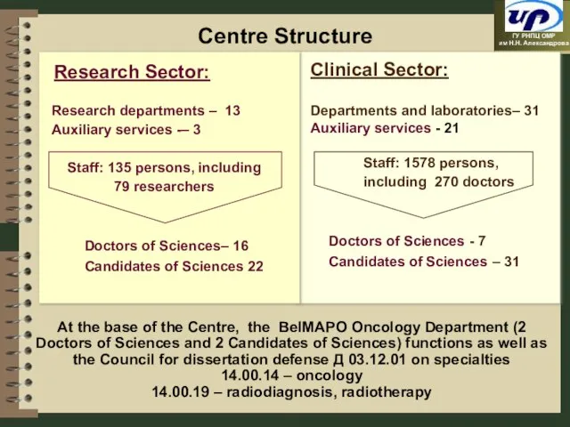
Слайд 10Centre Structure
At the base of the Centre, the BelMAPO Oncology Department (2
Centre Structure
At the base of the Centre, the BelMAPO Oncology Department (2
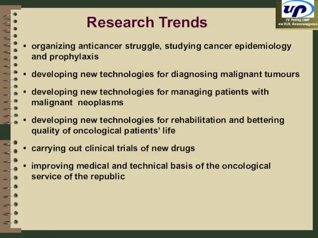
Слайд 11Research Trends
organizing anticancer struggle, studying cancer epidemiology and prophylaxis
developing new technologies for
Research Trends
organizing anticancer struggle, studying cancer epidemiology and prophylaxis
developing new technologies for
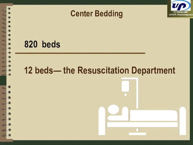
Слайд 12Center Bedding
820 beds
12 beds— the Resuscitation Department
Center Bedding
820 beds
12 beds— the Resuscitation Department
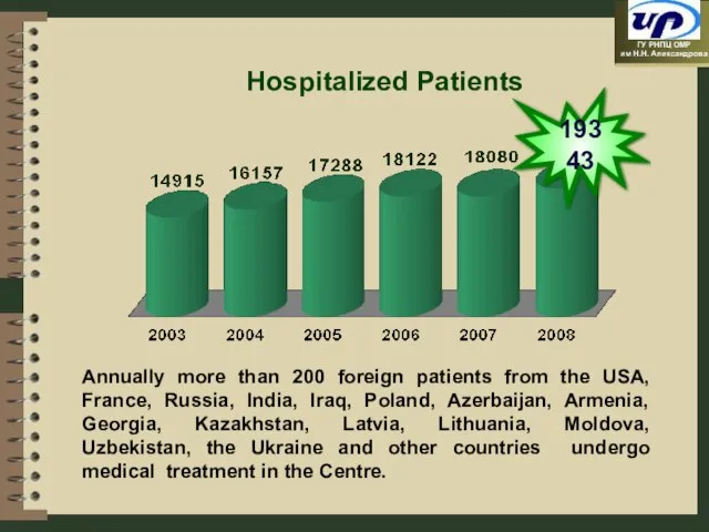
Слайд 13Hospitalized Patients
19343
Annually more than 200 foreign patients from the USA, France,
Hospitalized Patients
19343
Annually more than 200 foreign patients from the USA, France,
Слайд 14Diagnostic Base of
the Centre
Diagnostic Base of
the Centre
Слайд 15Practically, the whole specter of biochemical, clinical, immunohistological, radioisotopic, molecular and genetic
Practically, the whole specter of biochemical, clinical, immunohistological, radioisotopic, molecular and genetic
Слайд 16
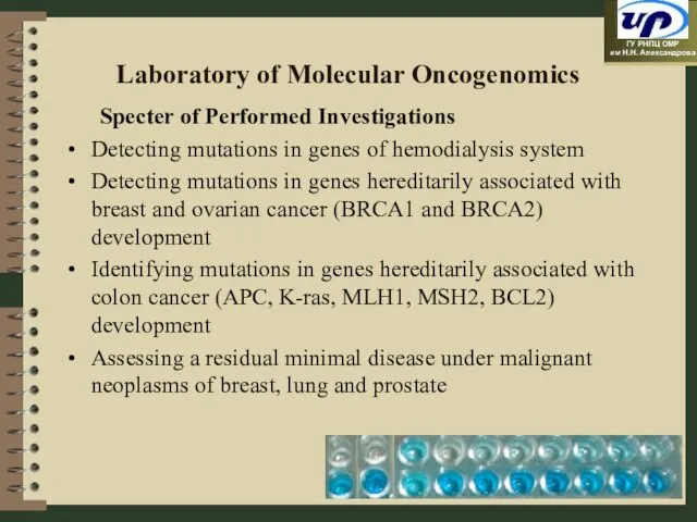
Laboratory of Molecular Oncogenomics
Specter of Performed Investigations
Detecting mutations in genes of
Laboratory of Molecular Oncogenomics
Specter of Performed Investigations
Detecting mutations in genes of
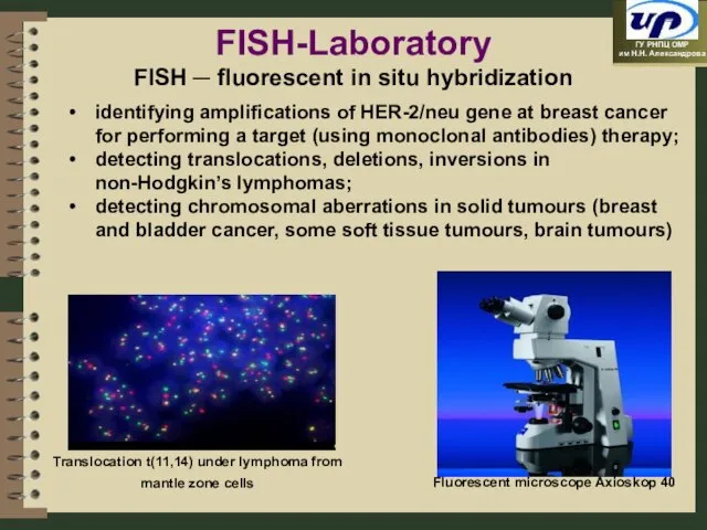
Слайд 17Translocation t(11,14) under lymphoma from mantle zone cells
Fluorescent microscope Axioskop 40
FISH-Laboratory
FISH
Translocation t(11,14) under lymphoma from mantle zone cells
Fluorescent microscope Axioskop 40
FISH-Laboratory
FISH
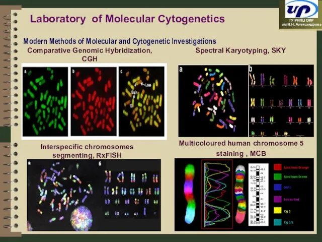
Слайд 18Laboratory of Molecular Cytogenetics
Laboratory of Molecular Cytogenetics
Слайд 19Morphological Methods of Investigation
Morphological Methods of Investigation
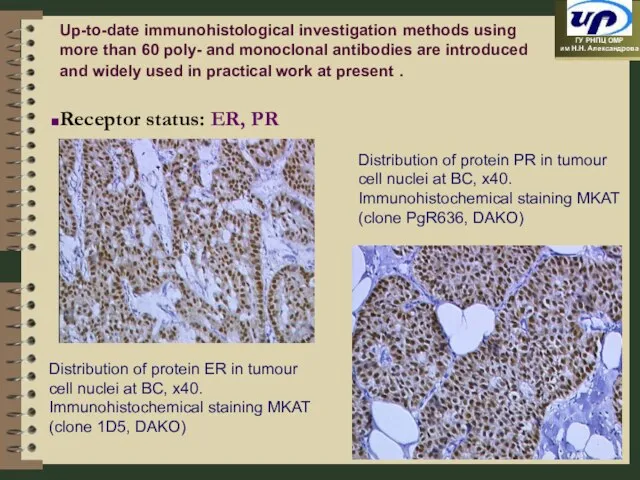
Слайд 20Distribution of protein ER in tumour cell nuclei at BC, x40.
Immunohistochemical staining
Distribution of protein ER in tumour cell nuclei at BC, x40.
Immunohistochemical staining
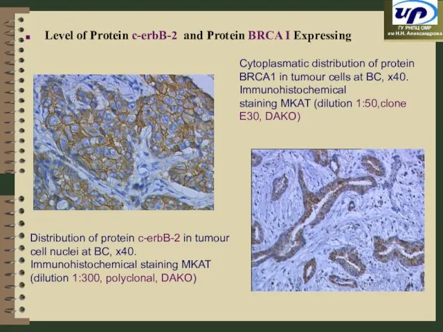
Слайд 21Distribution of protein c-erbB-2 in tumour cell nuclei at BC, x40.
Immunohistochemical staining
Distribution of protein c-erbB-2 in tumour cell nuclei at BC, x40.
Immunohistochemical staining
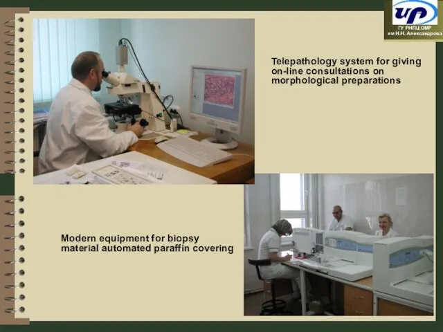
Слайд 22Telepathology system for giving on-line consultations on morphological preparations
Modern equipment for
Telepathology system for giving on-line consultations on morphological preparations
Modern equipment for
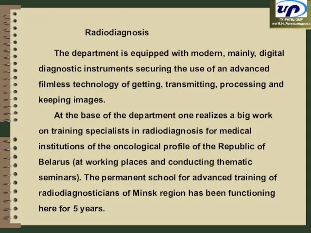
Слайд 23 Radiodiagnosis
The department is equipped with modern, mainly, digital diagnostic instruments securing the
Radiodiagnosis
The department is equipped with modern, mainly, digital diagnostic instruments securing the
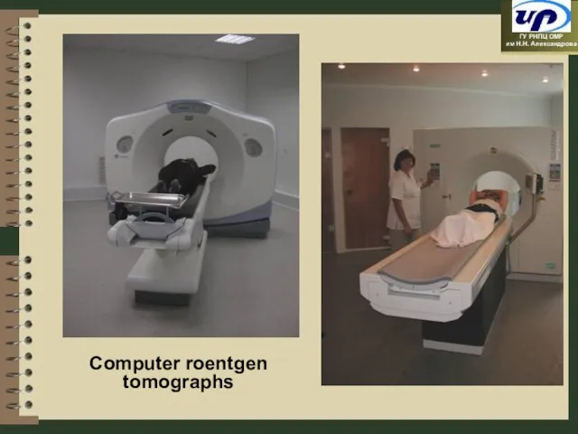
Слайд 24Computer roentgen tomographs
Computer roentgen tomographs
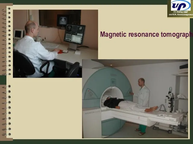
Слайд 25Magnetic resonance tomograph
Magnetic resonance tomograph
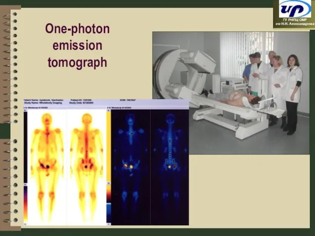
Слайд 26One-photon emission tomograph
One-photon emission tomograph
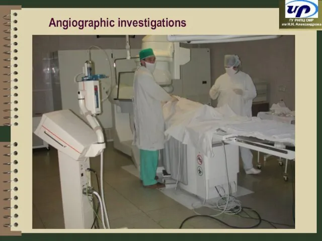
Слайд 27Angiographic investigations
Angiographic investigations
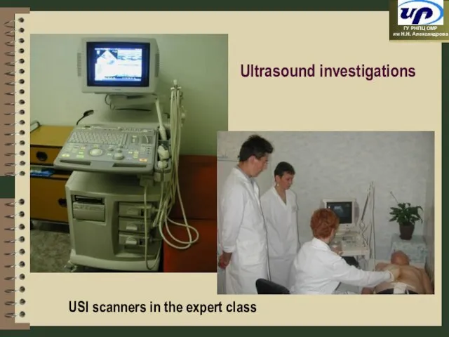
Слайд 28Ultrasound investigations
USI scanners in the expert class
Ultrasound investigations
USI scanners in the expert class
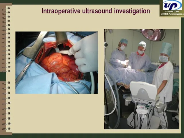
Слайд 29Intraoperative ultrasound investigation
Intraoperative ultrasound investigation
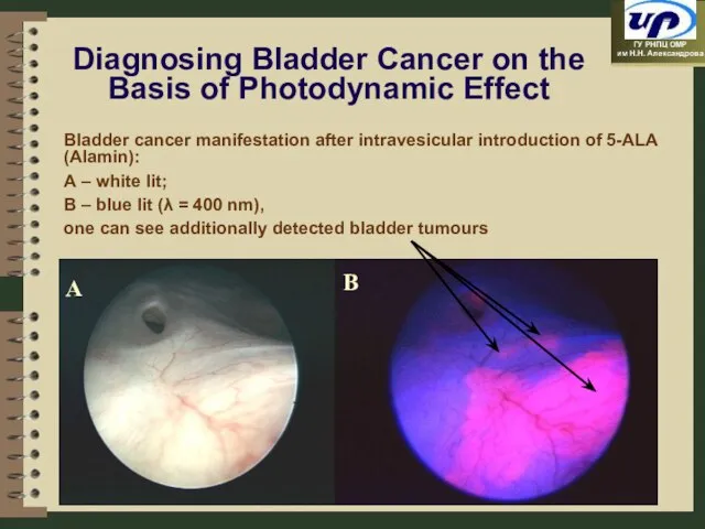
Слайд 30Diagnosing Bladder Cancer on the Basis of Photodynamic Effect
Bladder cancer manifestation after
Diagnosing Bladder Cancer on the Basis of Photodynamic Effect
Bladder cancer manifestation after
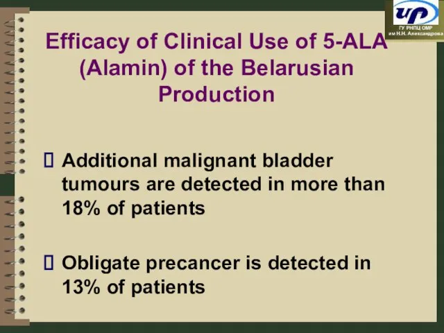
Слайд 31Efficacy of Clinical Use of 5-ALA (Alamin) of the Belarusian Production
Additional malignant
Efficacy of Clinical Use of 5-ALA (Alamin) of the Belarusian Production
Additional malignant
Слайд 32Highly Technological Methods of Treatment
Highly Technological Methods of Treatment
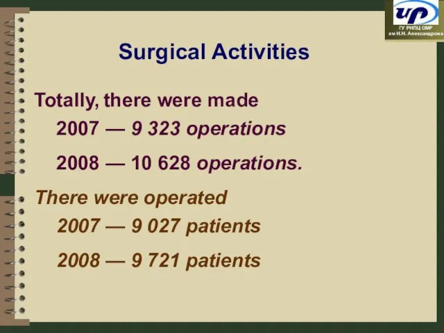
Слайд 33Surgical Activities
Totally, there were made
2007 — 9 323 operations
2008 — 10 628
Surgical Activities
Totally, there were made
2007 — 9 323 operations
2008 — 10 628
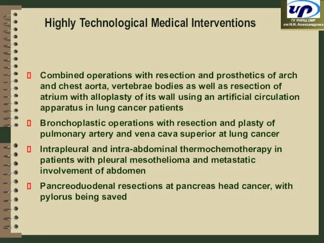
Слайд 34Highly Technological Medical Interventions
Combined operations with resection and prosthetics of arch and
Highly Technological Medical Interventions
Combined operations with resection and prosthetics of arch and
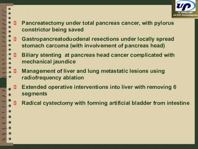
Слайд 35Pancreatectomy under total pancreas cancer, with pylorus constrictor being saved
Gastropancreatoduodenal resections under
Pancreatectomy under total pancreas cancer, with pylorus constrictor being saved
Gastropancreatoduodenal resections under
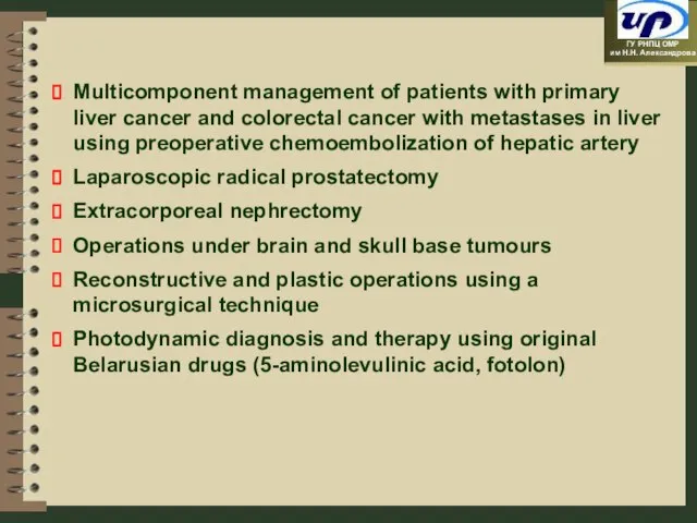
Слайд 36Multicomponent management of patients with primary liver cancer and colorectal cancer with
Multicomponent management of patients with primary liver cancer and colorectal cancer with
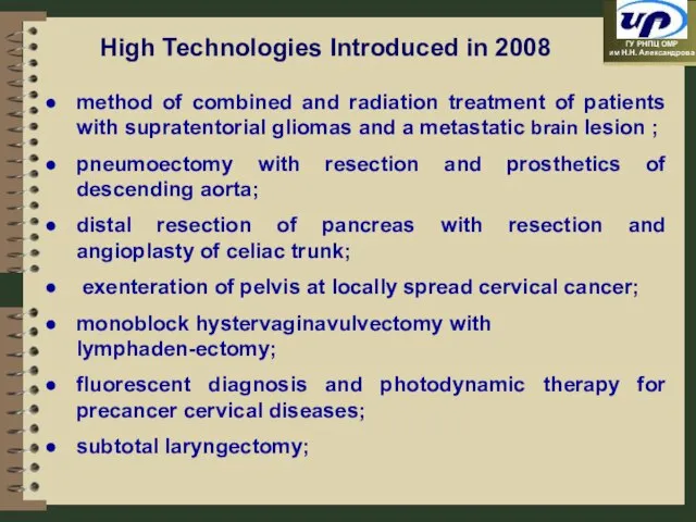
Слайд 37method of combined and radiation treatment of patients with supratentorial gliomas and
method of combined and radiation treatment of patients with supratentorial gliomas and
Слайд 38reconstruction of mandible using titanium implants;
use of the navigation system for diagnosing
reconstruction of mandible using titanium implants;
use of the navigation system for diagnosing
Слайд 39Radiofrequency Ablation of Tumours
Perfused elecrodes
Generator
Integra Elektrotom HITT®106
Radiofrequency Ablation of Tumours
Perfused elecrodes
Generator
Integra Elektrotom HITT®106
Слайд 40Radiofrequency Ablation of Hepatic Tumour
Liver CT with bolus contrasting (portal phase) before
Radiofrequency Ablation of Hepatic Tumour
Liver CT with bolus contrasting (portal phase) before
Слайд 41Radiofrequency Ablation of Pulmonary Neoplasm
Computer tomograms during the ablation (A) and 1
Radiofrequency Ablation of Pulmonary Neoplasm
Computer tomograms during the ablation (A) and 1
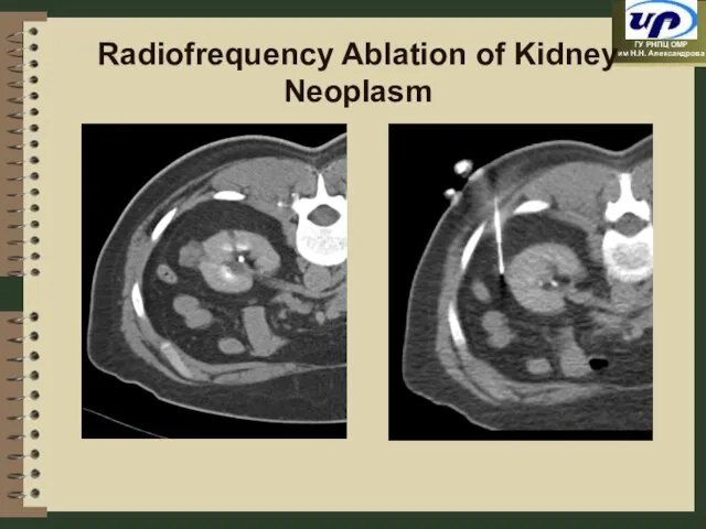
Слайд 42Radiofrequency Ablation of Kidney Neoplasm
Radiofrequency Ablation of Kidney Neoplasm
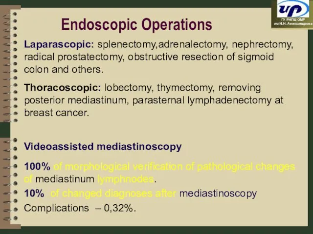
Слайд 43Endoscopic Operations
Laparascopic: splenectomy,adrenalectomy, nephrectomy, radical prostatectomy, obstructive resection of sigmoid colon and
Endoscopic Operations
Laparascopic: splenectomy,adrenalectomy, nephrectomy, radical prostatectomy, obstructive resection of sigmoid colon and
Слайд 44OPERATIONS UNDER HEAD AND NECK TUMOURS
OPERATIONS UNDER HEAD AND NECK TUMOURS

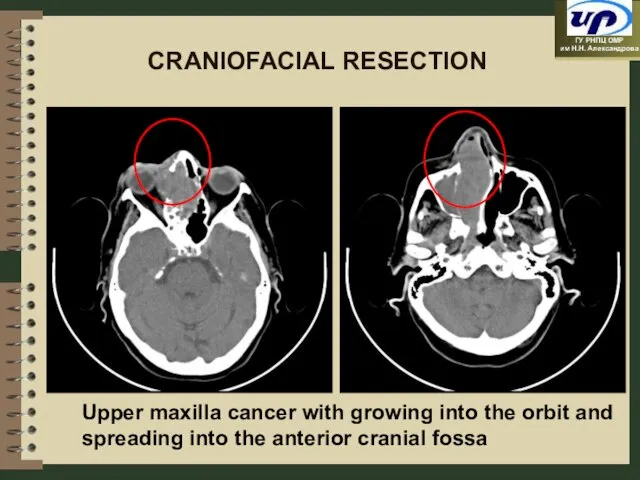
Слайд 45Upper maxilla cancer with growing into the orbit and spreading into the
Upper maxilla cancer with growing into the orbit and spreading into the
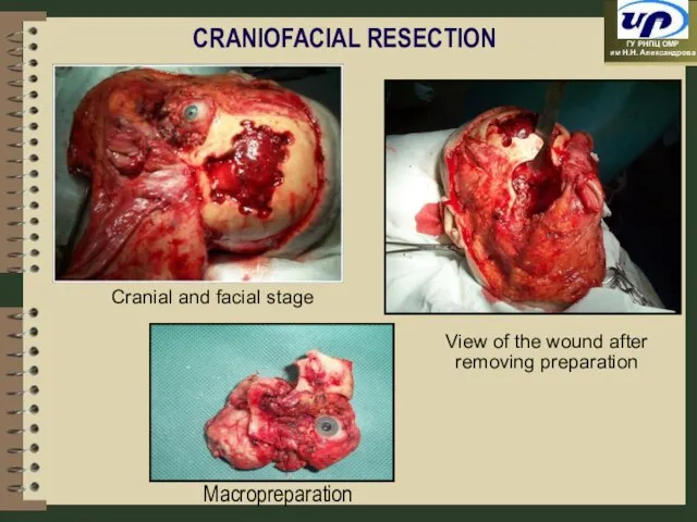
Слайд 46CRANIOFACIAL RESECTION
Cranial and facial stage
View of the wound after removing preparation
CRANIOFACIAL RESECTION
Cranial and facial stage
View of the wound after removing preparation
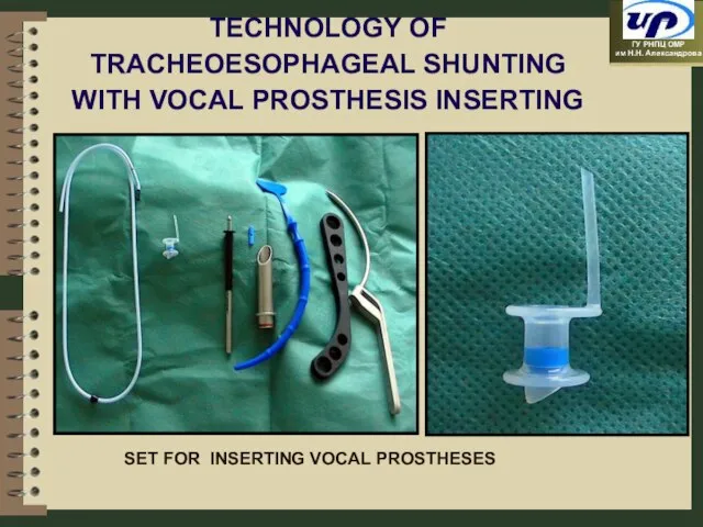
Слайд 47TECHNOLOGY OF TRACHEOESOPHAGEAL SHUNTING WITH VOCAL PROSTHESIS INSERTING
SET FOR INSERTING VOCAL PROSTHESES
TECHNOLOGY OF TRACHEOESOPHAGEAL SHUNTING WITH VOCAL PROSTHESIS INSERTING
SET FOR INSERTING VOCAL PROSTHESES
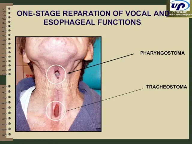
Слайд 48ONE-STAGE REPARATION OF VOCAL AND ESOPHAGEAL FUNCTIONS
PHARYNGOSTOMA
TRACHEOSTOMA
ONE-STAGE REPARATION OF VOCAL AND ESOPHAGEAL FUNCTIONS
PHARYNGOSTOMA
TRACHEOSTOMA
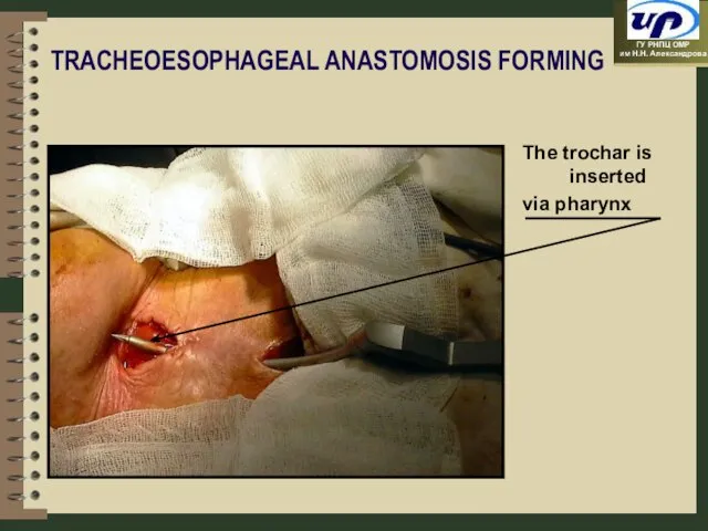
Слайд 49TRACHEOESOPHAGEAL ANASTOMOSIS FORMING
The trochar is inserted
via pharynx
TRACHEOESOPHAGEAL ANASTOMOSIS FORMING
The trochar is inserted
via pharynx
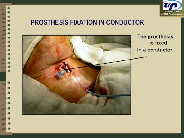
Слайд 50PROSTHESIS FIXATION IN CONDUCTOR
The prosthesis is fixed
in a conductor
PROSTHESIS FIXATION IN CONDUCTOR
The prosthesis is fixed
in a conductor
Слайд 51PHARYNX ANTERIOR WALL FORMING WITH
LOCAL TISSUES
Vocal prosthesis
PHARYNX ANTERIOR WALL FORMING WITH
LOCAL TISSUES
Vocal prosthesis

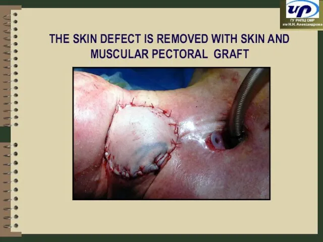
Слайд 52THE SKIN DEFECT IS REMOVED WITH SKIN AND MUSCULAR PECTORAL GRAFT
THE SKIN DEFECT IS REMOVED WITH SKIN AND MUSCULAR PECTORAL GRAFT
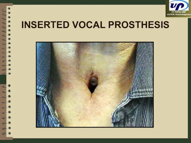
Слайд 53INSERTED VOCAL PROSTHESIS
INSERTED VOCAL PROSTHESIS
Слайд 54Plastic Operations in Patients with Breast Cancer
Plastic Operations in Patients with Breast Cancer
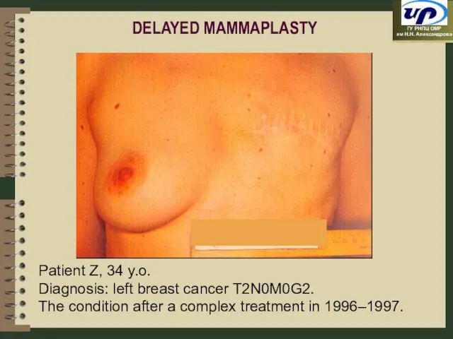
Слайд 55Patient Z, 34 y.o.
Diagnosis: left breast cancer T2N0M0G2.
The condition after a
Patient Z, 34 y.o.
Diagnosis: left breast cancer T2N0M0G2.
The condition after a
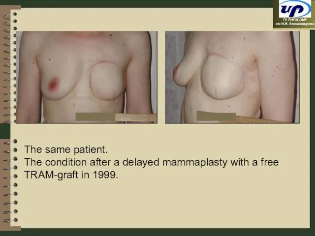
Слайд 56The same patient.
The condition after a delayed mammaplasty with a free TRAM-graft
The same patient.
The condition after a delayed mammaplasty with a free TRAM-graft
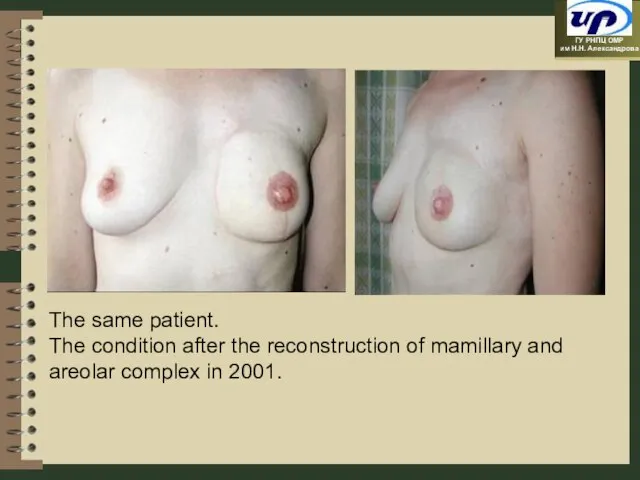
Слайд 57The same patient.
The condition after the reconstruction of mamillary and areolar complex
The same patient.
The condition after the reconstruction of mamillary and areolar complex
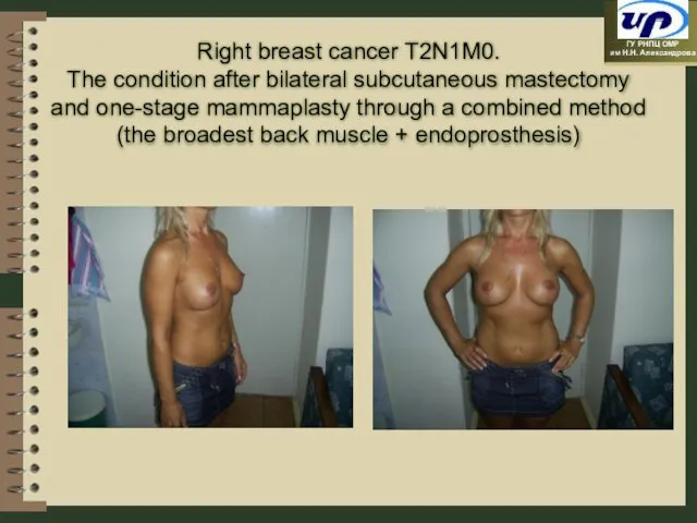
Слайд 58Right breast cancer Т2N1М0.
The condition after bilateral subcutaneous mastectomy and one-stage
Right breast cancer Т2N1М0.
The condition after bilateral subcutaneous mastectomy and one-stage
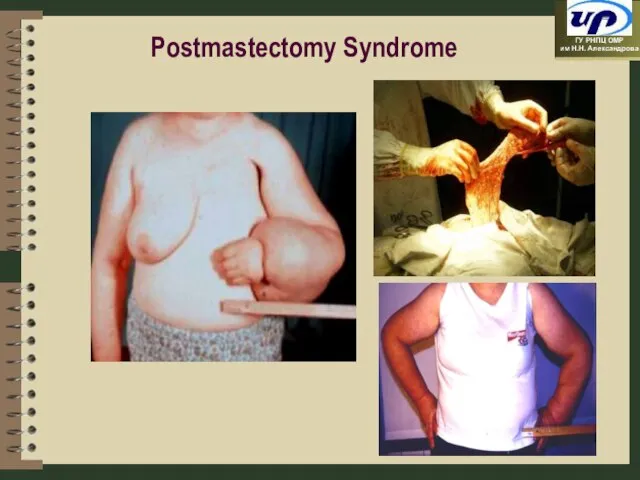
Слайд 59Postmastectomy Syndrome
Postmastectomy Syndrome
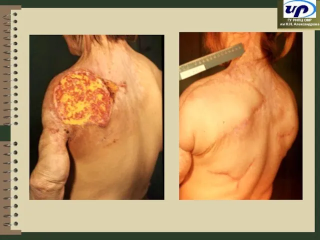
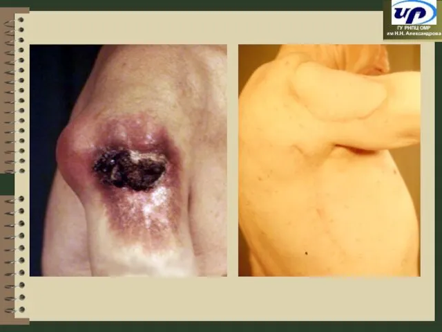
Слайд 60Plasty of Soft Tissues
Plasty of Soft Tissues
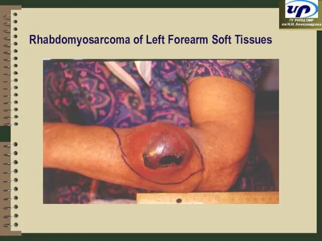
Слайд 63Rhabdomyosarcoma of Left Forearm Soft Tissues
Rhabdomyosarcoma of Left Forearm Soft Tissues
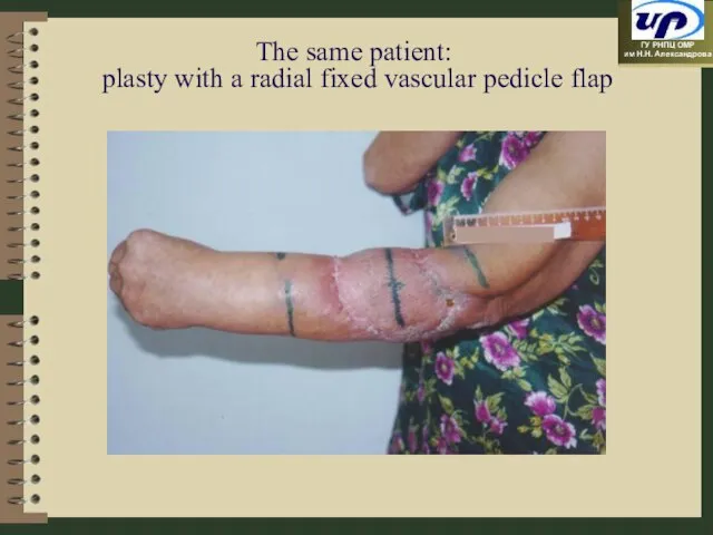
Слайд 64The same patient:
plasty with a radial fixed vascular pedicle flap
The same patient:
plasty with a radial fixed vascular pedicle flap
Слайд 65Operations Preserving Organs under Bone Tumours
Operations Preserving Organs under Bone Tumours
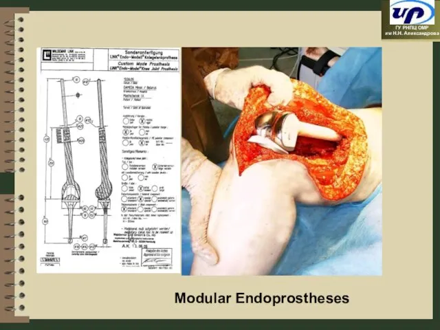
Слайд 66Modular Endoprostheses
Modular Endoprostheses
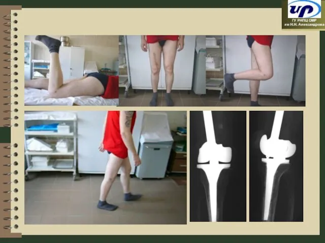
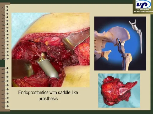
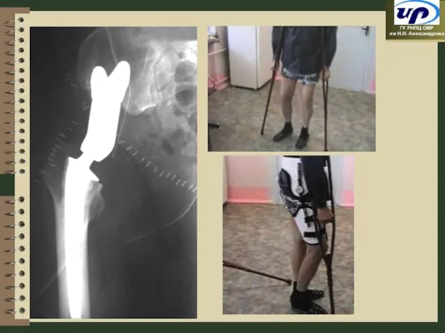
Слайд 69Endoprosthetics with saddle-like prosthesis
Endoprosthetics with saddle-like prosthesis
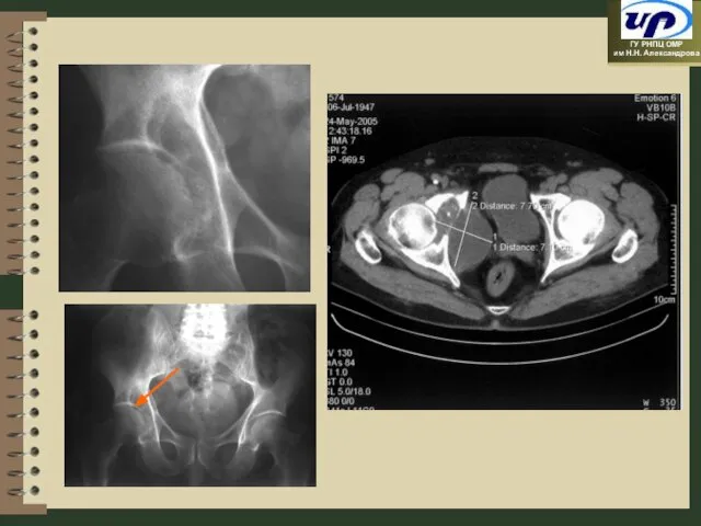
Слайд 71
Lung Cancer and Mediastrium Tumours
Lung Cancer and Mediastrium Tumours
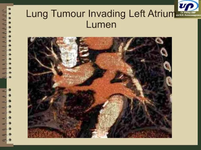
Слайд 72Lung Tumour Invading Left Atrium Lumen
Lung Tumour Invading Left Atrium Lumen
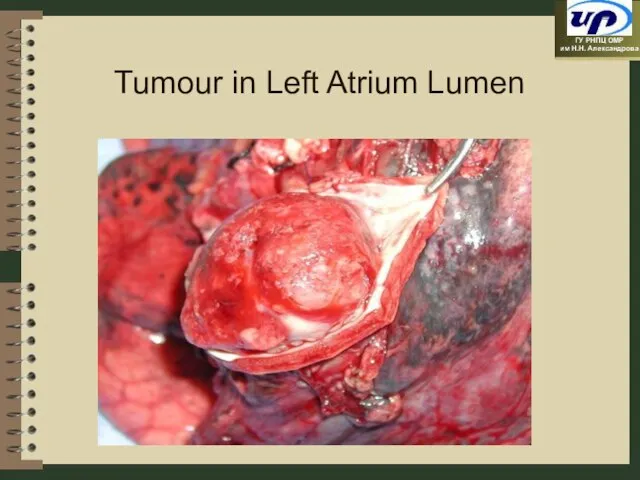
Слайд 73Tumour in Left Atrium Lumen
Tumour in Left Atrium Lumen
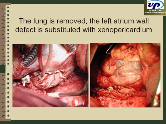
Слайд 74The lung is removed, the left atrium wall defect is substituted with
The lung is removed, the left atrium wall defect is substituted with
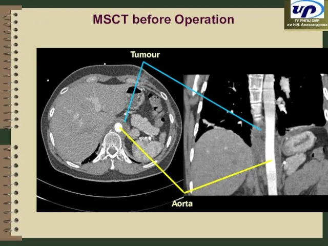
Слайд 75MSCT before Operation
MSCT before Operation
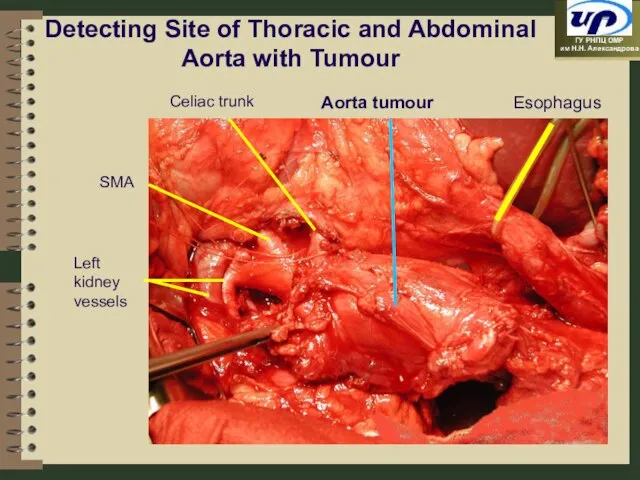
Слайд 76Detecting Site of Thoracic and Abdominal Aorta with Tumour
Esophagus
Celiac trunk
SMA
Left kidney vessels
Aorta
Detecting Site of Thoracic and Abdominal Aorta with Tumour
Esophagus
Celiac trunk
SMA
Left kidney vessels
Aorta
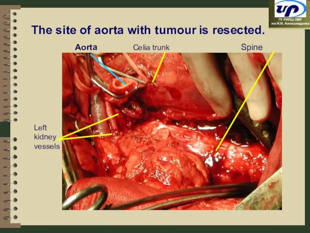
Слайд 77Spine
Left kidney vessels
Aorta
The site of aorta with tumour is resected.
Celia trunk
Spine
Left kidney vessels
Aorta
The site of aorta with tumour is resected.
Celia trunk
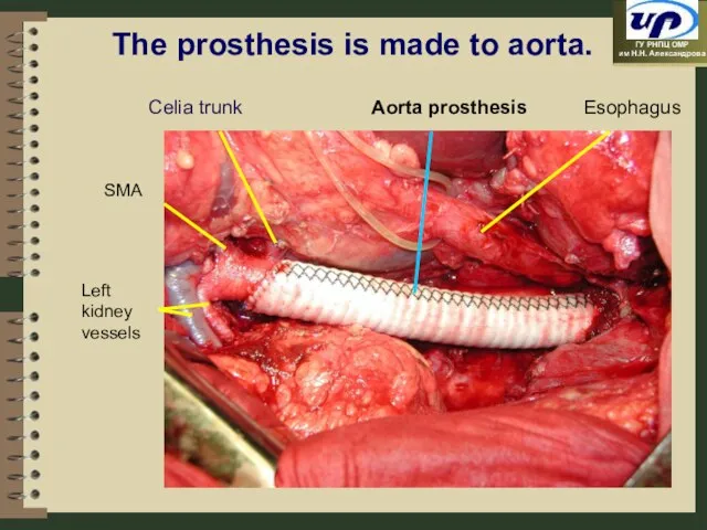
Слайд 78Esophagus
Left kidney vessels
Aorta prosthesis
The prosthesis is made to aorta.
Celia trunk
SMА
Esophagus
Left kidney vessels
Aorta prosthesis
The prosthesis is made to aorta.
Celia trunk
SMА
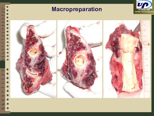
Слайд 79Macropreparation
Macropreparation
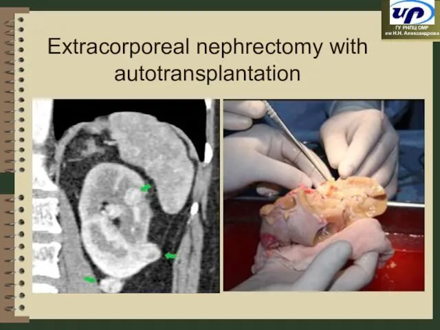
Слайд 80Extracorporeal nephrectomy with autotransplantation
Extracorporeal nephrectomy with autotransplantation
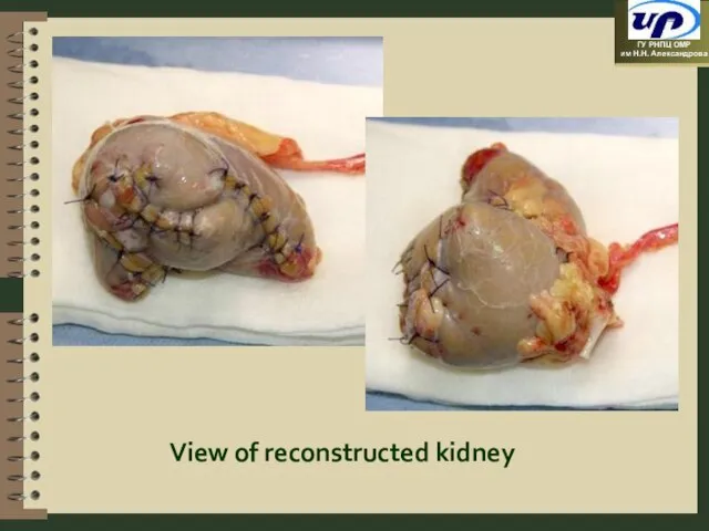
Слайд 81View of reconstructed kidney
View of reconstructed kidney
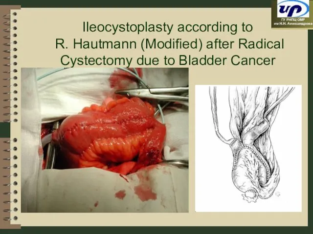
Слайд 82Ileocystoplasty according to R. Hautmann (Modified) after Radical Cystectomy due to Bladder
Ileocystoplasty according to R. Hautmann (Modified) after Radical Cystectomy due to Bladder
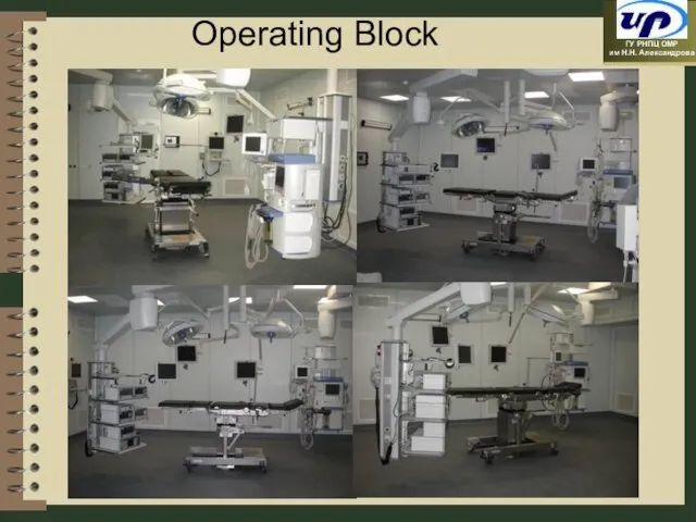
Слайд 83Operating Block
Operating Block
Слайд 84Resuscitation Department
Resuscitation Department
Слайд 85The apparatus “Artificial Kidney”
Procedure of extracorporeal detoxication using the apparatus «Multifiltrat»
The apparatus “Artificial Kidney”
Procedure of extracorporeal detoxication using the apparatus «Multifiltrat»
Слайд 86Radiotherapy
Radiotherapy
Слайд 87Material and Technical Basis of Remote Radiotherapy
Material and Technical Basis of Remote Radiotherapy
Слайд 88Material and Technical Basis of Short Focus and Contact Radiotherapy
Material and Technical Basis of Short Focus and Contact Radiotherapy
Слайд 89Material and Technical Basis for Preradiation Preparation
Material and Technical Basis for Preradiation Preparation
Слайд 90Material and Technical Basis (Auxiliary Devices and Dosimetry)
Material and Technical Basis (Auxiliary Devices and Dosimetry)
Слайд 91Linear accelerator with 2 photon energies and 6-8 energies of electrons with
Слайд 93High Technologies in Radiotherapy
Three-dimensional conformal radiotherapy
Radiotherapy with modulating dose intensity
Stereotaxic radiotherapy /
High Technologies in Radiotherapy
Three-dimensional conformal radiotherapy
Radiotherapy with modulating dose intensity
Stereotaxic radiotherapy /
Слайд 94Brachytherapy Using an Integrated X-ray and Topometric Complex (IBU)
Brachytherapy of prostate tumours
Brachytherapy Using an Integrated X-ray and Topometric Complex (IBU)
Brachytherapy of prostate tumours
Слайд 95Hyperthermia
Hyperthermia
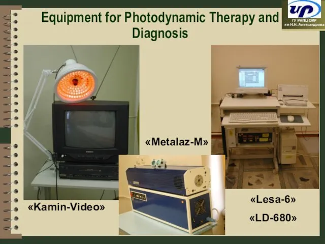
Слайд 96Equipment for Photodynamic Therapy and Diagnosis
«Metalaz-M»
«Kamin-Video»
«Lesa-6»
«LD-680»
Equipment for Photodynamic Therapy and Diagnosis
«Metalaz-M»
«Kamin-Video»
«Lesa-6»
«LD-680»
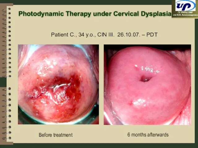
Слайд 976 months afterwards
Patient C., 34 y.o., CIN III. 26.10.07. – PDT
Before treatment
Photodynamic
6 months afterwards
Patient C., 34 y.o., CIN III. 26.10.07. – PDT
Before treatment
Photodynamic
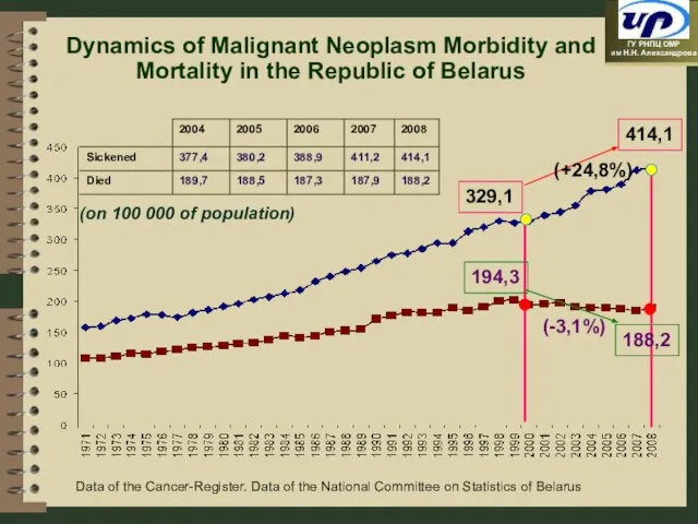
Слайд 98Dynamics of Malignant Neoplasm Morbidity and Mortality in the Republic of Belarus
194,3
(-3,1%)
329,1
414,1
188,2
(on
Dynamics of Malignant Neoplasm Morbidity and Mortality in the Republic of Belarus
194,3
(-3,1%)
329,1
414,1
188,2
(on
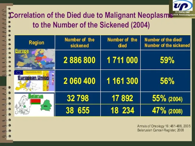
Слайд 99Europe
Belarus
European Union
Annals of Oncology 16: 481-488, 2005
Belarusian Cancer-Register, 2008
Correlation of the Died
Europe
Belarus
European Union
Annals of Oncology 16: 481-488, 2005
Belarusian Cancer-Register, 2008
Correlation of the Died
 Эффективность внедрения ФГОС в учреждениях НПО и СПО Удмуртской Республики
 Русский язык
 Путь к звездам
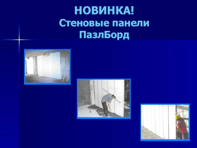 НОВИНКА!Стеновые панелиПазлБорд
 Управління персоналом та якістю проекту
 Новый год в Японии
 Microsoft Excel
 Мемориал - Дети войны, доиграем за вас!
 Русский классицизм и зарубежный модерн
 Презентация на тему Музей Прадо (Мадрид)
 Презентация на тему Эмоции и здоровье
  Сформировать представление о науке география
 CАЕ - инженерные расчеты
 Презентация на тему Базисные условия поставки Инкотермс - 2000
  ИХН
ИХН Набор баскетболистов
 Урок № 5
 Гиппократ. Описания свойств сангвиников, холериков, флегматиков
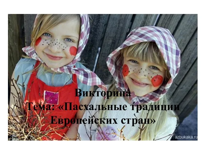 Пасхальные традиции Европейских стран
 Australian Coat of Arms
 to wake up wake up
  Библиография на тему Психология девиантного поведения
 Роль денег как средства обмена
 Практика внедрения систем менеджмента энергопотреблением на основе современных стандартов
 Презентация на тему Водоросли
  Образ жизни и длина теломер
 Системы и устройства передачи, приема и обработки сигналов
 Презентация на тему Художественная культура Античности