- Skin and soft tissue infections

Содержание
- 2. Повестка Раздел 1 Раздел 2 Раздел 3 Раздел 4 Раздел 5 Образец текста нижнего колонтитула 08.02.20XX
- 3. Введение В PowerPoint можно создавать презентации и делиться своими материалами с другими, где бы они ни
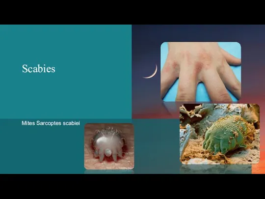
- 4. Scabies Mites Sarcoptes scabiei
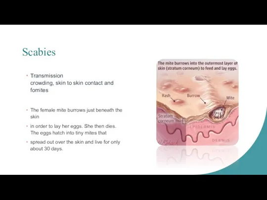
- 5. Scabies Transmission crowding, skin to skin contact and fomites The female mite burrows just beneath the
- 6. Scabies CLASSIC SCABIES Small erythematous papules “Knots on a rope” Pruritic Severe and worse at night
- 7. Scabies CRUSTED SCABIES Образец текста нижнего колонтитула 08.02.20XX Norwegian scabies Crusting, scaling fissuring affecting an older,
- 8. Scabies Diagnosis Clx – history and appearance of the rash Microscopy Treatment Permethrim Lindane Ivermectim 08.02.20XX
- 9. Pediculosis humanus capitis Pediculosis humanus corporis Pediculosis pubis Pediculosis ciliaris Head Lice Body Lice Pubic Lice
- 10. Head Lice Children, femals, Europian Direct contact or fomites Nits firmly “cemented” to human hair White
- 11. Body Lice Poverty, poor hygiene, crowding Direct contact and clothing Lays eggs in seams of clothing
- 12. Pubic Lice Sexual active, young adults and adolescents Sexual transmitted and fomites Contact with eyes can
- 13. Lice Symptoms Itchy Excoriation Hyperpigmentation Lymphadenopathy Bacteria transmitted by the body louse Rickettsia prowazekii Borrelia recurents
- 14. Lice Diagnosis Head lice or nits are usually on the scalp and nape of the neck
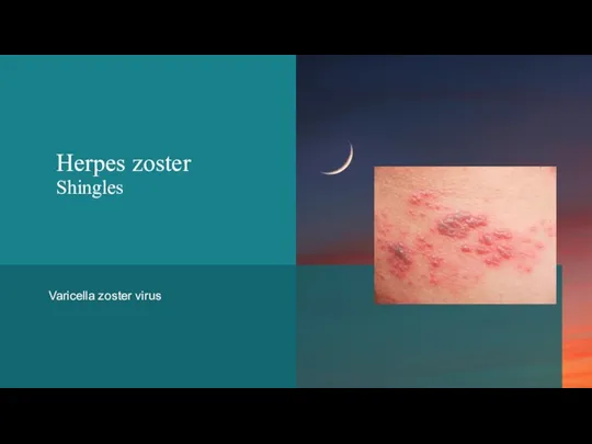
- 15. Varicella zoster virus Herpes zoster Shingles
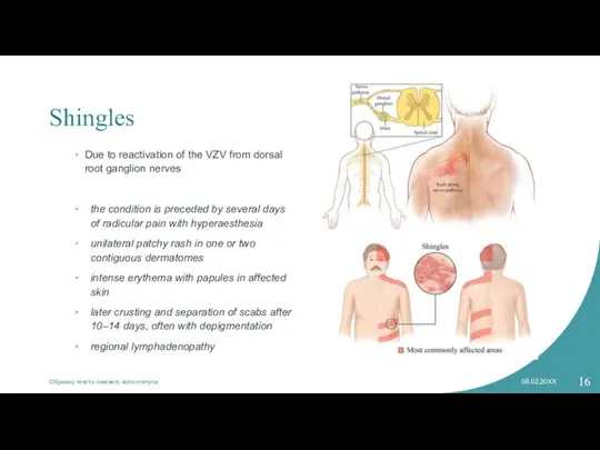
- 16. Shingles Due to reactivation of the VZV from dorsal root ganglion nerves the condition is preceded
- 17. Shingles Herpes Zoster oticus the trigeminal nerve Ramsay Hunt Syndrome Ipsilateral facial paralysis Ear pain Vesicles
- 18. Shingles Diagnostic RCR for detection of viral DNA Direct fluorescent antibody Tzanck swear Treatment Post-herpetic neuralgia
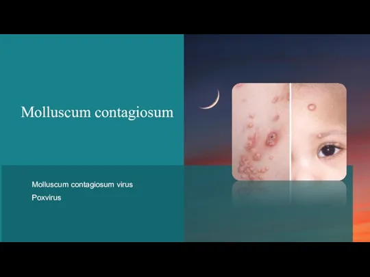
- 19. Molluscum contagiosum Molluscum contagiosum virus Poxvirus
- 20. Molluscum contagiosum Children and sexually active adults Painless Incubation period between 2-6 weeks Persist for months
- 21. Molluscum contagiosum Dome-shaped papules with umbilication 2-3mm in diameter Pink-white to flash colored Single or multiple
- 22. Molluscum contagiosum Diagnosis Clinical Histology Henderson – Peterson bodies Treatment Self-limiting Cryotherapy, Cantharidin, Curretage, Imiguimod, Topical
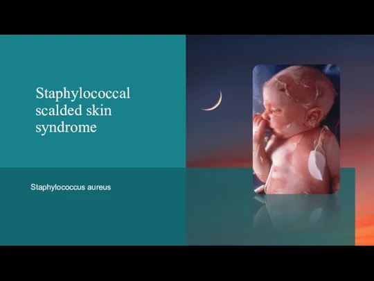
- 23. Staphylococcus aureus Staphylococcal scalded skin syndrome
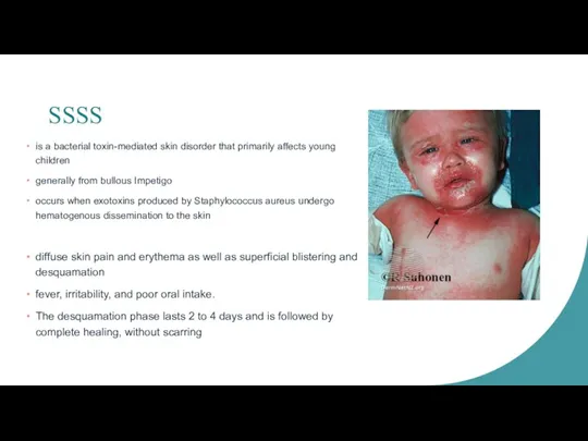
- 24. SSSS is a bacterial toxin-mediated skin disorder that primarily affects young children generally from bullous Impetigo
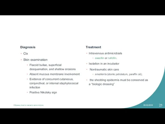
- 25. Diagnosis Clx Skin examination Flaccid bullae, superficial desquamation, and shallow erosions Absent mucous membrane involvement Evidence
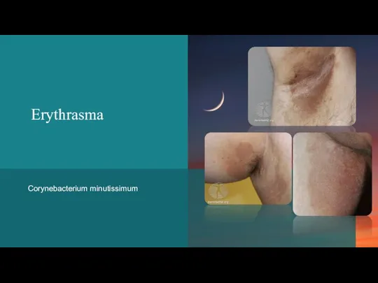
- 26. Erythrasma Corynebacterium minutissimum
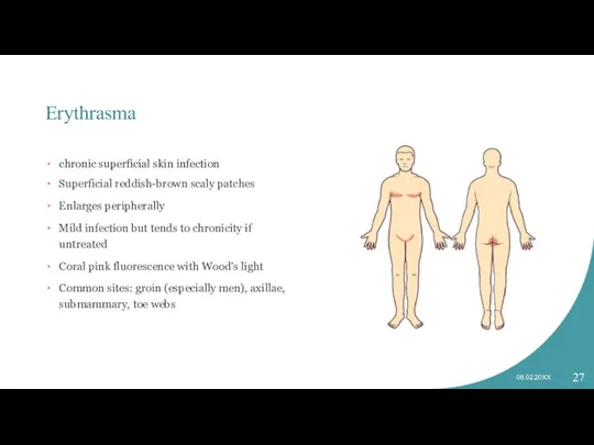
- 27. Erythrasma chronic superficial skin infection Superficial reddish-brown scaly patches Enlarges peripherally Mild infection but tends to
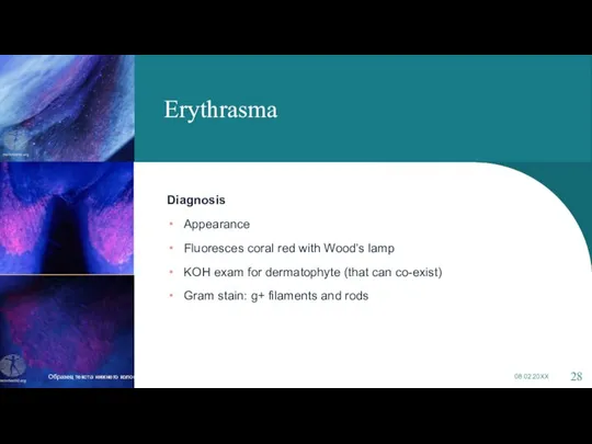
- 28. Erythrasma Diagnosis Appearance Fluoresces coral red with Wood’s lamp KOH exam for dermatophyte (that can co-exist)
- 29. Erythrasma Treatment Topical imidazole e.g. miconazole or erythromycin 2% gel Oral roxithromycin or erythromycin Loose fitting
- 30. Tinea versicolor Образец текста нижнего колонтитула 08.02.20XX
- 32. Скачать презентацию
Слайд 3Введение
В PowerPoint можно создавать презентации и делиться своими материалами с другими, где
Введение
В PowerPoint можно создавать презентации и делиться своими материалами с другими, где
Слайд 4Scabies
Mites Sarcoptes scabiei
Scabies
Mites Sarcoptes scabiei
Слайд 5Scabies
Transmission
crowding, skin to skin contact and fomites
The female mite burrows just beneath
Scabies
Transmission
crowding, skin to skin contact and fomites
The female mite burrows just beneath
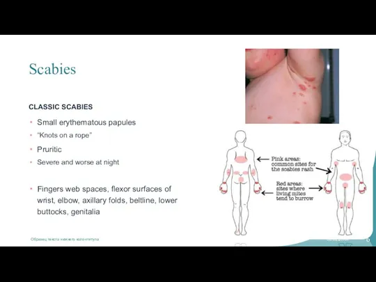
Слайд 6Scabies
CLASSIC SCABIES
Small erythematous papules
“Knots on a rope”
Pruritic
Severe and worse at night
Fingers web
Scabies
CLASSIC SCABIES
Small erythematous papules
“Knots on a rope”
Pruritic
Severe and worse at night
Fingers web
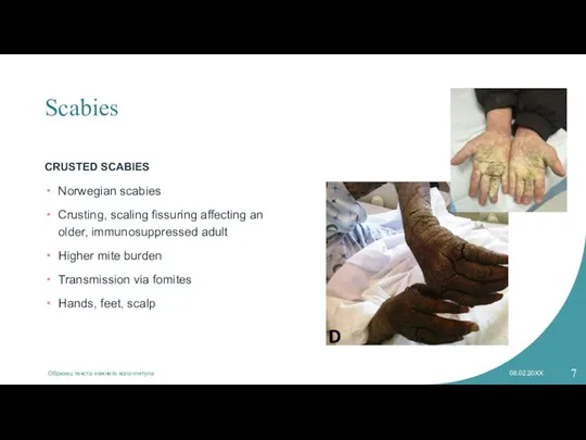
Слайд 7Scabies
CRUSTED SCABIES
Образец текста нижнего колонтитула
08.02.20XX
Norwegian scabies
Crusting, scaling fissuring affecting an older, immunosuppressed
Scabies
CRUSTED SCABIES
Образец текста нижнего колонтитула
08.02.20XX
Norwegian scabies
Crusting, scaling fissuring affecting an older, immunosuppressed
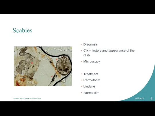
Слайд 8Scabies
Diagnosis
Clx – history and appearance of the rash
Microscopy
Treatment
Permethrim
Lindane
Ivermectim
08.02.20XX
Образец текста нижнего колонтитула
Scabies
Diagnosis
Clx – history and appearance of the rash
Microscopy
Treatment
Permethrim
Lindane
Ivermectim
08.02.20XX
Образец текста нижнего колонтитула
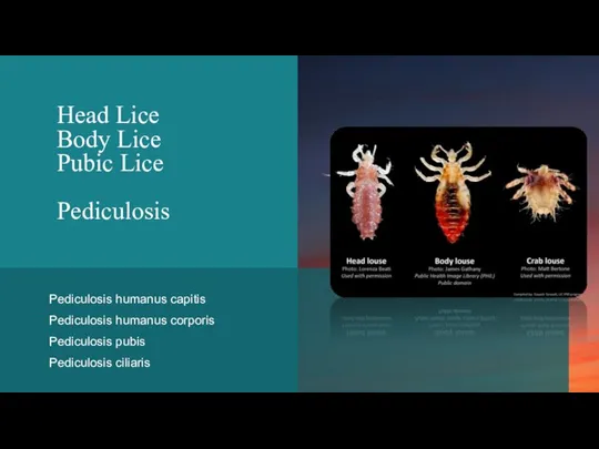
Слайд 9Pediculosis humanus capitis
Pediculosis humanus corporis
Pediculosis pubis
Pediculosis ciliaris
Head Lice
Body Lice
Pubic Lice
Pediculosis
Pediculosis humanus corporis
Pediculosis pubis
Pediculosis ciliaris
Head Lice
Body Lice
Pubic Lice
Pediculosis
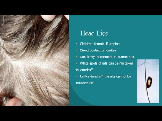
Слайд 10Head Lice
Children, femals, Europian
Direct contact or fomites
Nits firmly “cemented” to human hair
White
Head Lice
Children, femals, Europian
Direct contact or fomites
Nits firmly “cemented” to human hair
White
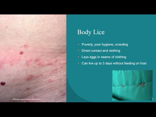
Слайд 11Body Lice
Poverty, poor hygiene, crowding
Direct contact and clothing
Lays eggs in seams of
Body Lice
Poverty, poor hygiene, crowding
Direct contact and clothing
Lays eggs in seams of
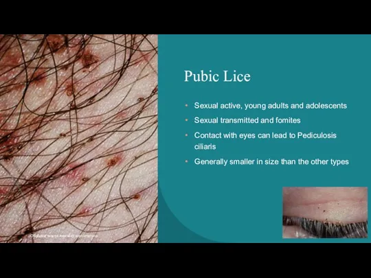
Слайд 12Pubic Lice
Sexual active, young adults and adolescents
Sexual transmitted and fomites
Contact with eyes
Pubic Lice
Sexual active, young adults and adolescents
Sexual transmitted and fomites
Contact with eyes
Слайд 13Lice
Symptoms
Itchy
Excoriation
Hyperpigmentation
Lymphadenopathy
Bacteria transmitted by the body louse
Rickettsia prowazekii
Borrelia recurents
Borrelia quintana
08.02.20XX
Образец текста нижнего колонтитула
Lice
Symptoms
Itchy
Excoriation
Hyperpigmentation
Lymphadenopathy
Bacteria transmitted by the body louse
Rickettsia prowazekii
Borrelia recurents
Borrelia quintana
08.02.20XX
Образец текста нижнего колонтитула

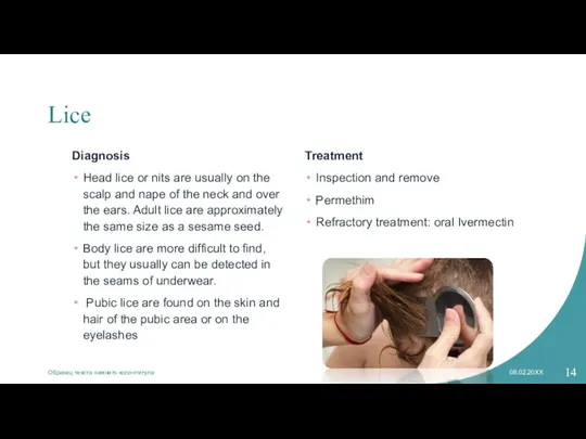
Слайд 14Lice
Diagnosis
Head lice or nits are usually on the scalp and nape of
Lice
Diagnosis
Head lice or nits are usually on the scalp and nape of
Слайд 15Varicella zoster virus
Herpes zoster
Shingles
Herpes zoster
Shingles
Слайд 16Shingles
Due to reactivation of the VZV from dorsal root ganglion nerves
the condition
Shingles
Due to reactivation of the VZV from dorsal root ganglion nerves
the condition
Слайд 17Shingles
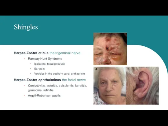
Herpes Zoster oticus the trigeminal nerve
Ramsay Hunt Syndrome
Ipsilateral facial paralysis
Ear pain
Vesicles in
Shingles
Herpes Zoster oticus the trigeminal nerve
Ramsay Hunt Syndrome
Ipsilateral facial paralysis
Ear pain
Vesicles in
Слайд 18Shingles
Diagnostic
RCR for detection of viral DNA
Direct fluorescent antibody
Tzanck swear
Treatment
<72 hrs – valacyclovir,
Shingles
Diagnostic
RCR for detection of viral DNA
Direct fluorescent antibody
Tzanck swear
Treatment
<72 hrs – valacyclovir,
Слайд 19
Molluscum contagiosum
Molluscum contagiosum virus
Poxvirus
Molluscum contagiosum
Molluscum contagiosum virus
Poxvirus
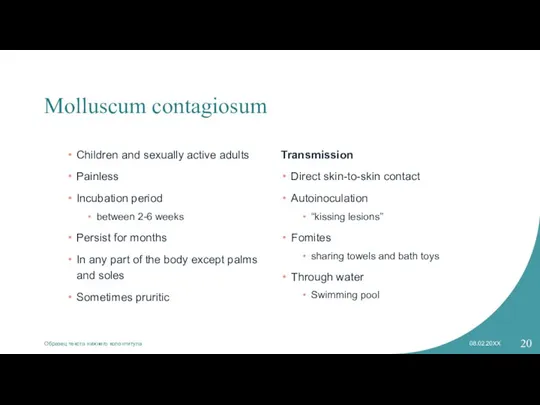
Слайд 20Molluscum contagiosum
Children and sexually active adults
Painless
Incubation period
between 2-6 weeks
Persist for months
Molluscum contagiosum
Children and sexually active adults
Painless
Incubation period
between 2-6 weeks
Persist for months
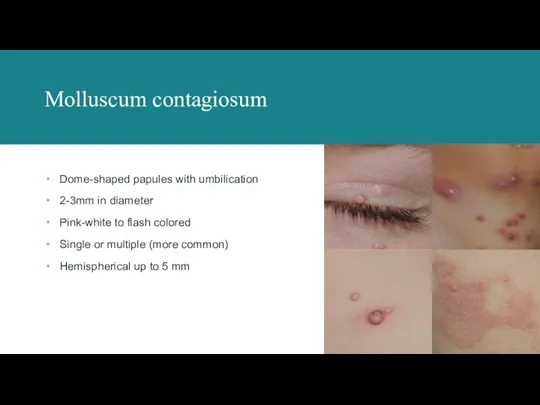
Слайд 21Molluscum contagiosum
Dome-shaped papules with umbilication
2-3mm in diameter
Pink-white to flash colored
Single or multiple
Molluscum contagiosum
Dome-shaped papules with umbilication
2-3mm in diameter
Pink-white to flash colored
Single or multiple
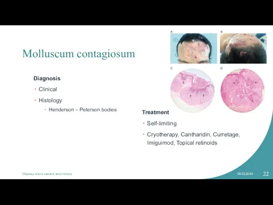
Слайд 22Molluscum contagiosum
Diagnosis
Clinical
Histology
Henderson – Peterson bodies
Treatment
Self-limiting
Cryotherapy, Cantharidin, Curretage, Imiguimod, Topical retinoids
08.02.20XX
Образец текста
Molluscum contagiosum
Diagnosis
Clinical
Histology
Henderson – Peterson bodies
Treatment
Self-limiting
Cryotherapy, Cantharidin, Curretage, Imiguimod, Topical retinoids
08.02.20XX
Образец текста
Слайд 23Staphylococcus aureus
Staphylococcal scalded skin syndrome
Staphylococcal scalded skin syndrome
Слайд 24SSSS
is a bacterial toxin-mediated skin disorder that primarily affects young children
generally from
SSSS
is a bacterial toxin-mediated skin disorder that primarily affects young children
generally from
Слайд 25Diagnosis
Clx
Skin examination
Flaccid bullae, superficial desquamation, and shallow erosions
Absent mucous membrane involvement
Evidence of
Diagnosis
Clx
Skin examination
Flaccid bullae, superficial desquamation, and shallow erosions
Absent mucous membrane involvement
Evidence of
Слайд 26
Erythrasma
Corynebacterium minutissimum
Erythrasma
Corynebacterium minutissimum
Слайд 27Erythrasma
chronic superficial skin infection
Superficial reddish-brown scaly patches
Enlarges peripherally
Mild infection but tends to
Erythrasma
chronic superficial skin infection
Superficial reddish-brown scaly patches
Enlarges peripherally
Mild infection but tends to
Слайд 28Erythrasma
Diagnosis
Appearance
Fluoresces coral red with Wood’s lamp
KOH exam for dermatophyte (that can co-exist)
Gram
Erythrasma
Diagnosis
Appearance
Fluoresces coral red with Wood’s lamp
KOH exam for dermatophyte (that can co-exist)
Gram
Слайд 29Erythrasma
Treatment
Topical imidazole e.g. miconazole or erythromycin 2% gel
Oral roxithromycin or erythromycin
Loose fitting
Erythrasma
Treatment
Topical imidazole e.g. miconazole or erythromycin 2% gel
Oral roxithromycin or erythromycin
Loose fitting
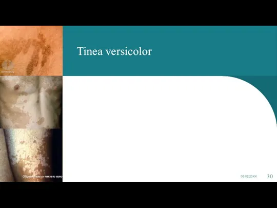
Слайд 30Tinea versicolor
Образец текста нижнего колонтитула
08.02.20XX
Tinea versicolor
Образец текста нижнего колонтитула
08.02.20XX
 Dressing of skins
 Ecology In the City оf Volzhsky
 Car - places in town
 Translate from Russian into English
 Презентация на тему Welcome to Wales
  Love / hate
 Success
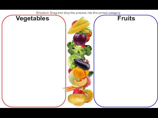 Fruit or vegetable
 Презентация на тему Достопримечательности Берлина
  Reading Comprehension. Episode 1
 Shops
Shops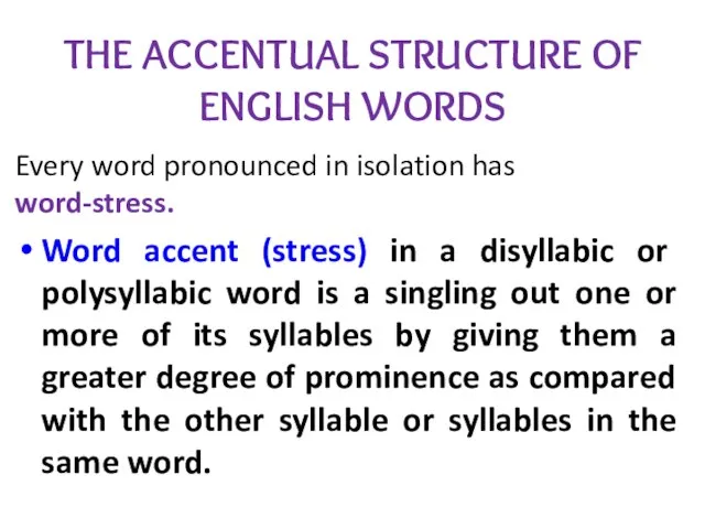 The accentual structure of english words
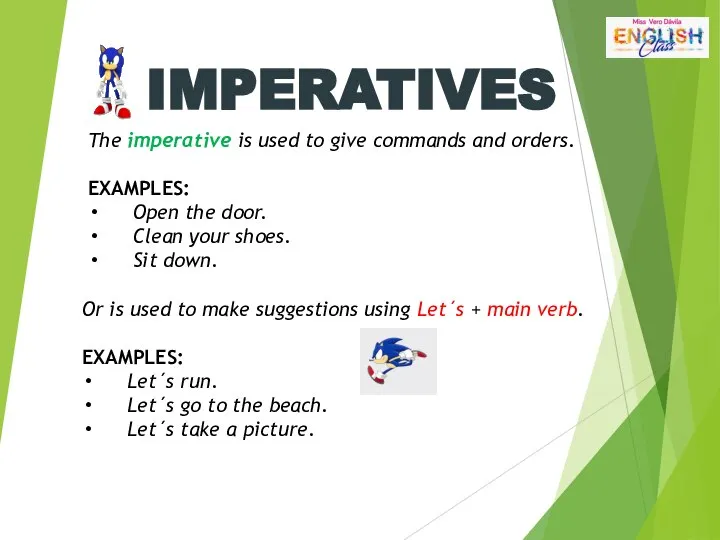 Imperatives. Grammar drills
 Truth or dare
 Какую букву рисует бабочка 2
 My itinerary for a trip around the USA
 Present Simple & Continuous sentences
 What are we going to do in several minutes
 Кабинет иностранного языка
 Real numbers
 English Listening Practice
 Весёлый чемодан. Часть 1
 Difference between Poland, Ukraine and Norway
 Novocherkassk College of Industrial Technologies and Management
 Угадай песню по картинкам
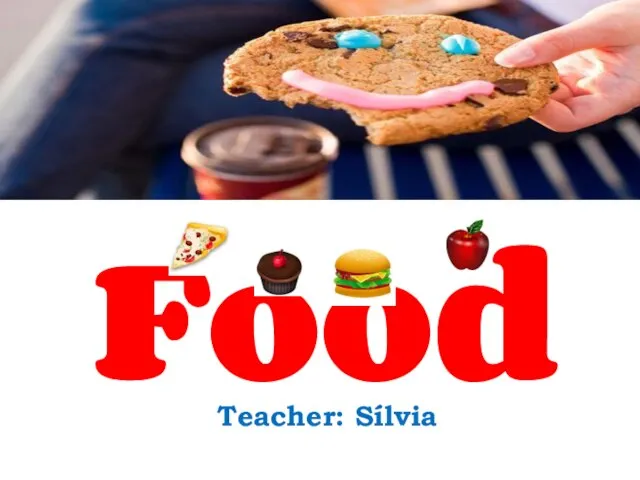 Food. Vocabulary
 Square. Rectangle
 Grammati̇cal category of case