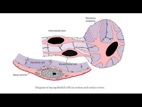
Слайд 2Diagram of myoepithelial cells in section and surface views.
Слайд 3Alveolar Cell from lactating mammary gland.
N, nucleus; TJ, tight junction; GJ,
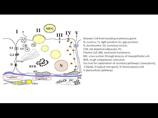
gap junction;
D, desmosome; SV, secretory vesicle;
FDA, fat-depleted adipocyte; PC,
Plasma Cell; BM, basement membrane;
ME, cross section through process of myoepithelial cell;
RER, rough endoplasmic reticulum.
See text for explanation of secretory pathways I (exocytosis),
II (lipid), III (apical transport), IV (transcytosis) and
V (paracellular pathway).

 Олигосахариды и их классификация
 Венерин башмачок - гордость Верховажского района
 Витамин Вс
 Адам эмбриологиясы. Дамудың кризистік кезеңдері. Онтогенез
 Жизнь диких животных зимой
 Дыхательная система
 Физиология обмена веществ и энергии. Терморегуляция
 Животные. Викторина
 История развития генетики в России
 Животные дома
 Презентация на тему Происхождение человека (9 класс)
  Anamorphic fungi бөлімінің фитопатогенді саңырауқұлақтарына сипаттама
 Выделительная система
 Регуляция работы сердца и кровеносных сосудов
 Опорно - двигательная система. Интересные факты
 Зимующие птицы нашего Крыма
 Теории возникновения жизни (таблицы к лекции)
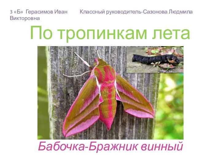 По тропинкам лета. Бабочка - Бражник винный. 3 класс
 Поиск способов усовершенствования методики растворимости гранулированных удобрений
 Факторы, влияющие на развитие и функционирование нервной системы
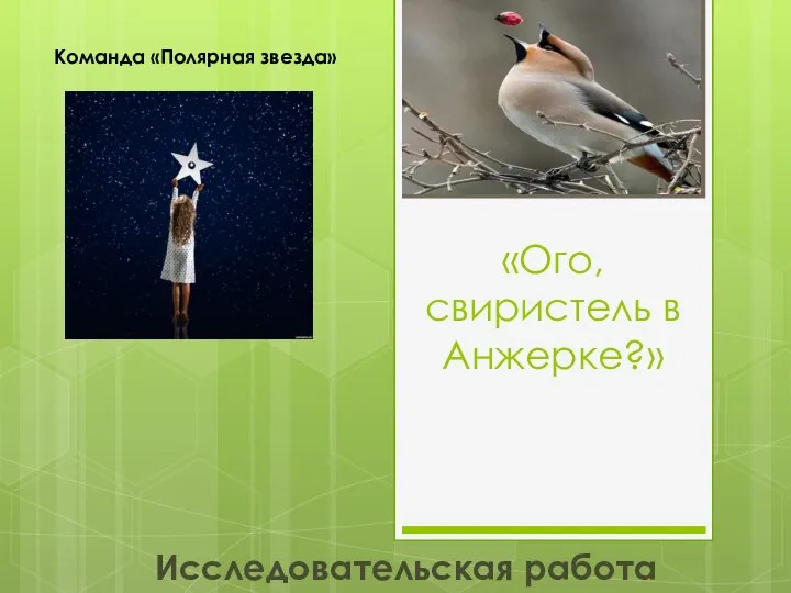 Свиристель в Анжерке. Исследовательская работа
 Индивидуальное развитие
 Странички позитива. Кошки
 Грибы Грузди
 Презентация на тему Почему табак называют ядом
  Презентация на тему Побег и почки
  Растения
 Строение и функции головного мозга