- Seminar B5 The cell: Organelles

Содержание
- 2. What is a Cell? A cell is the basic unit of life All organisms are made
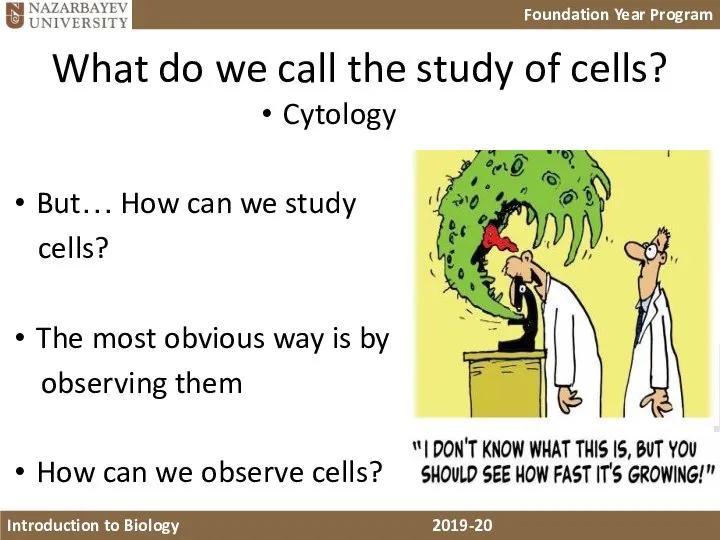
- 3. What do we call the study of cells? Cytology But… How can we study cells? The
- 4. Microscopy!!! What types of microscopy do you know?
- 5. Microscopy Terms Magnification is the increase in an object’s image size compared with its actual size.
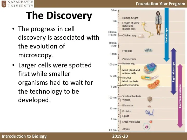
- 6. The Discovery The progress in cell discovery is associated with the evolution of microscopy. Larger cells
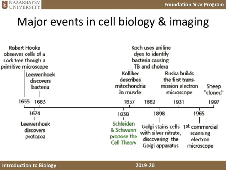
- 7. Major events in cell biology & imaging
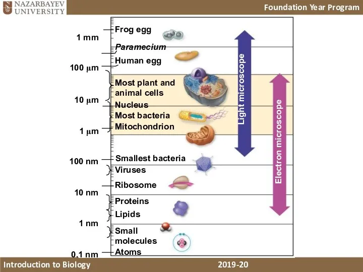
- 8. Frog egg Paramecium Human egg Most plant and animal cells Nucleus Most bacteria Mitochondrion Smallest bacteria
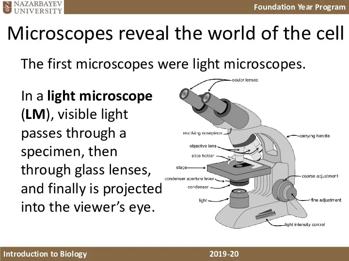
- 9. Microscopes reveal the world of the cell In a light microscope (LM), visible light passes through
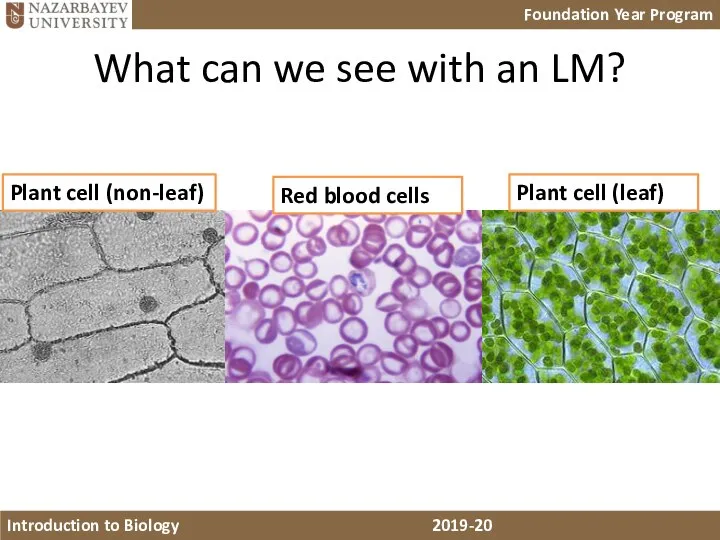
- 10. What can we see with an LM? Plant cell (non-leaf) Red blood cells Plant cell (leaf)
- 11. Electron microscopes Beginning in the 1930s, scientists started using a very powerful microscope called the electron
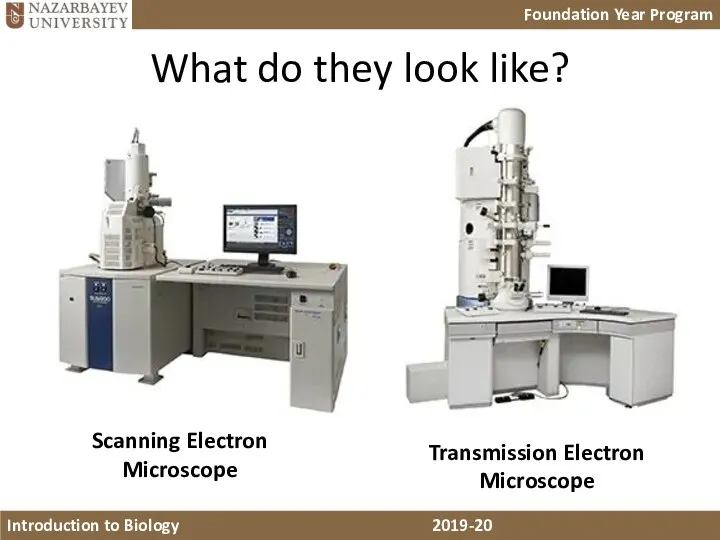
- 12. What do they look like? Scanning Electron Microscope Transmission Electron Microscope
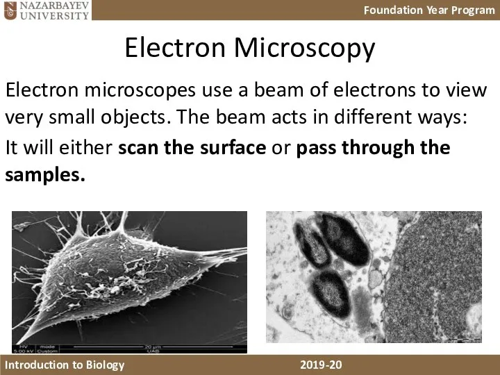
- 13. Electron Microscopy Electron microscopes use a beam of electrons to view very small objects. The beam
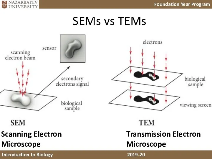
- 14. Scanning Electron Microscope Transmission Electron Microscope SEMs vs TEMs
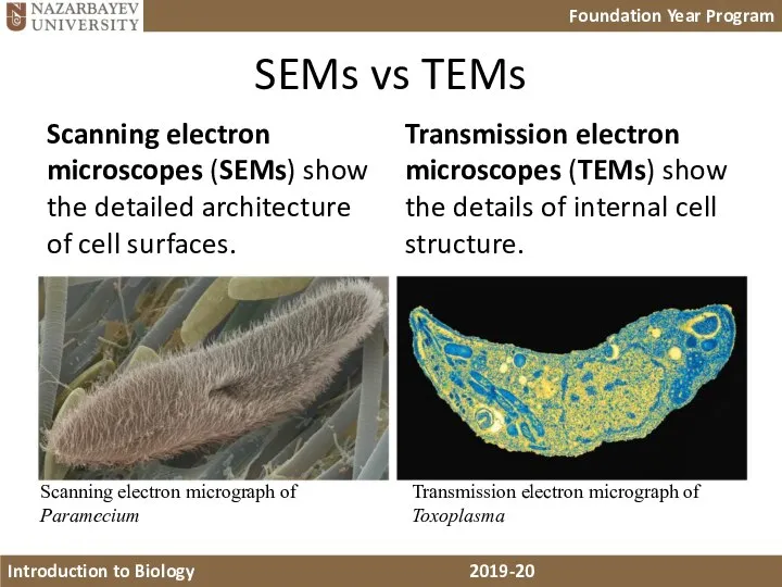
- 15. SEMs vs TEMs Scanning electron microscopes (SEMs) show the detailed architecture of cell surfaces. Transmission electron
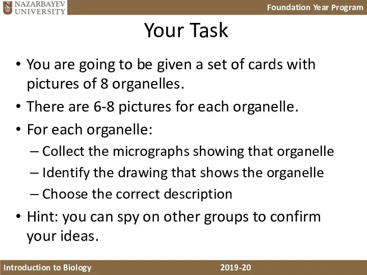
- 16. Your Task You are going to be given a set of cards with pictures of 8
- 17. Let’s check your conclusions!!
- 18. Nucleus
- 19. RER
- 20. SER
- 21. Golgi Body
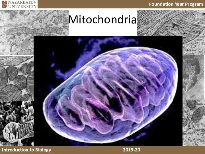
- 22. Mitochondria
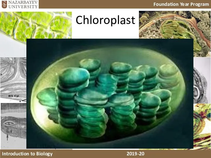
- 23. Chloroplast
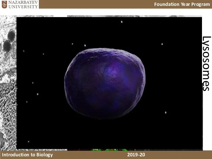
- 24. Lysosomes
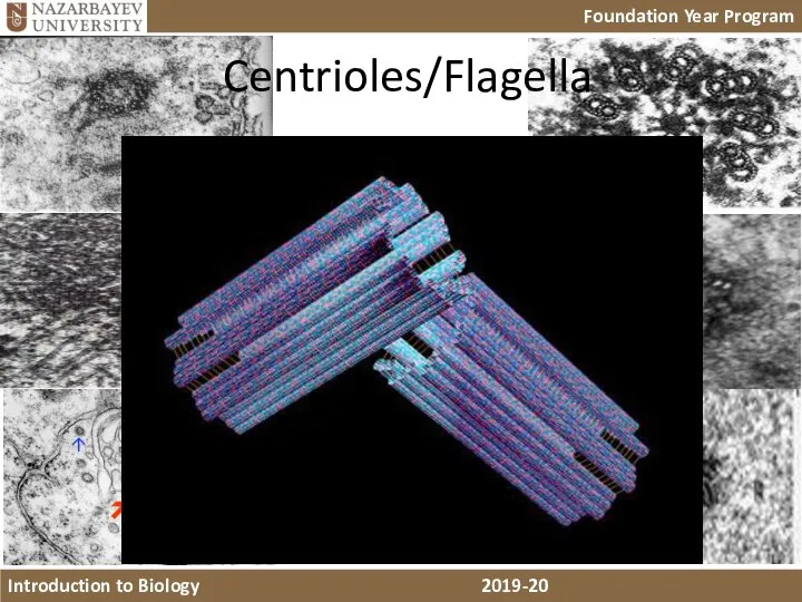
- 25. Centrioles/Flagella
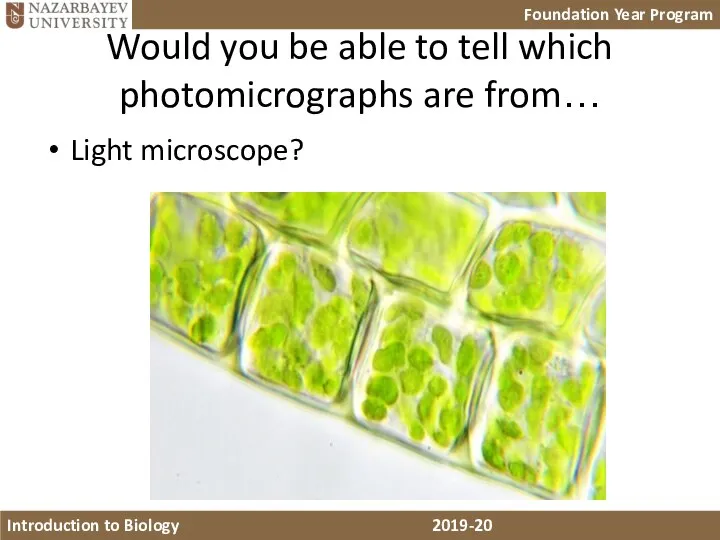
- 26. Would you be able to tell which photomicrographs are from… Light microscope?
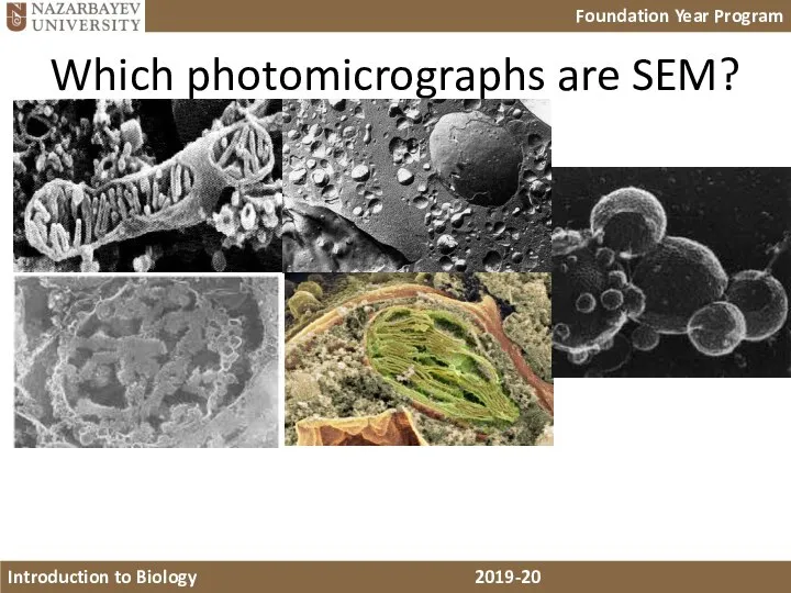
- 27. Which photomicrographs are SEM?
- 29. Скачать презентацию
Слайд 3What do we call the study of cells?
Cytology
But… How can we study
What do we call the study of cells?
Cytology
But… How can we study
Слайд 4Microscopy!!!
What types of microscopy do you know?
Microscopy!!!
What types of microscopy do you know?
Слайд 5Microscopy Terms
Magnification is the increase in an object’s image size compared with
Microscopy Terms
Magnification is the increase in an object’s image size compared with
Слайд 6The Discovery
The progress in cell discovery is associated with the evolution of
The Discovery
The progress in cell discovery is associated with the evolution of
Слайд 7Major events in cell biology & imaging
Major events in cell biology & imaging
Слайд 8Frog egg
Paramecium
Human egg
Most plant and
animal cells
Nucleus
Most bacteria
Mitochondrion
Smallest bacteria
Viruses
Ribosome
Proteins
Lipids
Small
molecules
Atoms
Light microscope
Electron microscope
1 mm
100 nm
10
Frog egg
Paramecium
Human egg
Most plant and
animal cells
Nucleus
Most bacteria
Mitochondrion
Smallest bacteria
Viruses
Ribosome
Proteins
Lipids
Small
molecules
Atoms
Light microscope
Electron microscope
1 mm
100 nm
10
Слайд 9Microscopes reveal the world of the cell
In a light microscope (LM), visible
Microscopes reveal the world of the cell
In a light microscope (LM), visible
Слайд 10What can we see with an LM?
Plant cell (non-leaf)
Red blood cells
What can we see with an LM?
Plant cell (non-leaf)
Red blood cells
Слайд 11Electron microscopes
Beginning in the 1930s, scientists started using a very powerful microscope
Electron microscopes
Beginning in the 1930s, scientists started using a very powerful microscope
Слайд 12What do they look like?
Scanning Electron Microscope
Transmission Electron Microscope
What do they look like?
Scanning Electron Microscope
Transmission Electron Microscope
Слайд 13Electron Microscopy
Electron microscopes use a beam of electrons to view very small
Electron Microscopy
Electron microscopes use a beam of electrons to view very small
Слайд 14Scanning Electron Microscope
Transmission Electron Microscope
SEMs vs TEMs
Scanning Electron Microscope
Transmission Electron Microscope
SEMs vs TEMs
Слайд 15SEMs vs TEMs
Scanning electron microscopes (SEMs) show the detailed architecture of cell
SEMs vs TEMs
Scanning electron microscopes (SEMs) show the detailed architecture of cell
Слайд 16 Your Task
You are going to be given a set of cards
Your Task
You are going to be given a set of cards
Слайд 17Let’s check your conclusions!!
Let’s check your conclusions!!
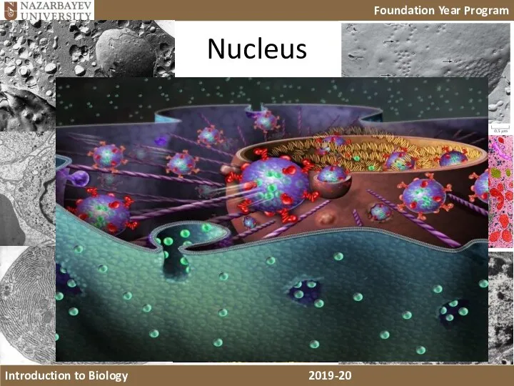
Слайд 18Nucleus
Nucleus
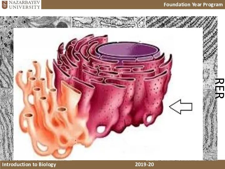
Слайд 19RER
RER
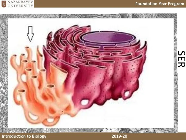
Слайд 20SER
SER
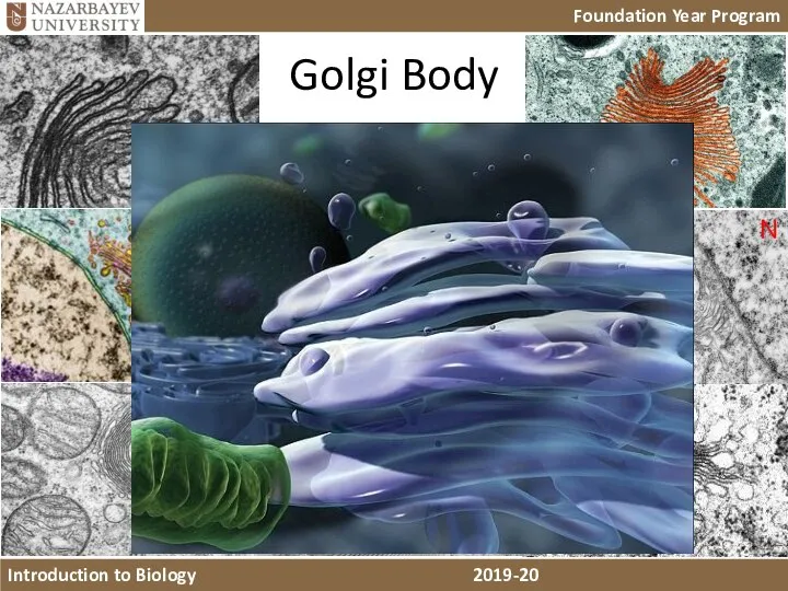
Слайд 21Golgi Body
Golgi Body
Слайд 22Mitochondria
Mitochondria
Слайд 23Chloroplast
Chloroplast
Слайд 24Lysosomes
Lysosomes
Слайд 25Centrioles/Flagella
Centrioles/Flagella
Слайд 26Would you be able to tell which photomicrographs are from…
Light microscope?
Would you be able to tell which photomicrographs are from…
Light microscope?
Слайд 27Which photomicrographs are SEM?
Which photomicrographs are SEM?
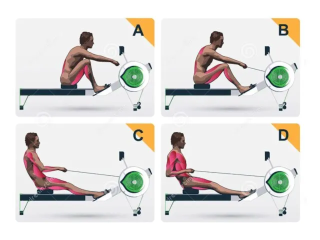 Работа мышц
 Кузнечик. Анатомия
 05. Тип Саркожгутиконосцы. Класс Саркодовые
 Презентация на тему Что такое систематика
  Розыскная служба
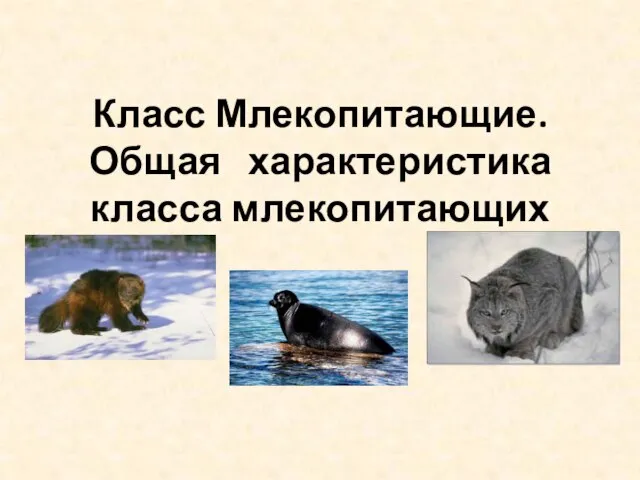 Общая характеристика Класса Млекопитающих
 Постэмбриональный период развития организмов
 Организм человека, как единая биологическая система
 Растения. Жизненные циклы растений
 Плесневые грибы и дрожжи
 Кровоснабжение глаза
 физиология
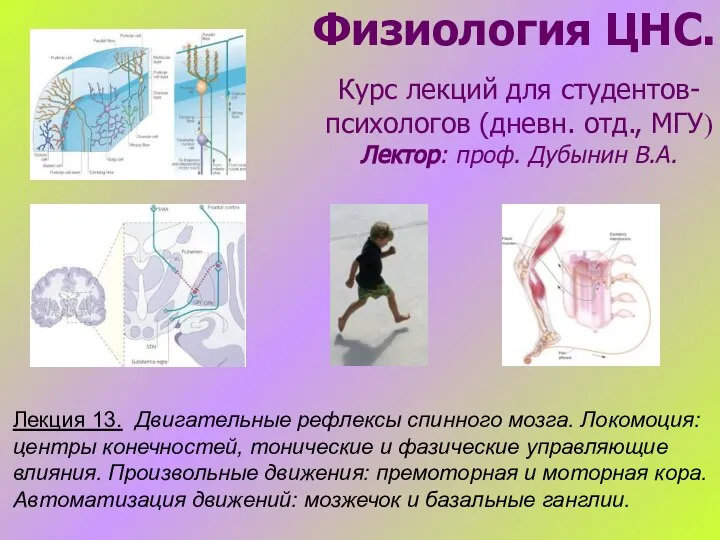 Двигательные рефлексы спинного мозга. Локомоция: центры конечностей, тонические и фазические управляющие влияния
 Презентация на тему Современные представления о возникновении жизни
  Зависимость дыхания от факторов среды и его связь с продуктивностью растений
 Береза - дерево чудес
 Тип моллюски, или мягкотелые
 Цепи питания
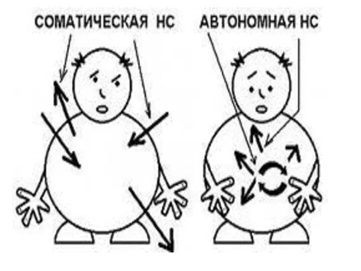 Нервная система
 Валеология. Центральная проблема валеологии
 1666861337135100
 Строение клетки
 Ферменты. Часть 1
 Видоизменения листьев
 Значение и охрана ланцетников
 Мой джунгарский хомячок
 Зеленые водоросли
 Плауны, хвощи, папоротники