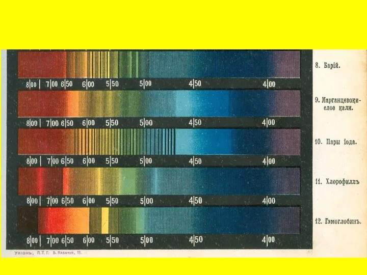
- Radiation (излучения)

Содержание
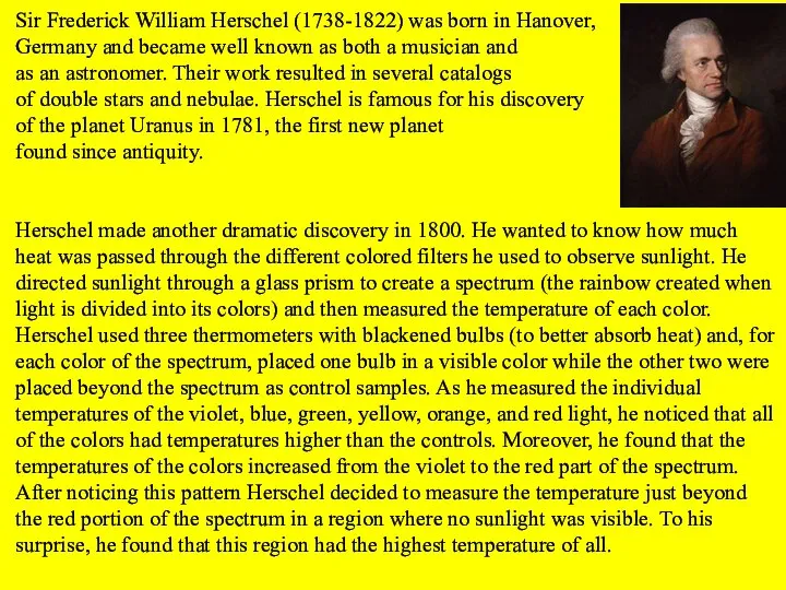
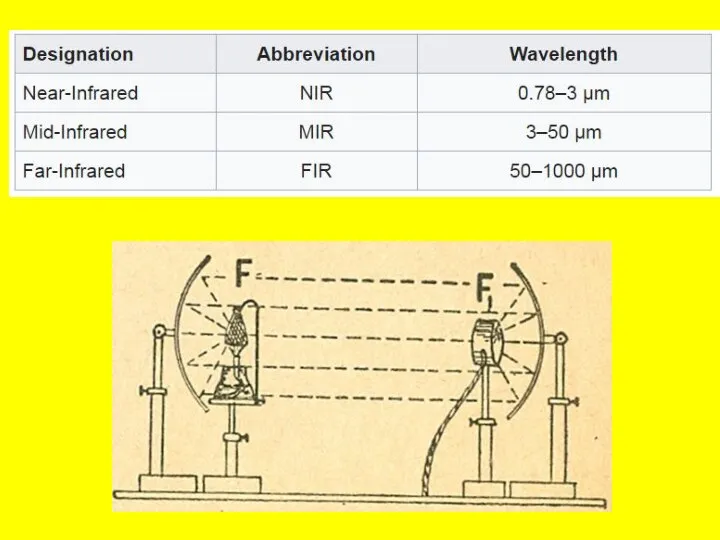
- 3. Sir Frederick William Herschel (1738-1822) was born in Hanover, Germany and became well known as both
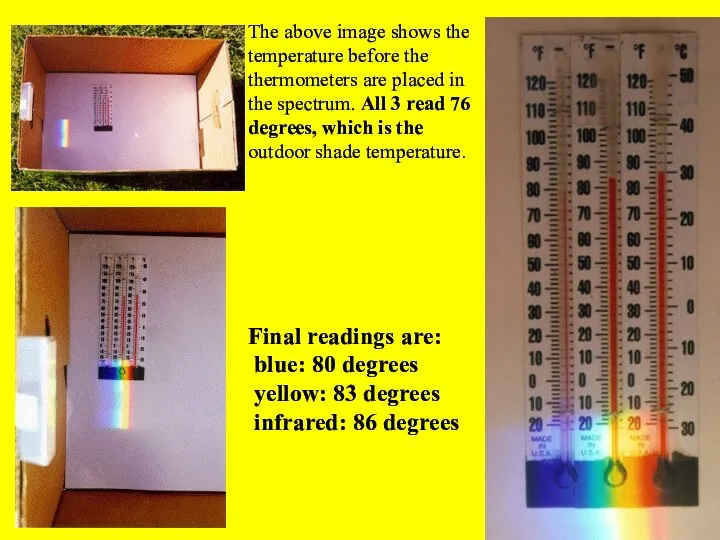
- 5. The above image shows the temperature before the thermometers are placed in the spectrum. All 3
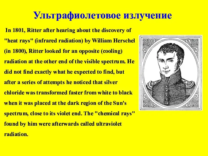
- 7. In 1801, Ritter after hearing about the discovery of "heat rays" (infrared radiation) by William Herschel
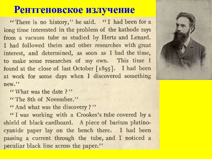
- 9. Рентгеновское излучение
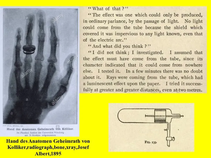
- 10. Hand des Anatomen Geheimrath von Kolliker,radiograph,bone,xray,Josef Albert,1895
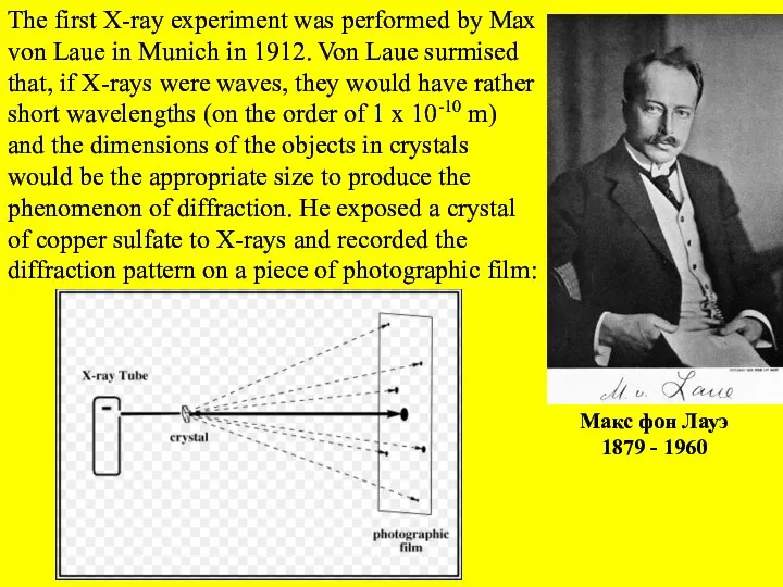
- 14. Макс фон Лауэ 1879 - 1960 The first X-ray experiment was performed by Max von Laue
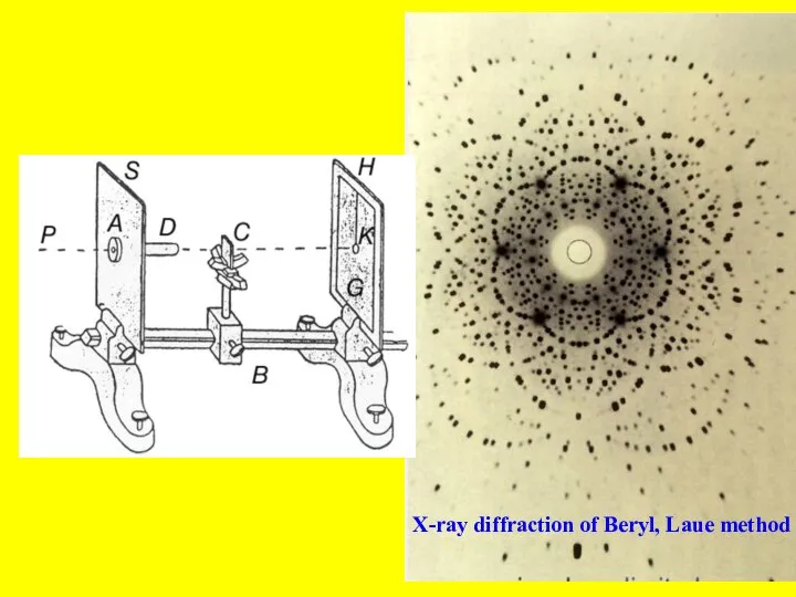
- 15. X-ray diffraction of Beryl, Laue method
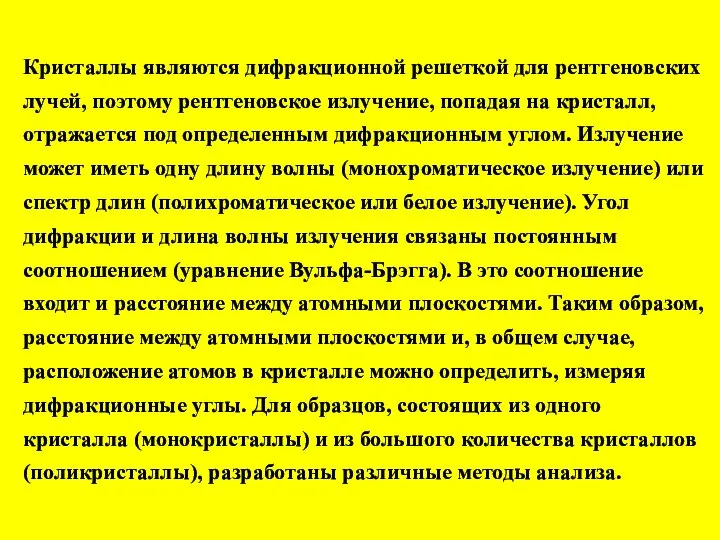
- 16. Кристаллы являются дифракционной решеткой для рентгеновских лучей, поэтому рентгеновское излучение, попадая на кристалл, отражается под определенным
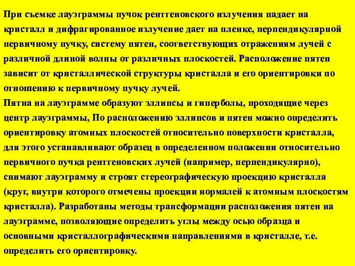
- 17. При съемке лауэграммы пучок рентгеновского излучения падает на кристалл и дифрагированное излучение дает на пленке, перпендикулярной
- 19. Скачать презентацию
Слайд 3Sir Frederick William Herschel (1738-1822) was born in Hanover,
Germany and became well
Sir Frederick William Herschel (1738-1822) was born in Hanover,
Germany and became well
Слайд 5The above image shows the temperature before the thermometers are placed in
The above image shows the temperature before the thermometers are placed in
Слайд 7 In 1801, Ritter after hearing about the discovery of "heat rays"
In 1801, Ritter after hearing about the discovery of "heat rays"
Слайд 9Рентгеновское излучение
Рентгеновское излучение
Слайд 10Hand des Anatomen Geheimrath von Kolliker,radiograph,bone,xray,Josef Albert,1895
Hand des Anatomen Geheimrath von Kolliker,radiograph,bone,xray,Josef Albert,1895

Слайд 14Макс фон Лауэ
1879 - 1960
The first X-ray experiment was performed by Max
Макс фон Лауэ
1879 - 1960
The first X-ray experiment was performed by Max
Слайд 15X-ray diffraction of Beryl, Laue method
X-ray diffraction of Beryl, Laue method
Слайд 16Кристаллы являются дифракционной решеткой для рентгеновских лучей, поэтому рентгеновское излучение, попадая на
Кристаллы являются дифракционной решеткой для рентгеновских лучей, поэтому рентгеновское излучение, попадая на
Слайд 17При съемке лауэграммы пучок рентгеновского излучения падает на кристалл и дифрагированное излучение
При съемке лауэграммы пучок рентгеновского излучения падает на кристалл и дифрагированное излучение
 Алгоритмизация информации на уроках физики
 Ядерный реактор. АЭС
 задачи на равноускоренное движение (1)
 Нагревание водяным паром
 Критерии прочности намоточных труб при растяжении, кручении и сложном напряженном состоянии
 Решение задач. Сила Ампера
 Излучение и поглощение света атомами . Виды спектров,спектральный анализ.
 Эмиссия и катоды
 Сверхпроводимость. Свойства
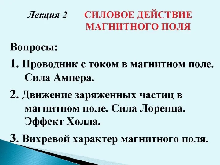 Силовое действие магнитного поля
 Цвет. Основы цветоведения
 Экзотические типы радиоактивного распада
 Презентация на тему Основные акустические опасные факторы воздействия на человека
  Переменный электрический ток
 Переменный ток
 Лазер, устройство лазера
 Основы термодинамики
 Амперметр. Измерение силы тока
 Путешествие в прошлое пылесоса
 Klassifikatsia_dvigateley_vnutrennego_sgorania
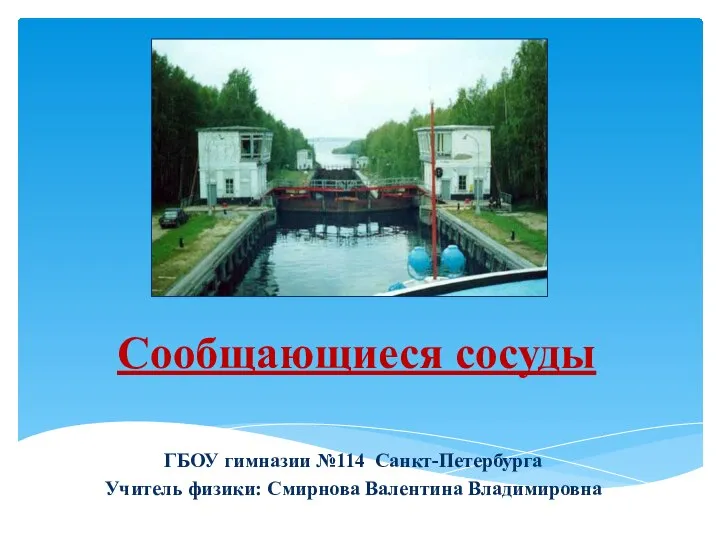 Сообщающиеся сосуды
 Конфигурации (проводка) моторов HI-FINITY
 Физика и технология наноструктур
 Законы постоянного тока
 pril1_2 (1)
 Презентация на тему Тепловые машины и их КПД
  Охранная сигнализация
 Устойчивость и точность систем управления