- Autopsy report Patient N, 76 уears

Содержание
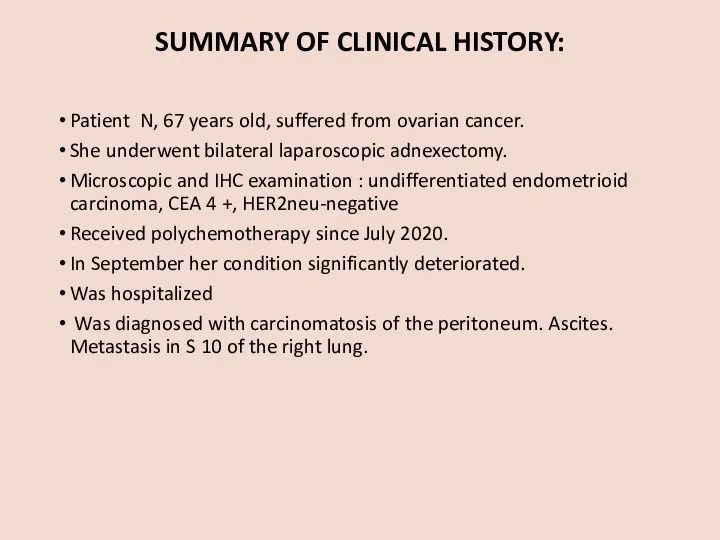
- 2. SUMMARY OF CLINICAL HISTORY: Patient N, 67 years old, suffered from ovarian cancer. She underwent bilateral
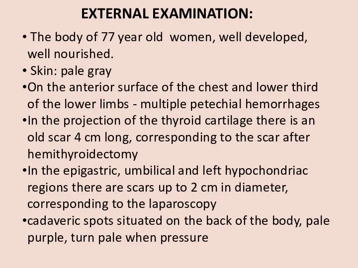
- 3. EXTERNAL EXAMINATION: The body of 77 year old women, well developed, well nourished. Skin: pale gray
- 4. INTERNAL EXAMINATION
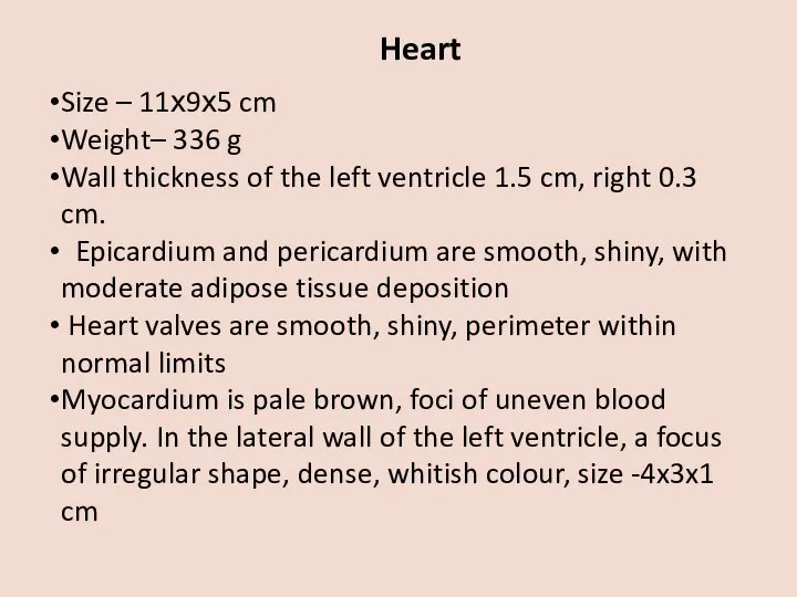
- 5. Heart Size – 11х9х5 cm Weight– 336 g Wall thickness of the left ventricle 1.5 cm,
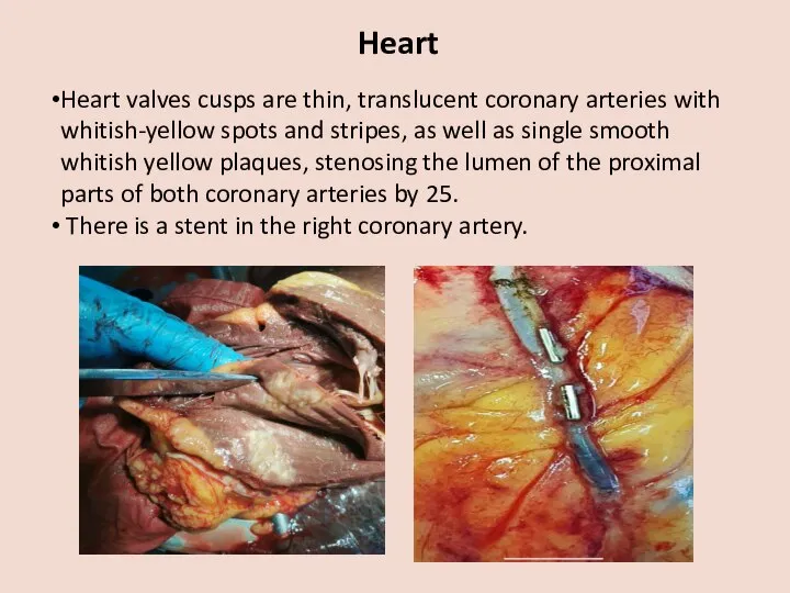
- 6. Heart Heart valves cusps are thin, translucent coronary arteries with whitish-yellow spots and stripes, as well
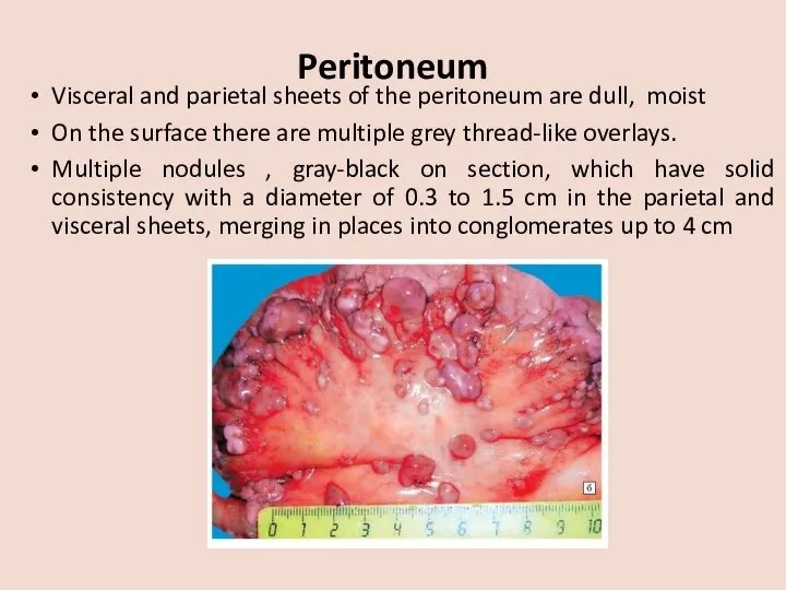
- 7. Peritoneum Visceral and parietal sheets of the peritoneum are dull, moist On the surface there are
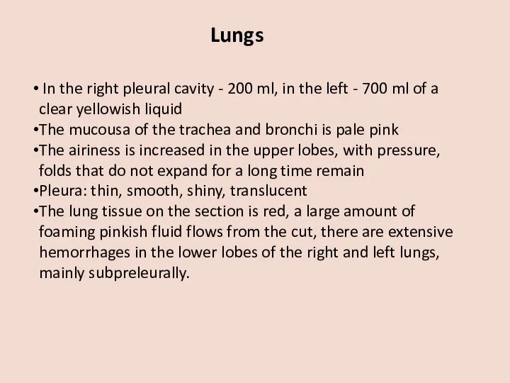
- 8. Lungs In the right pleural cavity - 200 ml, in the left - 700 ml of
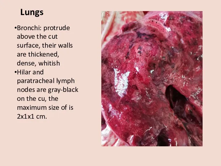
- 9. Lungs Bronchi: protrude above the cut surface, their walls are thickened, dense, whitish Hilar and paratracheal
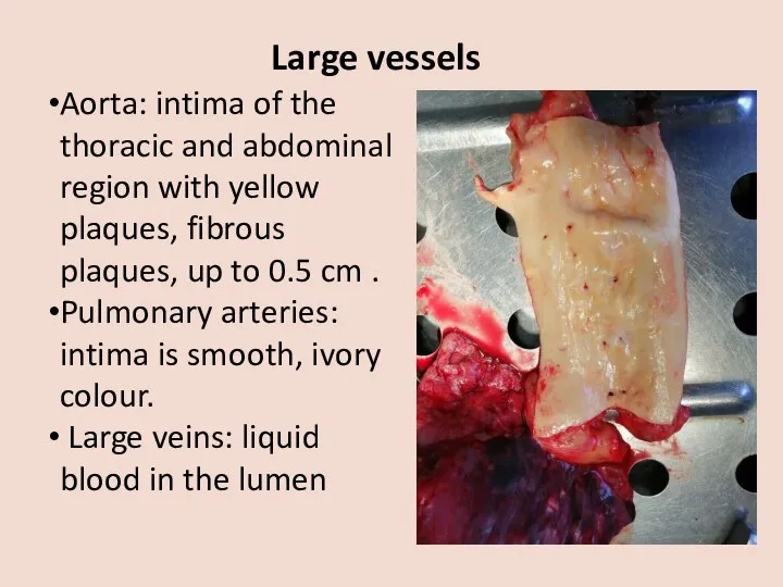
- 10. Large vessels Aorta: intima of the thoracic and abdominal region with yellow plaques, fibrous plaques, up
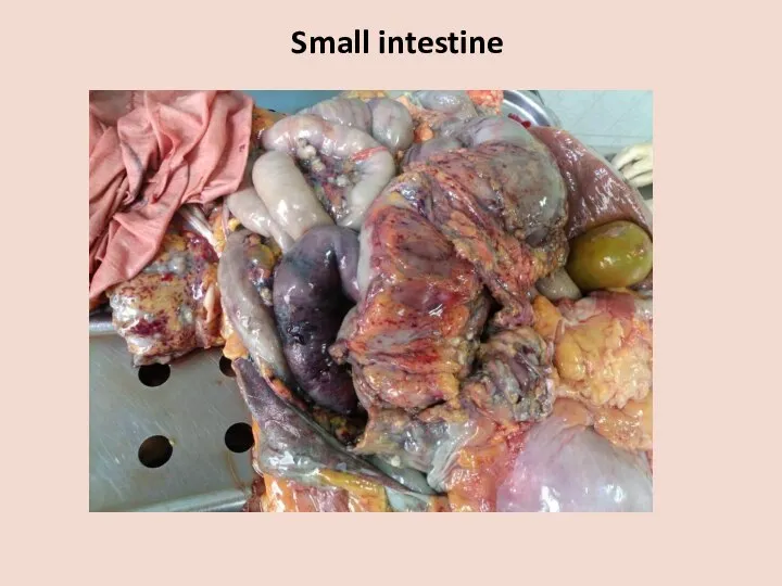
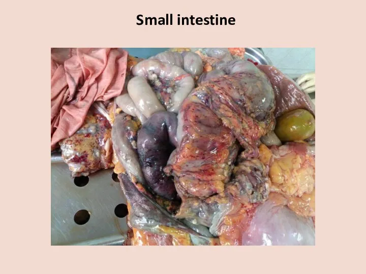
- 11. Gastrointestinal tract Esophagus: mucousa is gray, with longitudinal foldings Veins of the lower third are not
- 12. Small intestine
- 13. Small intestine
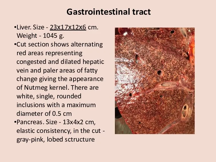
- 14. Gastrointestinal tract Liver. Size - 23х17х12х6 cm. Weight - 1045 g. Cut section shows alternating red
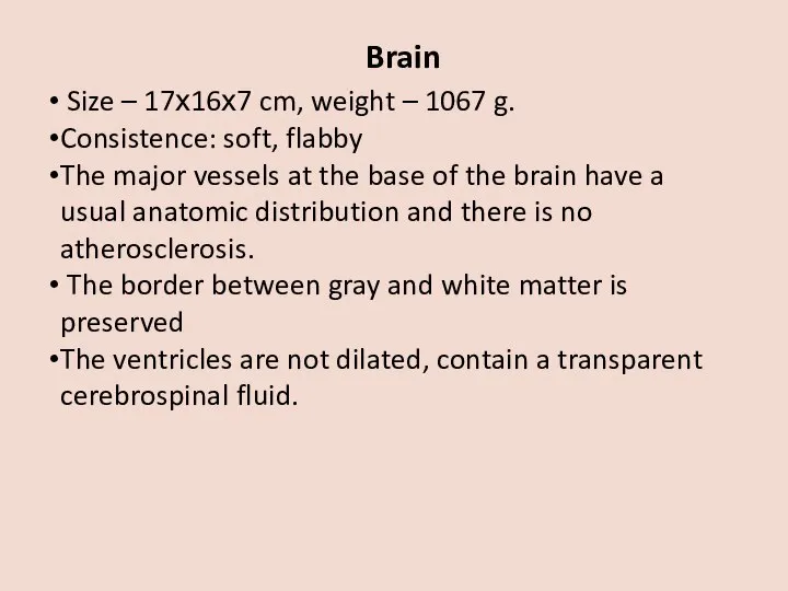
- 15. Brain Size – 17х16х7 cm, weight – 1067 g. Consistence: soft, flabby The major vessels at
- 16. Kidneys Size: left kidney - 10,5х5х3 cm, right kidney – 9,5x4x3 cm Weight: left kidney -
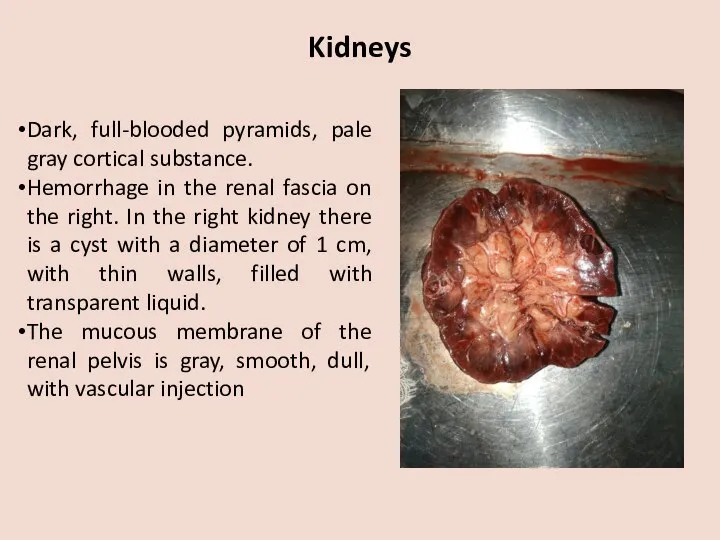
- 17. Kidneys Dark, full-blooded pyramids, pale gray cortical substance. Hemorrhage in the renal fascia on the right.
- 18. Organs of the urogenital system Bladder. The mucous membrane is grey, has foldings. Uterus - 7x3x2cm,
- 19. Splin Size - 11x8x3 cm Weight - 112 gWith a smooth capsule Flabby consistency Dark cherry
- 20. Endocrine system Thyroid. Size: the right lobe – 1,5x1x1 cm, the left lobe – is resected,
- 21. Microscopic examination
- 22. Peritoneal lesions, H&E, х100
- 23. Peritoneal lesions, H&E, х200
- 24. Peritoneal lesions, H&E , х00
- 25. Peritoneal lesions, H&E , х400
- 26. Mesenteric lymph node, H&E , х400
- 27. Heart, H&E, х200
- 28. Heart, H&E, х400
- 29. Heart, H&E, х200
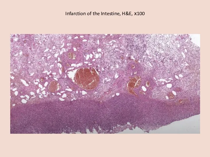
- 30. Infarction of the Intestine, H&E, х100
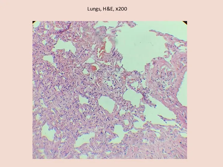
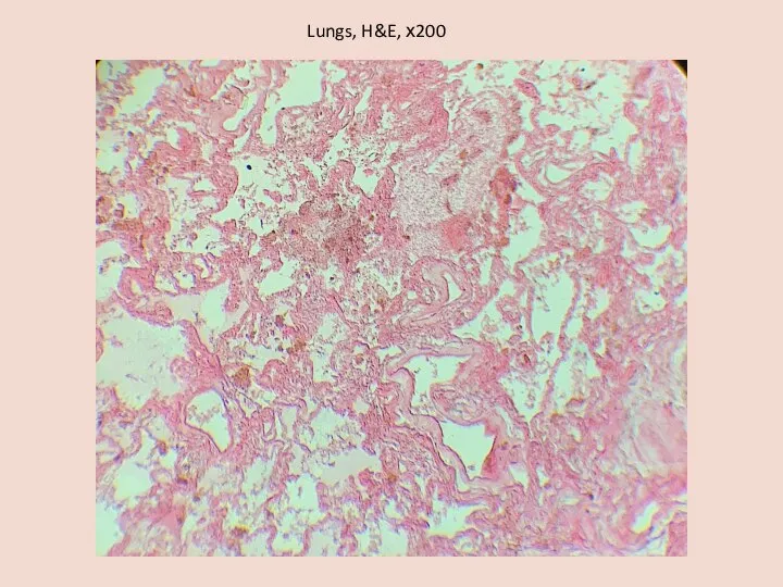
- 31. Lungs, H&E, х200
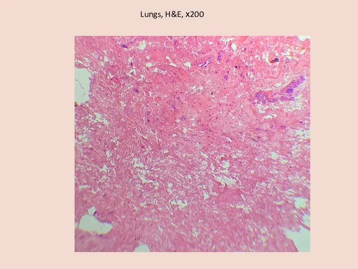
- 32. Lungs, H&E, х200
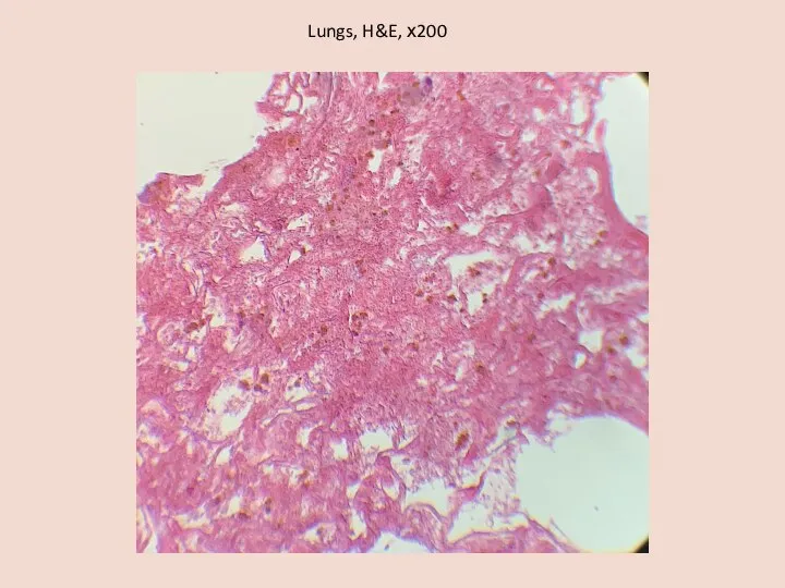
- 33. Lungs, H&E, х200
- 34. Lungs, H&E, х200
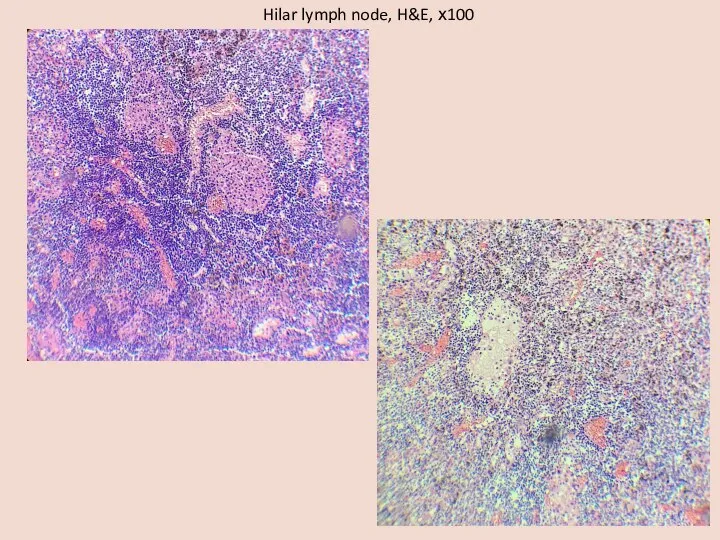
- 35. Hilar lymph node, H&E, х100
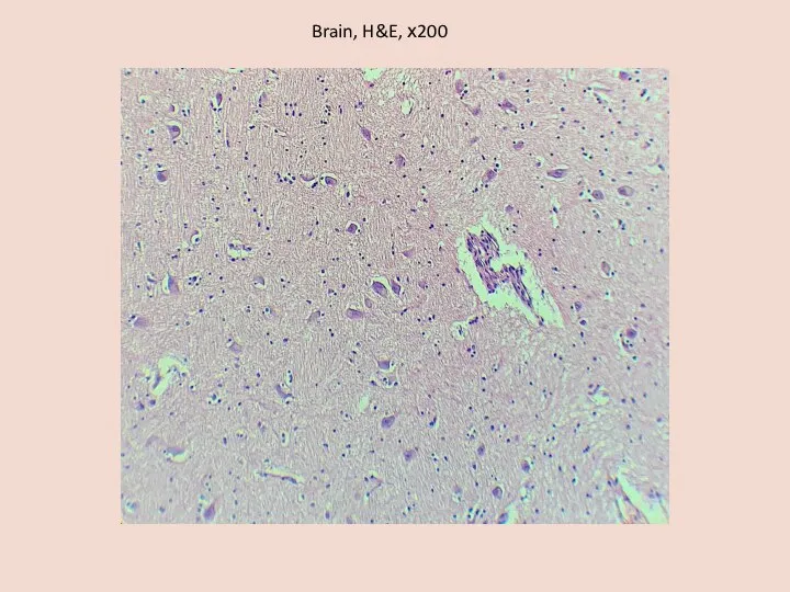
- 36. Brain, H&E, х200
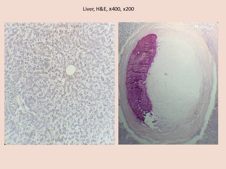
- 37. Liver, H&E, х400, x200
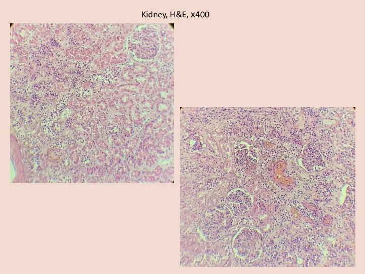
- 38. Kidney, H&E, х400
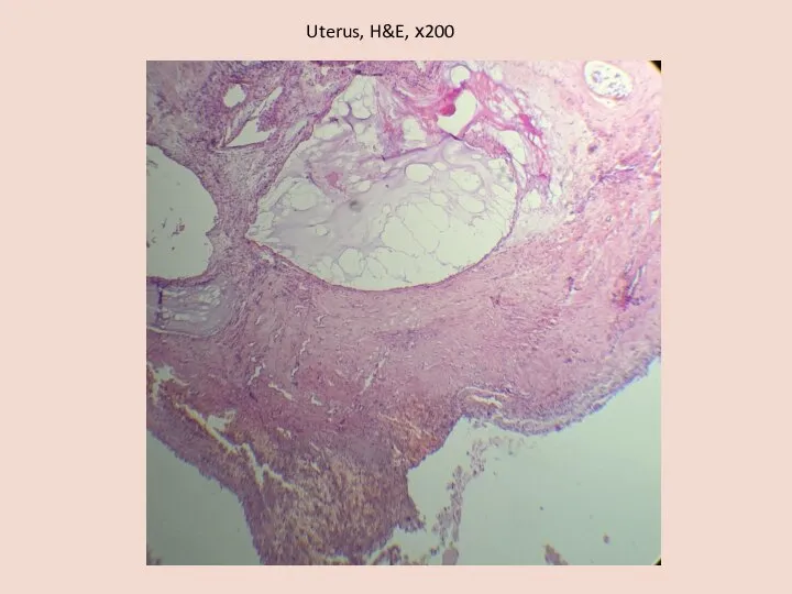
- 39. Uterus, H&E, х200
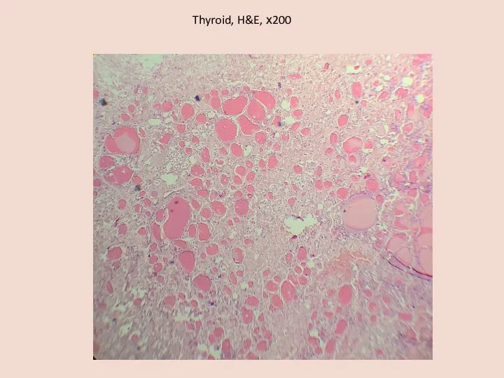
- 40. Thyroid, H&E, х200
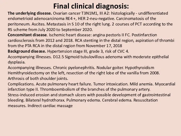
- 41. Final clinical diagnosis: The underlying disease. Ovarian cancer T3N1M1, III A2: histologically - undifferentiated endometrioid adenocarcinoma
- 42. Pathological diagnosis (primary): The underlying disease. Ovarian cancer with parietal and visceral peritoneum carcinomatosis, liver metastases
- 43. Accompanying illnesses. Chronic obstructive mucous bronchitis. Diffuse reticular pneumosclerosis. Chronic obstructive pulmonary emphysema. Large focal cardiosclerosis
- 44. CLINICOPATHOLOGIC CORRELATION Patient N, 67 years old, suffered from ovarian cancer with carcinomatosis of the peritoneum
- 45. Pathological diagnosis after histological examination: . The histological examination confirmed the pathological diagnosis. Clarified the nature
- 47. Скачать презентацию
Слайд 3EXTERNAL EXAMINATION:
The body of 77 year old women, well developed,
EXTERNAL EXAMINATION:
The body of 77 year old women, well developed,
Слайд 4INTERNAL EXAMINATION
INTERNAL EXAMINATION
Слайд 5Heart
Size – 11х9х5 cm
Weight– 336 g
Wall thickness of the left ventricle 1.5
Heart
Size – 11х9х5 cm
Weight– 336 g
Wall thickness of the left ventricle 1.5
Слайд 6Heart
Heart valves cusps are thin, translucent coronary arteries with whitish-yellow spots
Heart
Heart valves cusps are thin, translucent coronary arteries with whitish-yellow spots
Слайд 7Peritoneum
Visceral and parietal sheets of the peritoneum are dull, moist
On the
Peritoneum
Visceral and parietal sheets of the peritoneum are dull, moist
On the
Слайд 8Lungs
In the right pleural cavity - 200 ml, in the left
Lungs
In the right pleural cavity - 200 ml, in the left
Слайд 9Lungs
Bronchi: protrude above the cut surface, their walls are thickened, dense, whitish
Hilar
Lungs
Bronchi: protrude above the cut surface, their walls are thickened, dense, whitish
Hilar
Слайд 10Large vessels
Aorta: intima of the thoracic and abdominal region with yellow plaques,
Large vessels
Aorta: intima of the thoracic and abdominal region with yellow plaques,
Слайд 11Gastrointestinal tract
Esophagus: mucousa is gray, with longitudinal foldings
Veins of the lower third
Gastrointestinal tract
Esophagus: mucousa is gray, with longitudinal foldings
Veins of the lower third
Слайд 12 Small intestine
Small intestine
Слайд 13 Small intestine
Small intestine
Слайд 14Gastrointestinal tract
Liver. Size - 23х17х12х6 cm. Weight - 1045 g.
Cut section shows
Gastrointestinal tract
Liver. Size - 23х17х12х6 cm. Weight - 1045 g.
Cut section shows
Слайд 15Brain
Size – 17х16х7 cm, weight – 1067 g.
Consistence: soft, flabby
The
Brain
Size – 17х16х7 cm, weight – 1067 g.
Consistence: soft, flabby
The
Слайд 16Kidneys
Size: left kidney - 10,5х5х3 cm, right kidney – 9,5x4x3 cm
Weight:
Kidneys
Size: left kidney - 10,5х5х3 cm, right kidney – 9,5x4x3 cm
Weight:
Слайд 17Kidneys
Dark, full-blooded pyramids, pale gray cortical substance.
Hemorrhage in the renal fascia
Kidneys
Dark, full-blooded pyramids, pale gray cortical substance.
Hemorrhage in the renal fascia
Слайд 18Organs of the urogenital system
Bladder. The mucous membrane is grey, has
Organs of the urogenital system
Bladder. The mucous membrane is grey, has
Слайд 19Splin
Size - 11x8x3 cm
Weight - 112 gWith a smooth capsule
Splin
Size - 11x8x3 cm
Weight - 112 gWith a smooth capsule
Слайд 20Endocrine system
Thyroid. Size: the right lobe – 1,5x1x1 cm, the left lobe
Endocrine system
Thyroid. Size: the right lobe – 1,5x1x1 cm, the left lobe
Слайд 21Microscopic examination
Microscopic examination
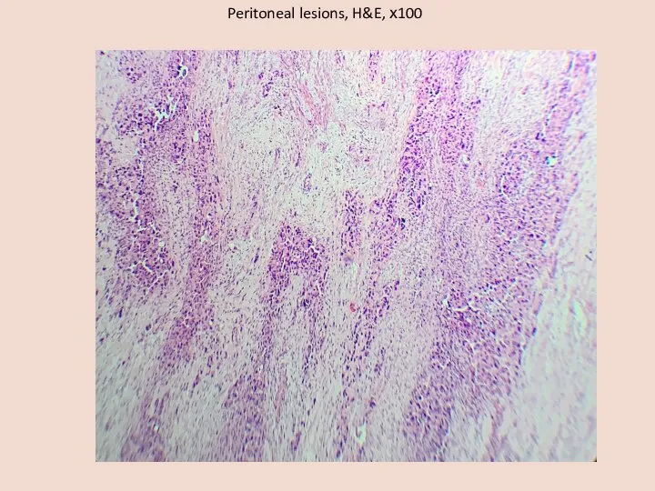
Слайд 22Peritoneal lesions, H&E, х100
Peritoneal lesions, H&E, х100
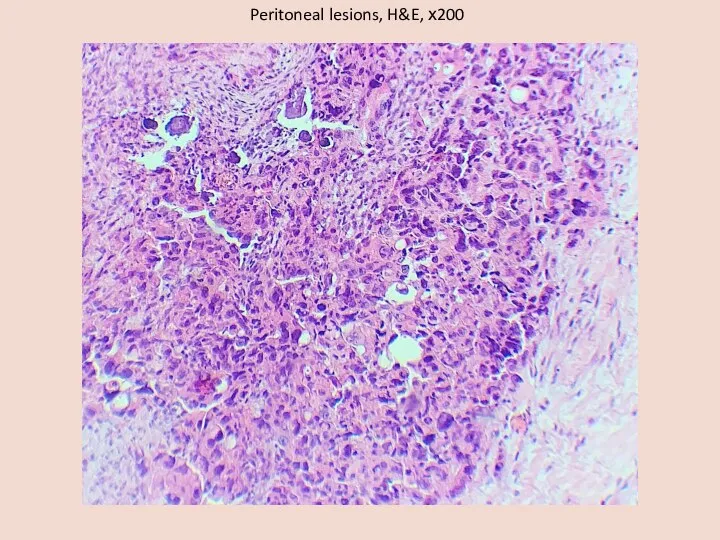
Слайд 23Peritoneal lesions, H&E, х200
Peritoneal lesions, H&E, х200
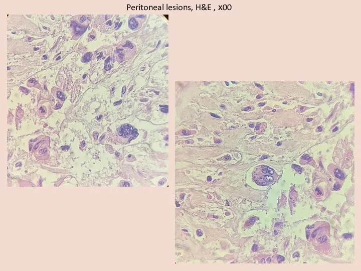
Слайд 24Peritoneal lesions, H&E , х00
Peritoneal lesions, H&E , х00
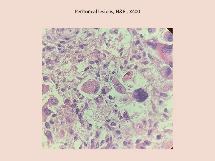
Слайд 25Peritoneal lesions, H&E , х400
Peritoneal lesions, H&E , х400
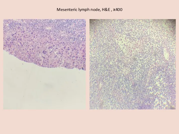
Слайд 26Mesenteric lymph node, H&E , х400
Mesenteric lymph node, H&E , х400
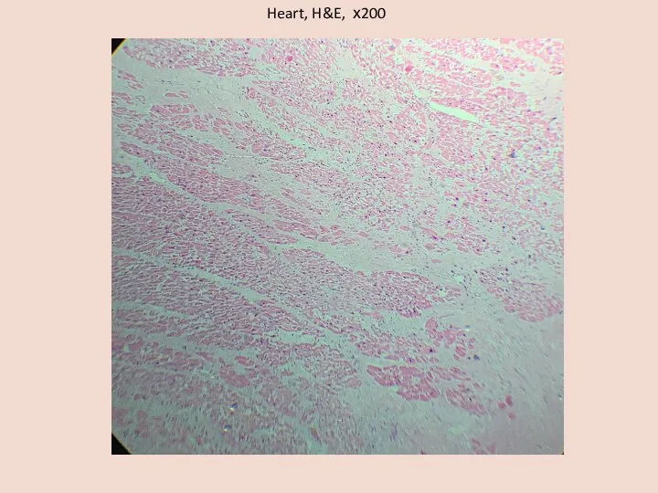
Слайд 27Heart, H&E, х200
Heart, H&E, х200
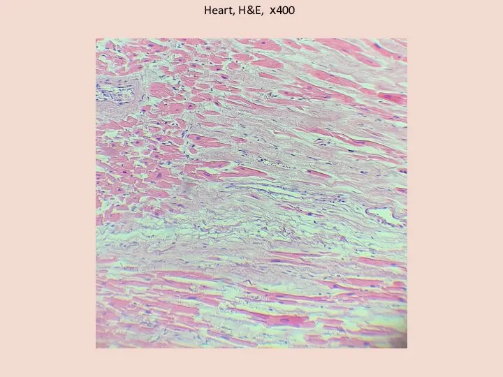
Слайд 28Heart, H&E, х400
Heart, H&E, х400
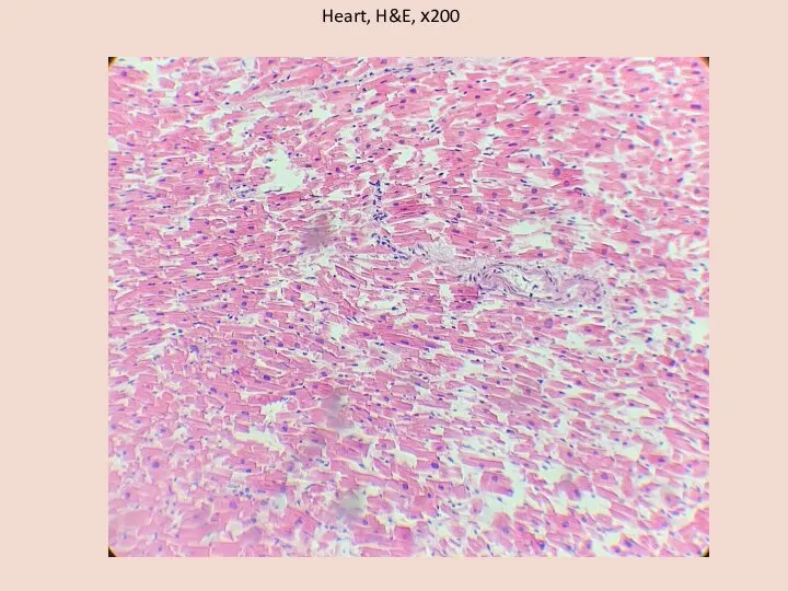
Слайд 29Heart, H&E, х200
Heart, H&E, х200
Слайд 30Infarction of the Intestine, H&E, х100
Infarction of the Intestine, H&E, х100
Слайд 31Lungs, H&E, х200
Lungs, H&E, х200
Слайд 32Lungs, H&E, х200
Lungs, H&E, х200
Слайд 33Lungs, H&E, х200
Lungs, H&E, х200
Слайд 34Lungs, H&E, х200
Lungs, H&E, х200
Слайд 35Hilar lymph node, H&E, х100
Hilar lymph node, H&E, х100
Слайд 36Brain, H&E, х200
Brain, H&E, х200
Слайд 37Liver, H&E, х400, x200
Liver, H&E, х400, x200
Слайд 38Kidney, H&E, х400
Kidney, H&E, х400
Слайд 39Uterus, H&E, х200
Uterus, H&E, х200
Слайд 40Thyroid, H&E, х200
Thyroid, H&E, х200
Слайд 41Final clinical diagnosis:
The underlying disease. Ovarian cancer T3N1M1, III A2: histologically
Final clinical diagnosis:
The underlying disease. Ovarian cancer T3N1M1, III A2: histologically
Слайд 42Pathological diagnosis (primary):
The underlying disease. Ovarian cancer with parietal and visceral peritoneum
Pathological diagnosis (primary):
The underlying disease. Ovarian cancer with parietal and visceral peritoneum
Слайд 43Accompanying illnesses. Chronic obstructive mucous bronchitis. Diffuse reticular pneumosclerosis. Chronic obstructive pulmonary
Accompanying illnesses. Chronic obstructive mucous bronchitis. Diffuse reticular pneumosclerosis. Chronic obstructive pulmonary
Слайд 44CLINICOPATHOLOGIC CORRELATION
Patient N, 67 years old, suffered from ovarian cancer with
CLINICOPATHOLOGIC CORRELATION
Patient N, 67 years old, suffered from ovarian cancer with

Слайд 45Pathological diagnosis after histological examination:
.
The histological examination confirmed the pathological diagnosis. Clarified
Pathological diagnosis after histological examination:
.
The histological examination confirmed the pathological diagnosis. Clarified
 Аускультация сердца
 Использование локального отрицательного давления в хирургии
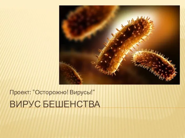 Вирус бешенства
 Лекарственные препараты, влияющие на функции крови
 Эпилепсия
 Қр дәрілік заттарға сараптаманы жүргізу ережесі мен тәртібі
 Лабораторные показатели липидного обмена
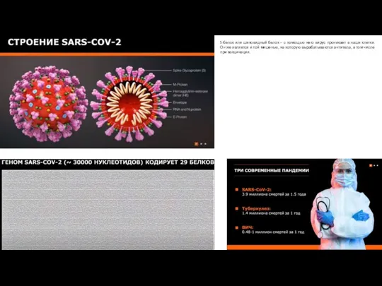 Электронные микрофотографии SARS-COV-2
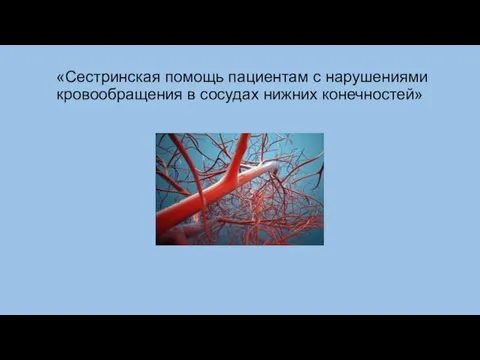 Сестринская помощь пациентам с нарушениями кровообращения в сосудах нижних конечностей
 Зат және энергия алмасуына кіріспе
 Қыздар құпиясы
 Схема физической реабилитации при повреждениях коленного и голеностопного суставов
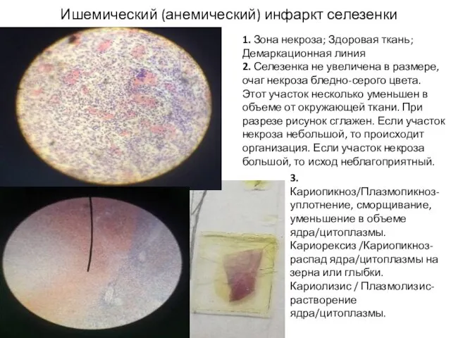 Ишемический (анемический) инфаркт селезенки
 Донорство у тебя в крови
 Влияние алкоголя на организм матери и плода
 Профилактика деменций
 Педиатрия как наука
 Уничтожение России лишением рассудка и онкологией
 Неврастения. Неврозы
 Zalecenia federacji fpz w związku ze strategią walki z pandemią Covi019
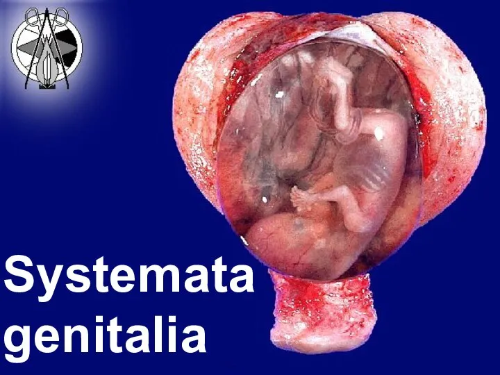 Понятие пола
 Кировский медицинский колледж
 Раны. Что это такое, и как с ними поступать?
 Плоскостопие
 Курение во время беременности и его последствия
 Врожденные хирургические патологии лица и полости рта
 Crimea state medical university
 Ауылда және қалада ұйымдастырылған балалар мен жасөспірімдер контингенттеріне емдікпрофилактикалық қызмет ету