- Cell Surface Membrane

Содержание
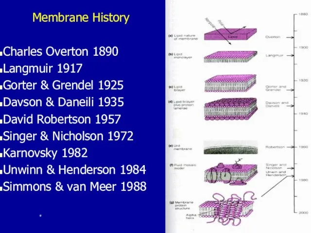
- 2. Membrane History Charles Overton 1890 Langmuir 1917 Gorter & Grendel 1925 Davson & Daneili 1935 David
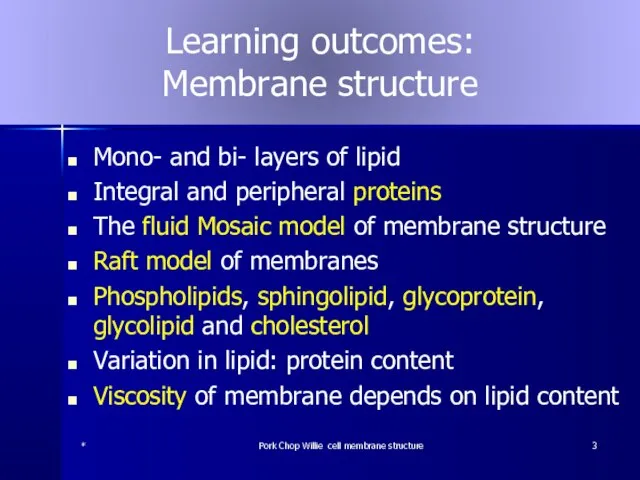
- 3. Learning outcomes: Membrane structure Mono- and bi- layers of lipid Integral and peripheral proteins The fluid
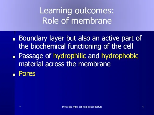
- 4. Learning outcomes: Role of membrane Boundary layer but also an active part of the biochemical functioning
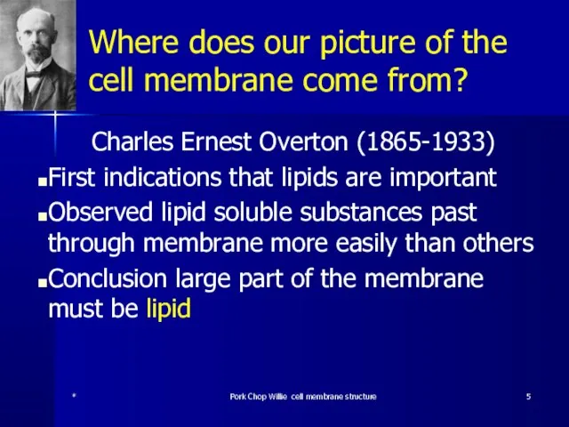
- 5. Where does our picture of the cell membrane come from? Charles Ernest Overton (1865-1933) First indications
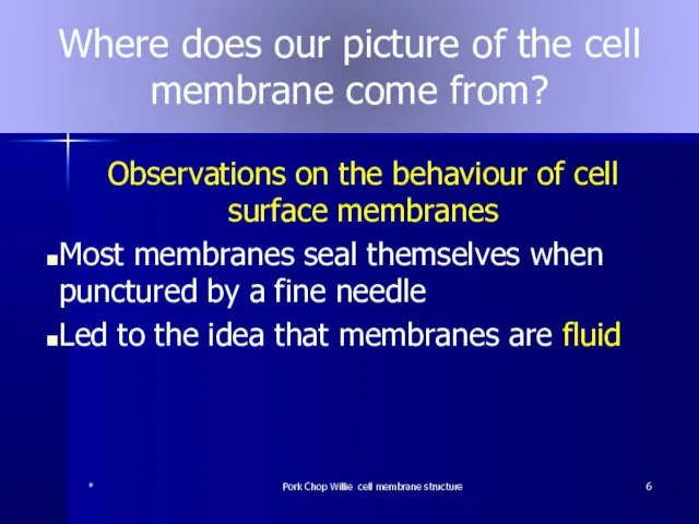
- 6. Where does our picture of the cell membrane come from? Observations on the behaviour of cell
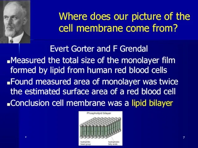
- 7. Where does our picture of the cell membrane come from? Evert Gorter and F Grendal Measured
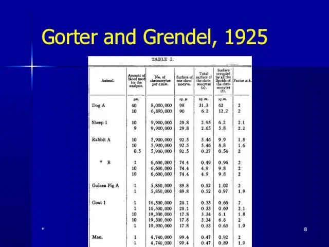
- 8. Gorter and Grendel, 1925 * Pork Chop Willie cell membrane structure
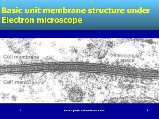
- 9. Basic unit membrane structure under Electron microscope * Pork Chop Willie cell membrane structure
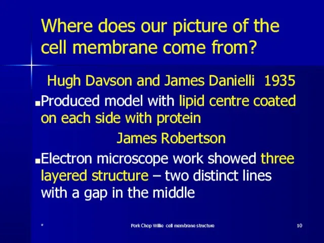
- 10. Where does our picture of the cell membrane come from? Hugh Davson and James Danielli 1935
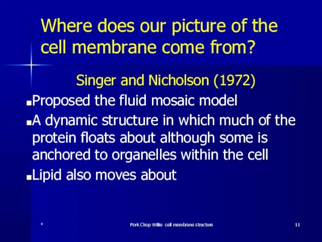
- 11. Where does our picture of the cell membrane come from? Singer and Nicholson (1972) Proposed the
- 12. * Pork Chop Willie cell membrane structure
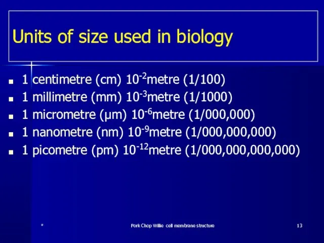
- 13. Units of size used in biology 1 centimetre (cm) 10-2metre (1/100) 1 millimetre (mm) 10-3metre (1/1000)
- 14. Cell Surface Membrane Structure Under the electron microscope bilayer structure is revealed Two distinct lines 7nm
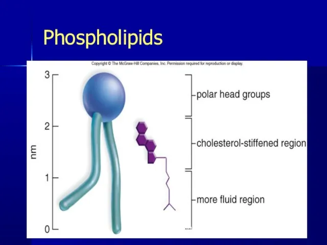
- 15. Phospholipids Lipid molecule three fatty acid molecules and a glycerol Phospholipid only two fatty acids, a
- 16. Lipid molecule * Pork Chop Willie cell membrane structure
- 17. Phospholipid * Pork Chop Willie cell membrane structure
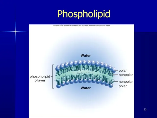
- 18. Phospholipid bilayer Phophate head of the molecule is polar; one end is slightly positive and the
- 19. Phospholipid bilayer 2 Fats and water don’t mix When added to water phospholipids arrange themselves to
- 20. Phospholipid bilayer 3 They form a layer on the surface with their hydrophobic tails directed out
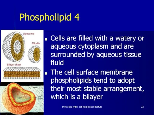
- 21. Phospholipids in water form a monolayer on the surface or spherical micelles * Pork Chop Willie
- 22. Phospholipid 4 Cells are filled with a watery or aqueous cytoplasm and are surrounded by aqueous
- 23. Phospholipid Pork Chop Willie cell membrane structure
- 24. Phospholipid 5 This arrangement avoids the hydrophobic fatty acid tails having any contact with water on
- 25. Phospholipids * Pork Chop Willie cell membrane structure
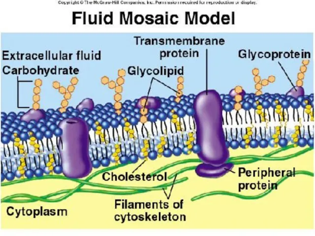
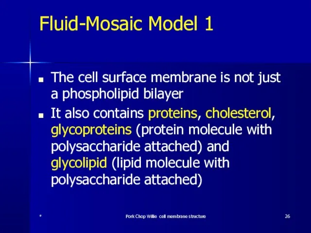
- 26. Fluid-Mosaic Model 1 The cell surface membrane is not just a phospholipid bilayer It also contains
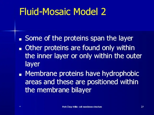
- 27. Fluid-Mosaic Model 2 Some of the proteins span the layer Other proteins are found only within
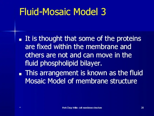
- 28. Fluid-Mosaic Model 3 It is thought that some of the proteins are fixed within the membrane
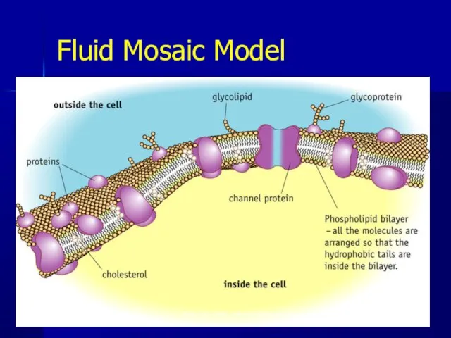
- 29. Fluid Mosaic Model * Pork Chop Willie cell membrane structure
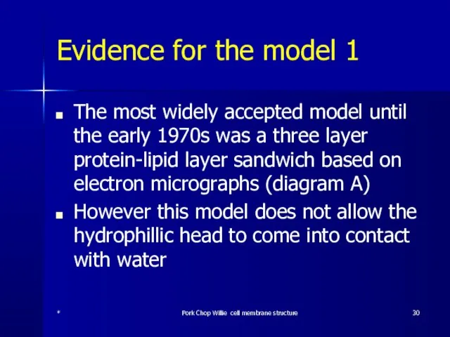
- 30. Evidence for the model 1 The most widely accepted model until the early 1970s was a
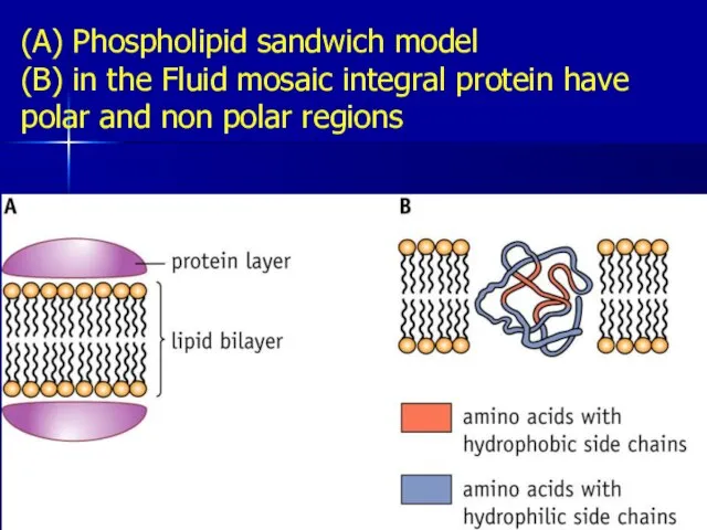
- 31. (A) Phospholipid sandwich model (B) in the Fluid mosaic integral protein have polar and non polar
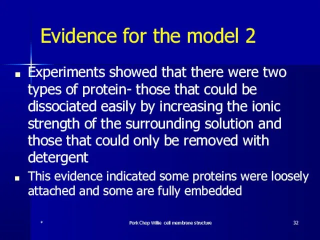
- 32. Evidence for the model 2 Experiments showed that there were two types of protein- those that
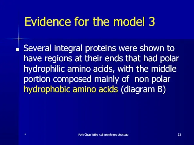
- 33. Evidence for the model 3 Several integral proteins were shown to have regions at their ends
- 34. (A) Phospholipid sandwich model (B) in the Fluid mosaic integral protein have polar and non polar
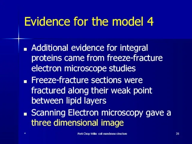
- 35. Evidence for the model 4 Additional evidence for integral proteins came from freeze-fracture electron microscope studies
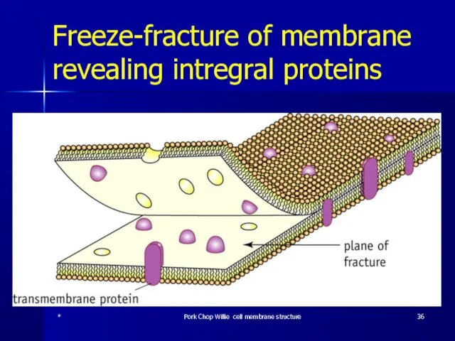
- 36. Freeze-fracture of membrane revealing intregral proteins * Pork Chop Willie cell membrane structure
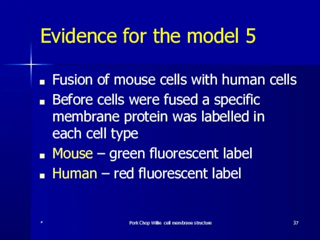
- 37. Evidence for the model 5 Fusion of mouse cells with human cells Before cells were fused
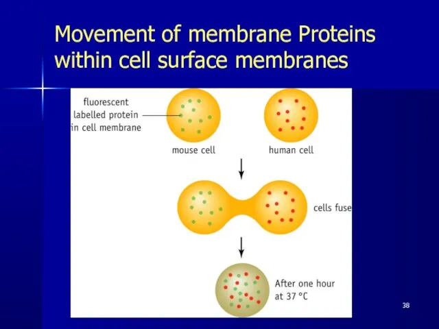
- 38. Movement of membrane Proteins within cell surface membranes Pork Chop Willie cell membrane structure
- 39. Membrane Protein Diversity
- 40. Functions of Membrane Proteins Channel Proteins: Tubular Allow passage of molecules through membrane Carrier Proteins: Combine
- 41. More unsaturated phospholipids – more fluid The more phospholipids containing unsaturated fatty acids the more fluid
- 42. Cholesterol Cholesterol reduces the fluidity of the membrane by preventing movement of the phospholipids * Pork
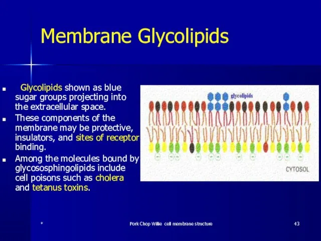
- 43. Membrane Glycolipids Glycolipids shown as blue sugar groups projecting into the extracellular space. These components of
- 44. Sphingolipid Structural lipid of which the parent structure is sphingosine rather than glycerol. Synthesised in the
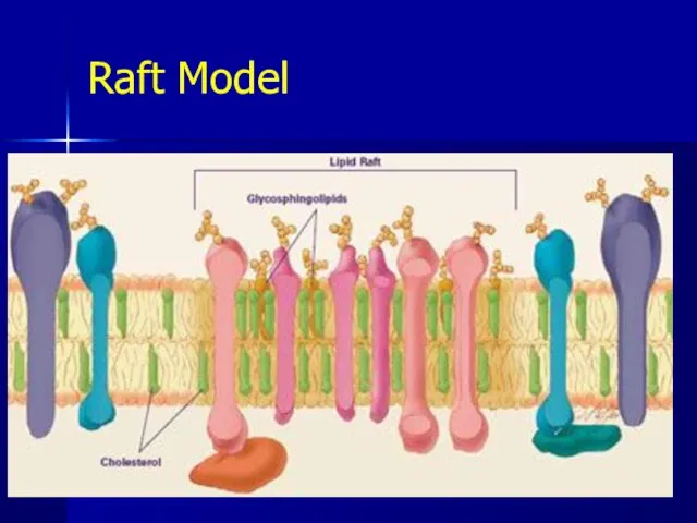
- 45. Raft Model * Pork Chop Willie cell membrane structure
- 46. Raft Model Lipid rafts are possible island like structure present in cellular membranes. They are enriched
- 47. Raft Model The existence of lipid rafts in cell membrane has not yet been approved completely
- 48. * Pork Chop Willie cell membrane structure Q1 According to the fluid-mosaic model for the plasma
- 49. * Pork Chop Willie cell membrane structure A1 According to the fluid-mosaic model for the plasma
- 50. Q2 Phospholipids have their hydrophilic polar heads facing the __________ and _____________fluid. The hydrophobic nonpolar tails
- 51. A2 Phospholipids have their hydrophilic polar heads facing the intracellular and extracellular fluid. The hydrophobic nonpolar
- 52. Q3 The proteins found in the plasma membrane may be _________ proteins, which are found within
- 53. A3 The proteins found in the plasma membrane may be integral proteins, which are found within
- 54. Q 4 State two roles of cholesterol in the membrane (2 marks) * Pork Chop Willie
- 55. A 4 State two roles of cholesterol in the membrane (2 marks) Regulates membrane fluidity; Mechanical
- 56. Q5 There are many types of proteins in a membrane. Describe the role of two (2
- 58. Скачать презентацию
Слайд 2Membrane History
Charles Overton 1890
Langmuir 1917
Gorter & Grendel 1925
Davson & Daneili 1935
David Robertson
Membrane History
Charles Overton 1890
Langmuir 1917
Gorter & Grendel 1925
Davson & Daneili 1935
David Robertson
Слайд 3Learning outcomes:
Membrane structure
Mono- and bi- layers of lipid
Integral and peripheral proteins
The
Learning outcomes:
Membrane structure
Mono- and bi- layers of lipid
Integral and peripheral proteins
The
Слайд 4Learning outcomes:
Role of membrane
Boundary layer but also an active part of the
Learning outcomes:
Role of membrane
Boundary layer but also an active part of the
Слайд 5Where does our picture of the cell membrane come from?
Charles Ernest Overton
Where does our picture of the cell membrane come from?
Charles Ernest Overton
Слайд 6Where does our picture of the cell membrane come from?
Observations on the
Where does our picture of the cell membrane come from?
Observations on the
Слайд 7Where does our picture of the
cell membrane come from?
Evert Gorter and
Where does our picture of the
cell membrane come from?
Evert Gorter and
Слайд 8Gorter and Grendel, 1925
*
Pork Chop Willie cell membrane structure
Gorter and Grendel, 1925
*
Pork Chop Willie cell membrane structure
Слайд 9Basic unit membrane structure under Electron microscope
*
Pork Chop Willie cell membrane structure
Basic unit membrane structure under Electron microscope
*
Pork Chop Willie cell membrane structure
Слайд 10Where does our picture of the cell membrane come from?
Hugh Davson and
Where does our picture of the cell membrane come from?
Hugh Davson and
Слайд 11Where does our picture of the cell membrane come from?
Singer and Nicholson
Where does our picture of the cell membrane come from?
Singer and Nicholson
Слайд 12*
Pork Chop Willie cell membrane structure
*
Pork Chop Willie cell membrane structure
Слайд 13Units of size used in biology
1 centimetre (cm) 10-2metre (1/100)
1 millimetre (mm)
Units of size used in biology
1 centimetre (cm) 10-2metre (1/100)
1 millimetre (mm)
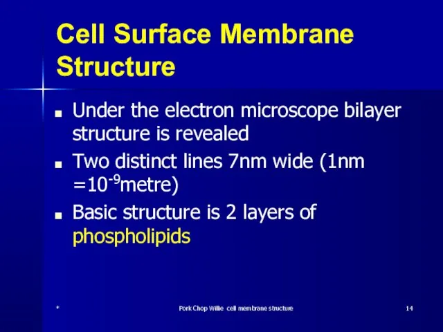
Слайд 14Cell Surface Membrane Structure
Under the electron microscope bilayer structure is revealed
Two distinct
Cell Surface Membrane Structure
Under the electron microscope bilayer structure is revealed
Two distinct
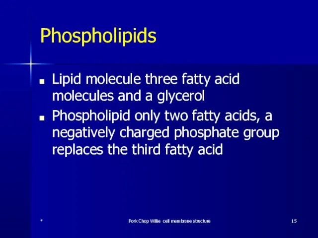
Слайд 15Phospholipids
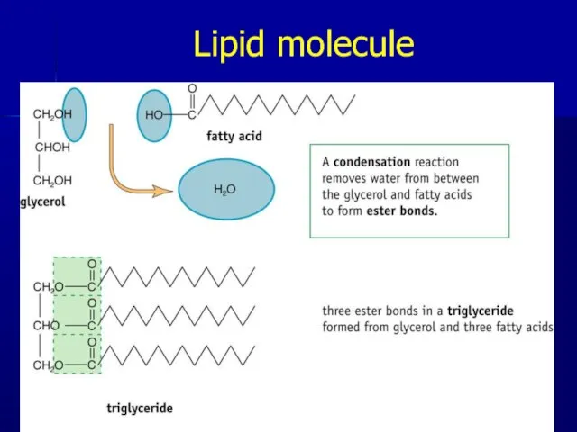
Lipid molecule three fatty acid molecules and a glycerol
Phospholipid only two fatty
Phospholipids
Lipid molecule three fatty acid molecules and a glycerol
Phospholipid only two fatty
Слайд 16Lipid molecule
*
Pork Chop Willie cell membrane structure
Lipid molecule
*
Pork Chop Willie cell membrane structure
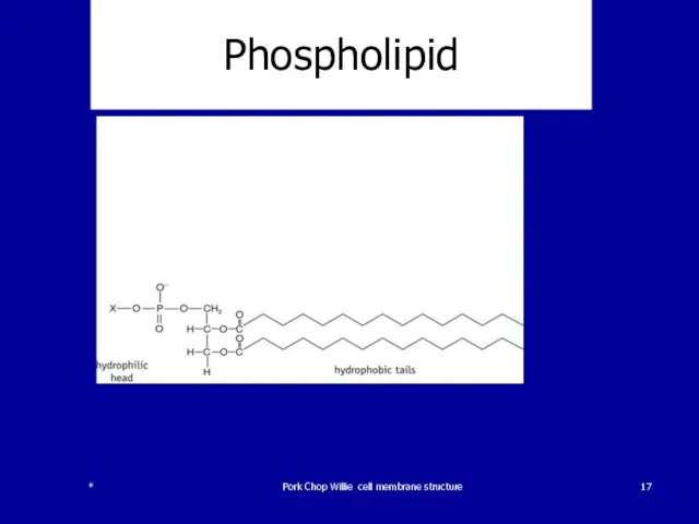
Слайд 17Phospholipid
*
Pork Chop Willie cell membrane structure
Phospholipid
*
Pork Chop Willie cell membrane structure
Слайд 18Phospholipid bilayer
Phophate head of the molecule is polar; one end is
Phospholipid bilayer
Phophate head of the molecule is polar; one end is
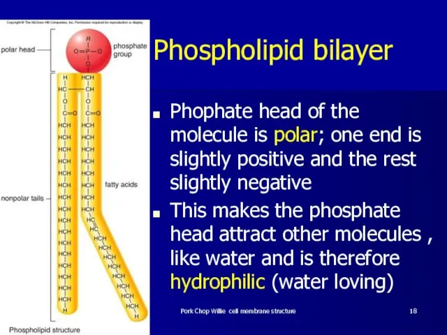
Слайд 19Phospholipid bilayer 2
Fats and water don’t mix
When added to water phospholipids arrange
Phospholipid bilayer 2
Fats and water don’t mix
When added to water phospholipids arrange
Слайд 20Phospholipid bilayer 3
They form a layer on the surface with their hydrophobic
Phospholipid bilayer 3
They form a layer on the surface with their hydrophobic
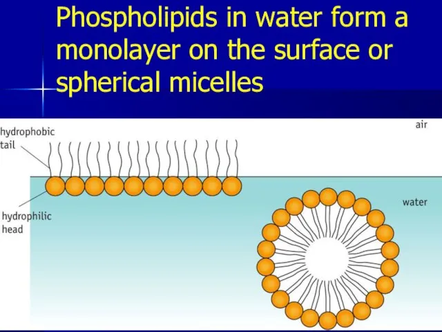
Слайд 21Phospholipids in water form a monolayer on the surface or spherical micelles
*
Pork
Phospholipids in water form a monolayer on the surface or spherical micelles
*
Pork
Слайд 22Phospholipid 4
Cells are filled with a watery or aqueous cytoplasm and are
Phospholipid 4
Cells are filled with a watery or aqueous cytoplasm and are
Слайд 23Phospholipid
Pork Chop Willie cell membrane structure
Phospholipid
Pork Chop Willie cell membrane structure
Слайд 24Phospholipid 5
This arrangement avoids the hydrophobic fatty acid tails having any contact
Phospholipid 5
This arrangement avoids the hydrophobic fatty acid tails having any contact
Слайд 25Phospholipids
*
Pork Chop Willie cell membrane structure
Phospholipids
*
Pork Chop Willie cell membrane structure
Слайд 26Fluid-Mosaic Model 1
The cell surface membrane is not just a phospholipid bilayer
It
Fluid-Mosaic Model 1
The cell surface membrane is not just a phospholipid bilayer
It
Слайд 27Fluid-Mosaic Model 2
Some of the proteins span the layer
Other proteins are found
Fluid-Mosaic Model 2
Some of the proteins span the layer
Other proteins are found
Слайд 28Fluid-Mosaic Model 3
It is thought that some of the proteins are fixed
Fluid-Mosaic Model 3
It is thought that some of the proteins are fixed
Слайд 29Fluid Mosaic Model
*
Pork Chop Willie cell membrane structure
Fluid Mosaic Model
*
Pork Chop Willie cell membrane structure
Слайд 30Evidence for the model 1
The most widely accepted model until the early
Evidence for the model 1
The most widely accepted model until the early
Слайд 31(A) Phospholipid sandwich model
(B) in the Fluid mosaic integral protein have
(A) Phospholipid sandwich model (B) in the Fluid mosaic integral protein have

Слайд 32Evidence for the model 2
Experiments showed that there were two types of
Evidence for the model 2
Experiments showed that there were two types of
Слайд 33Evidence for the model 3
Several integral proteins were shown to have regions
Evidence for the model 3
Several integral proteins were shown to have regions
Слайд 34(A) Phospholipid sandwich model
(B) in the Fluid mosaic integral protein have
(A) Phospholipid sandwich model (B) in the Fluid mosaic integral protein have
Слайд 35Evidence for the model 4
Additional evidence for integral proteins came from freeze-fracture
Evidence for the model 4
Additional evidence for integral proteins came from freeze-fracture
Слайд 36Freeze-fracture of membrane revealing intregral proteins
*
Pork Chop Willie cell membrane structure
Freeze-fracture of membrane revealing intregral proteins
*
Pork Chop Willie cell membrane structure
Слайд 37Evidence for the model 5
Fusion of mouse cells with human cells
Before cells
Evidence for the model 5
Fusion of mouse cells with human cells
Before cells
Слайд 38Movement of membrane Proteins within cell surface membranes
Pork Chop Willie cell membrane
Movement of membrane Proteins within cell surface membranes
Pork Chop Willie cell membrane
Слайд 39Membrane Protein Diversity
Membrane Protein Diversity
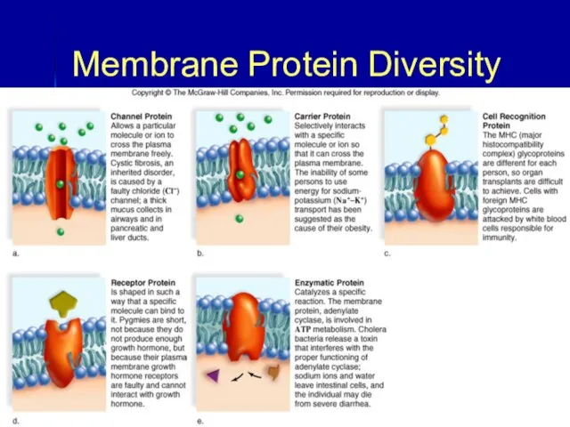
Слайд 40Functions of Membrane Proteins
Channel Proteins:
Tubular
Allow passage of molecules through membrane
Carrier Proteins:
Combine with
Functions of Membrane Proteins
Channel Proteins:
Tubular
Allow passage of molecules through membrane
Carrier Proteins:
Combine with
Слайд 41More unsaturated phospholipids – more fluid
The more phospholipids containing unsaturated fatty acids
More unsaturated phospholipids – more fluid
The more phospholipids containing unsaturated fatty acids
Слайд 42Cholesterol
Cholesterol reduces the fluidity of the membrane by preventing movement of the
Cholesterol
Cholesterol reduces the fluidity of the membrane by preventing movement of the
Слайд 43Membrane Glycolipids
Glycolipids shown as blue sugar groups projecting into the extracellular
Membrane Glycolipids
Glycolipids shown as blue sugar groups projecting into the extracellular
Слайд 44Sphingolipid
Structural lipid of which the parent structure is sphingosine rather than
Sphingolipid
Structural lipid of which the parent structure is sphingosine rather than
Слайд 45Raft Model
*
Pork Chop Willie cell membrane structure
Raft Model
*
Pork Chop Willie cell membrane structure
Слайд 46Raft Model
Lipid rafts are possible island like structure present in cellular membranes.
Raft Model
Lipid rafts are possible island like structure present in cellular membranes.
Слайд 47Raft Model
The existence of lipid rafts in cell membrane has not yet
Raft Model
The existence of lipid rafts in cell membrane has not yet
Слайд 48*
Pork Chop Willie cell membrane structure
Q1
According to the fluid-mosaic model for the
*
Pork Chop Willie cell membrane structure
Q1
According to the fluid-mosaic model for the
Слайд 49*
Pork Chop Willie cell membrane structure
A1
According to the fluid-mosaic model for the
*
Pork Chop Willie cell membrane structure
A1
According to the fluid-mosaic model for the
Слайд 50Q2
Phospholipids have their hydrophilic polar heads facing the __________ and _____________fluid. The
Q2
Phospholipids have their hydrophilic polar heads facing the __________ and _____________fluid. The
Слайд 51A2
Phospholipids have their hydrophilic polar heads facing the intracellular and extracellular fluid.
A2
Phospholipids have their hydrophilic polar heads facing the intracellular and extracellular fluid.
Слайд 52Q3
The proteins found in the plasma membrane may be _________ proteins, which
Q3
The proteins found in the plasma membrane may be _________ proteins, which
Слайд 53A3
The proteins found in the plasma membrane may be integral proteins, which
A3
The proteins found in the plasma membrane may be integral proteins, which
Слайд 54Q 4
State two roles of cholesterol in the membrane (2 marks)
*
Pork Chop
Q 4
State two roles of cholesterol in the membrane (2 marks)
*
Pork Chop
Слайд 55A 4
State two roles of cholesterol in the membrane (2 marks)
Regulates membrane
A 4
State two roles of cholesterol in the membrane (2 marks)
Regulates membrane
Слайд 56Q5
There are many types of proteins in a membrane. Describe the role
Q5
There are many types of proteins in a membrane. Describe the role
 Понятие трудового договора, его стороны и значение. Содержание трудового договора, порядок заключения и расторжения
 Автоматизация р
 All kinds of animals
 Экономика государства
 Компьютерные презентации
 Селекция2
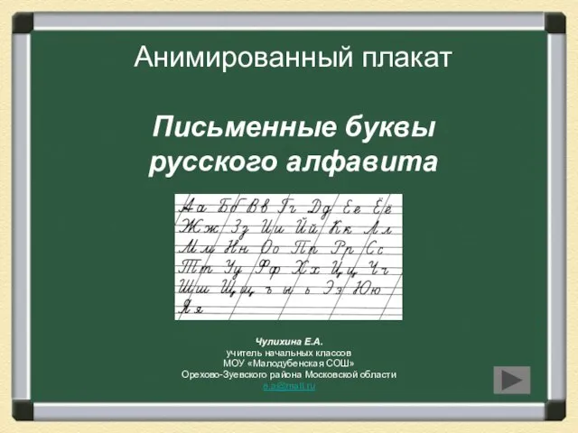 Презентация на тему Письменные буквы русского алфавита
  Суриков Сергей Григорьевич Ученик 9 б класса МБОУ СОШ № 9
  Художественное ремесло
 АСГОР «РГК»
 Знакомство с Богом
 Четыре живописца
 Терроризм как опаснейшее социально-политическое явление сегодняшнего мира
 Филиппины
  Программа поддержки многодетных семей в РФ
 Группа развития
 Презентация на тему Природные зоны Африки
  Тема доклада: «Обязательные виды страхования - драйвер роста или тупиковый путь развития?»
 Профориентационная работа в условиях школы-интерната
 Подводная угадайка
 Hafta 3-2Menderes Dönemi
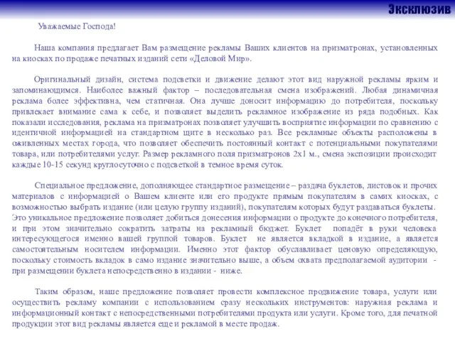 Эксклюзив
 ВРЕМЕНА ГОДА SEASONS
  Состав рабочих групп проектов окружного методического совета
 Презентация на тему Семейство Губоцветные
 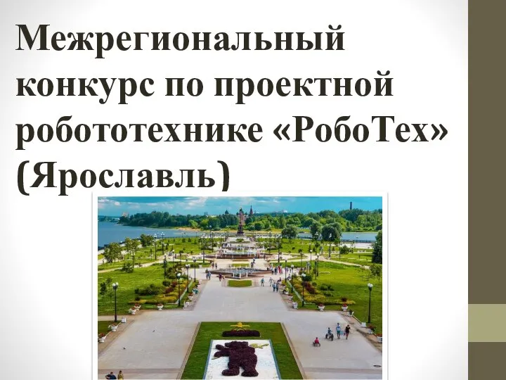 Межрегиональный конкурс по проектной робототехнике РобоТех (Ярославль)
 О результатах государственной (итоговой) аттестации выпускников IX классов общеобразовательных учреждений, организуемой региона
 презентация