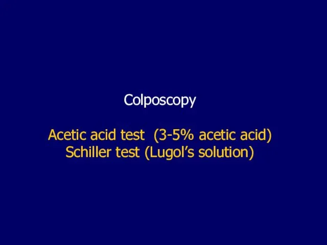
- Colposcopy Acetic acid test

Содержание
- 2. Colposcopy Acetic acid test (3-5% acetic acid) Schiller test (Lugol’s solution)
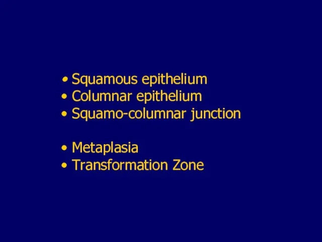
- 5. Squamous epithelium Columnar epithelium Squamo-columnar junction Metaplasia Transformation Zone
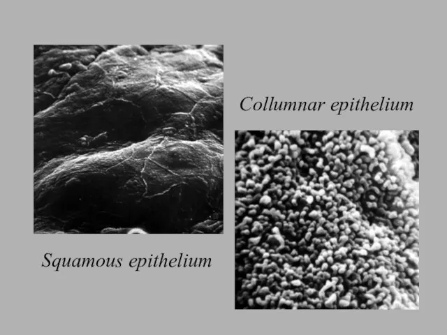
- 6. Squamous epithelium Collumnar epithelium
- 7. Squamo-collumnar junction- SCJ
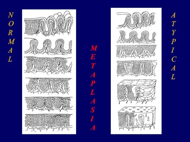
- 11. Metaplasia a physiological and benign process whereby the columnar epithelium is gradually replaced by squamous epithelium
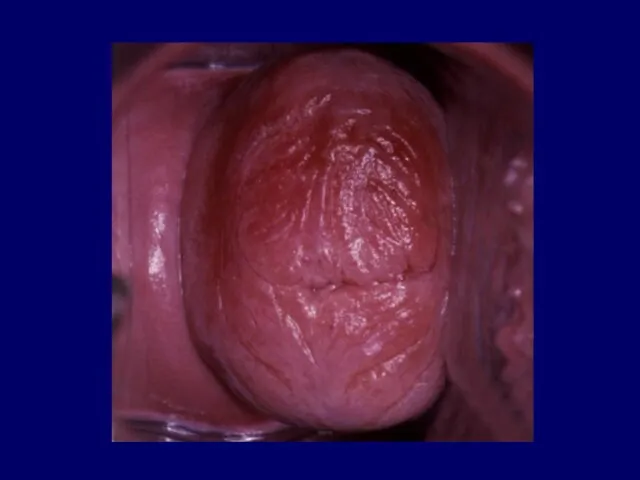
- 13. The result of normal metaplasia is a normal Transformation zone
- 15. Immature metaplastic cells are susceptible to the development of atypical cellular changes
- 16. The process of transformation from normal cells to atypical cells occurs under the influence of Human
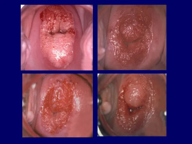
- 17. If atypical metaplasia takes place an abnormal Transformation zone develops
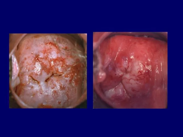
- 19. N O R M A L M E T A P L A S I A
- 20. In colposcopy, it is essential to asses whether Transformation zone is normal or abnormal
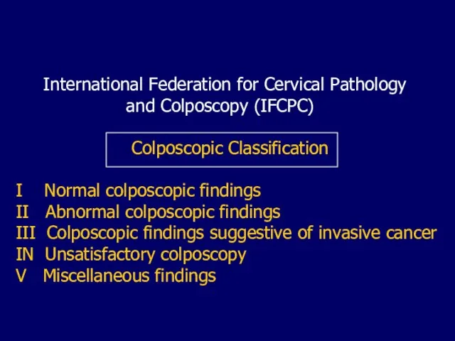
- 22. International Federation for Cervical Pathology and Colposcopy (IFCPC) Colposcopic Classification I Normal colposcopic findings II Abnormal
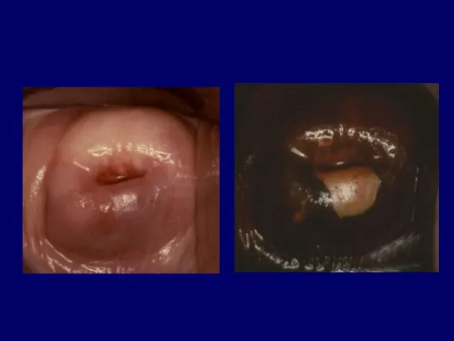
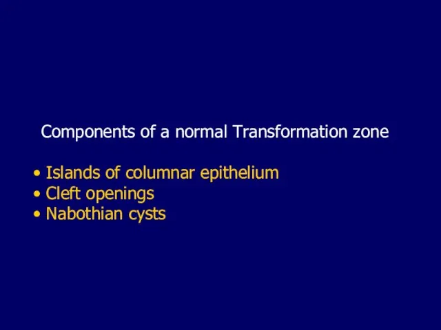
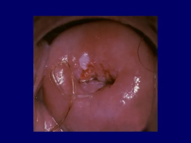
- 23. Components of a normal Transformation zone Islands of columnar epithelium Cleft openings Nabothian cysts
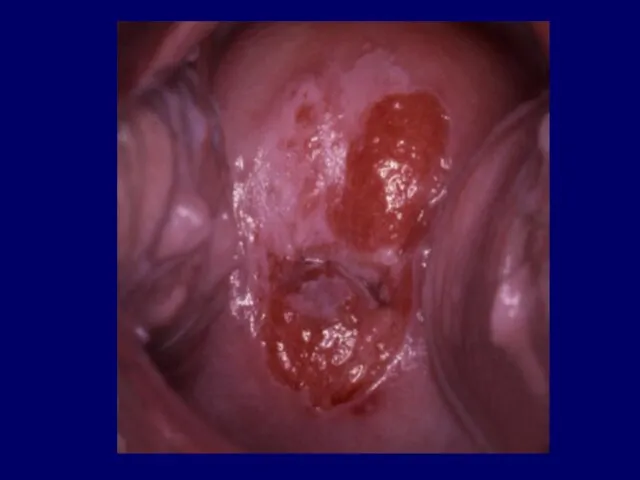
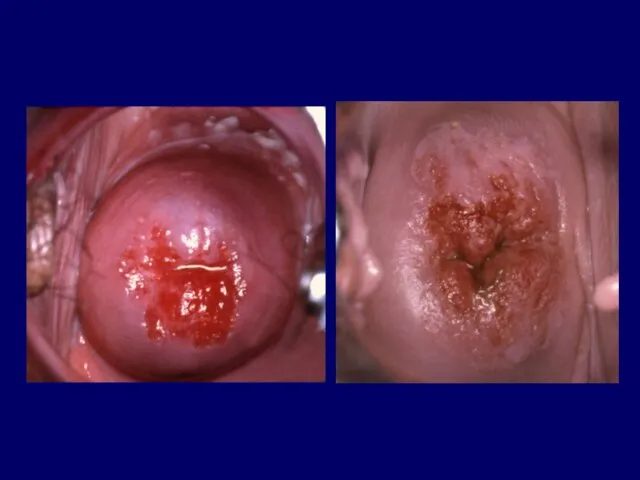
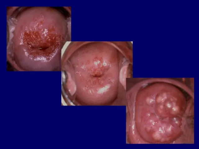
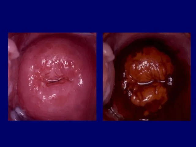
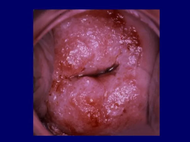
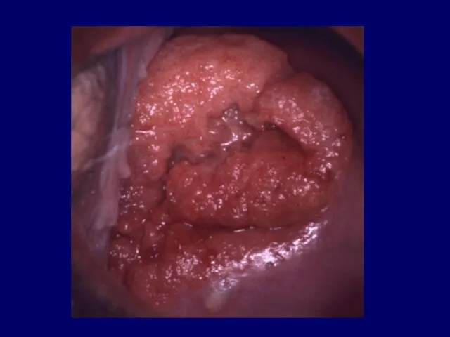
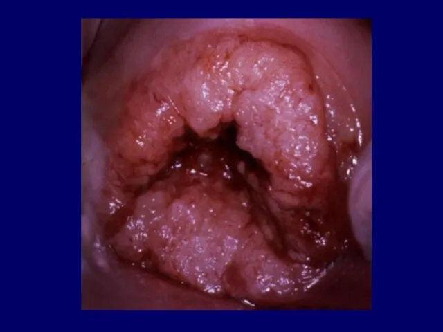
- 25. The abnormal Transformation zone is manifested as a wide spectrum of epithelial and vascular findings
- 26. Abnormal transformation zone is presented by abnormal (atypical) colposcopic findings
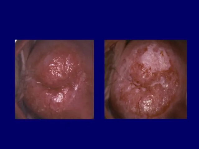
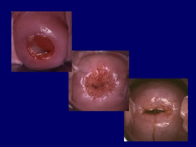
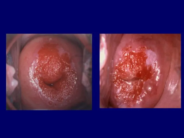
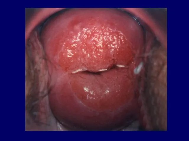
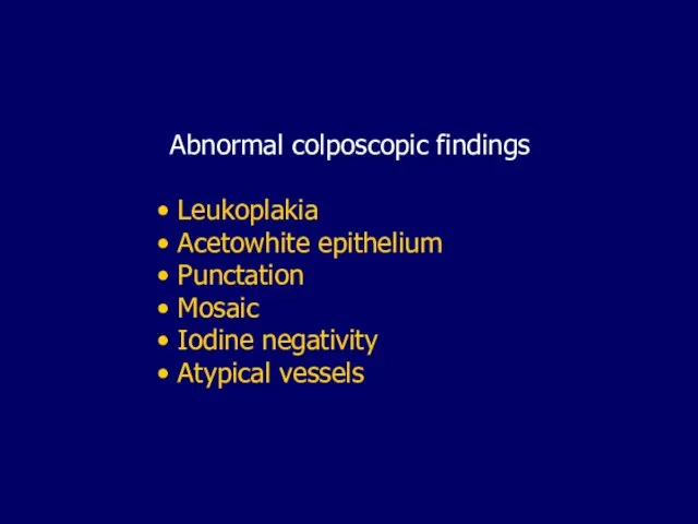
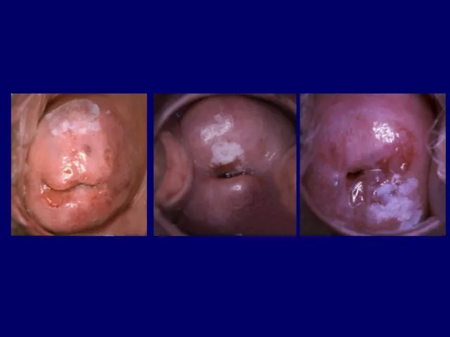
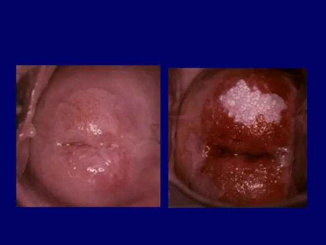
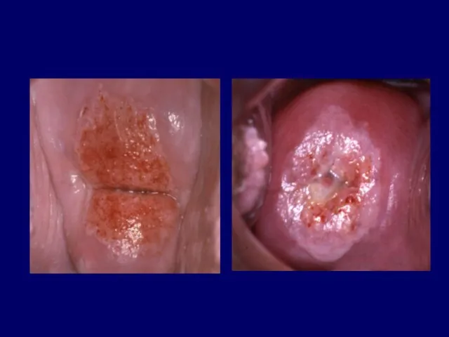
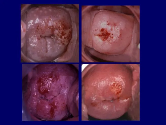
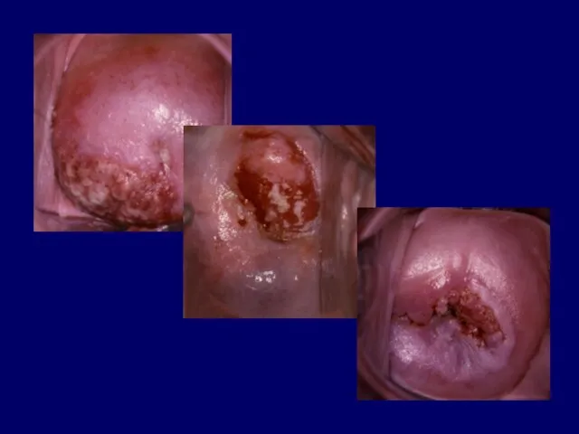
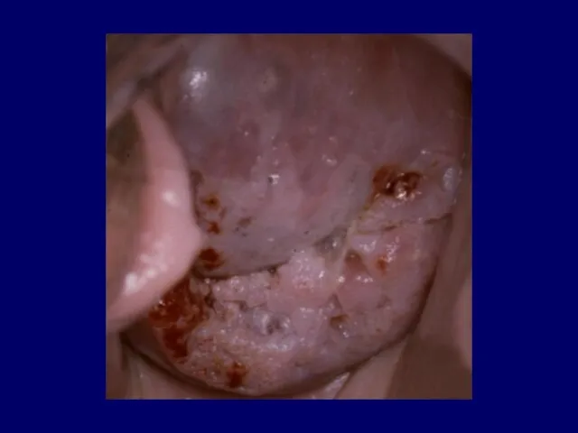
- 27. Abnormal colposcopic findings Leukoplakia Acetowhite epithelium Punctation Mosaic Iodine negativity Atypical vessels
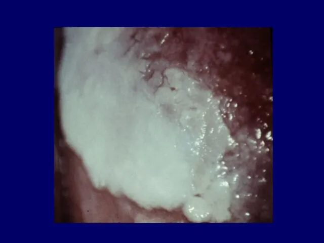
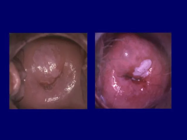
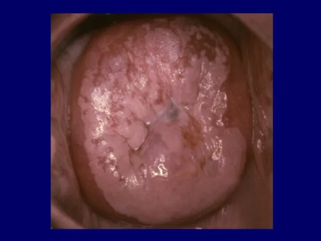
- 28. Leukoplakia or white plaque is visible grossly as a white often raised area that is not
- 33. Leukoplakia HPV infection Keratinizing CIN Keratinizing cancer Chronic trauma Radiotherapy Immature metaplasia
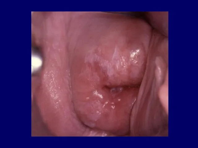
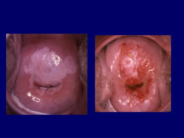
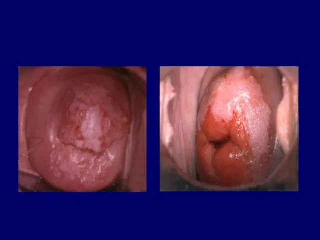
- 34. Acetowhite epitehlium Appears grossly normal but turns white after application of 3% to 5% acetic acid
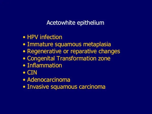
- 37. Acetowhite epithelium HPV infection Immature squamous metaplasia Regenerative or reparative changes Congenital Transformation zone Inflammation CIN
- 38. Any cells with an enlarged nucleus such as metaplatic cells or cells traumatized by infection or
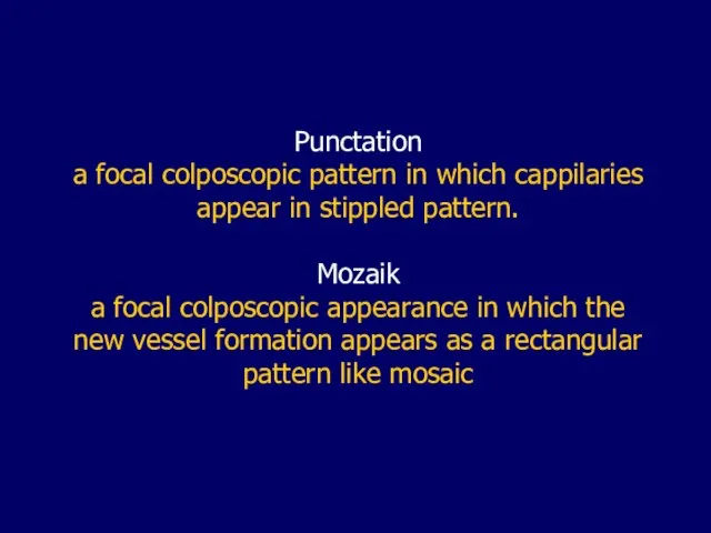
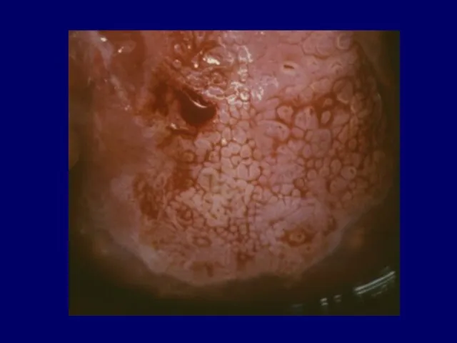
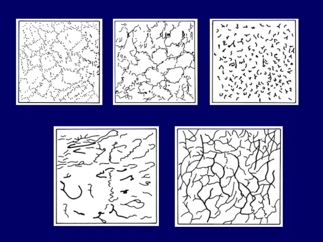
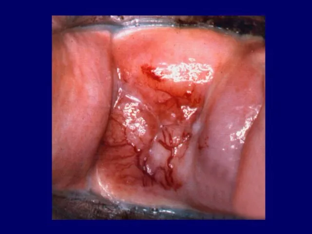
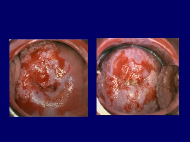
- 39. Punctation a focal colposcopic pattern in which cappilaries appear in stippled pattern. Mozaik a focal colposcopic
- 40. Punctation colposcopic finding reflecting the capillaries in the stromal papillae that are seen end-on and penetrate
- 43. Mosaic colposcopic finding reflecting the islands of squamous epithelium, encircled by blood vessels in a basket-like
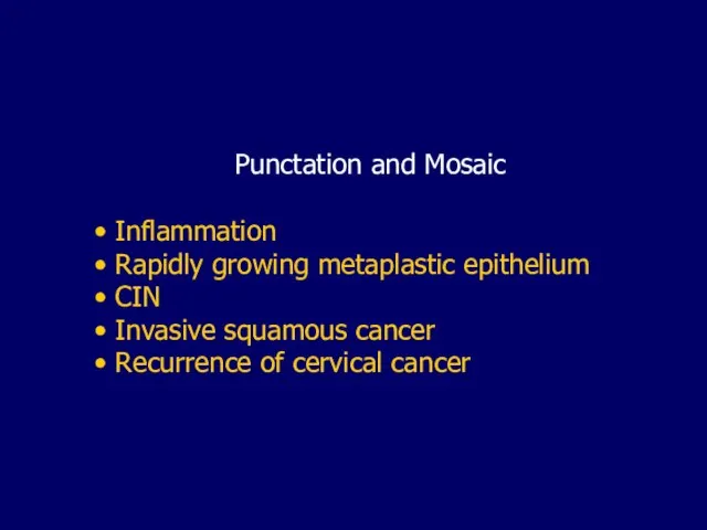
- 46. Punctation and Mosaic Inflammation Rapidly growing metaplastic epithelium CIN Invasive squamous cancer Recurrence of cervical cancer
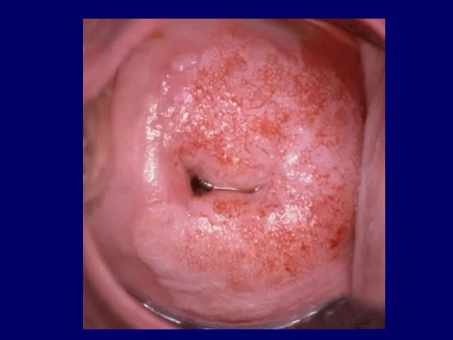
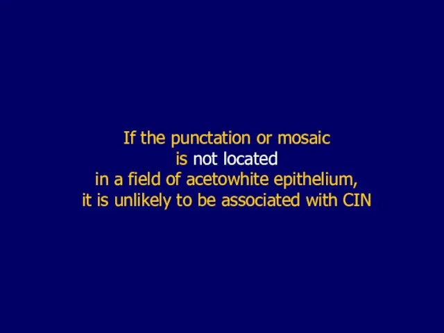
- 47. If the punctation or mosaic is not located in a field of acetowhite epithelium, it is
- 48. Iodine negativity Immature metaplasia Cervical intraepithelial neoplasia Low estrogen status (atrophy)
- 49. Atypical vessels Irregular vessels with an abrupt and interrupted course Appearing as commas, corkscrw capillaries or
- 50. Atypical vessels are the hallmark of invasion, but can be associated with other conditions such as
- 54. Development of abnormal colposcopic features may be the result of: Immature physiologic metaplasia Papilloma virus infection
- 55. Colposcopic index (score) a grading system used to evaluate the severity of the colposocpic findings
- 56. A number of scoring systems have been introduced: Coppleson & Pixley Burghardt Rubin & Barbo Reid
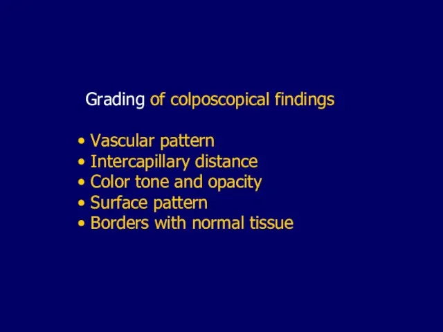
- 57. Grading of colposcopical findings Vascular pattern Intercapillary distance Color tone and opacity Surface pattern Borders with
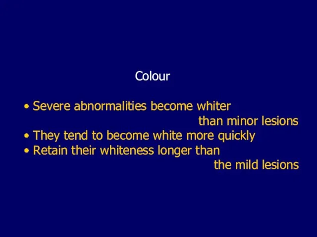
- 58. Colour Severe abnormalities become whiter than minor lesions They tend to become white more quickly Retain
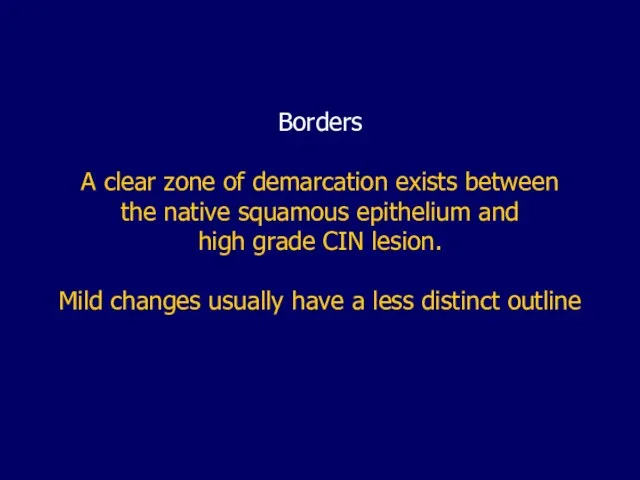
- 60. Borders A clear zone of demarcation exists between the native squamous epithelium and high grade CIN
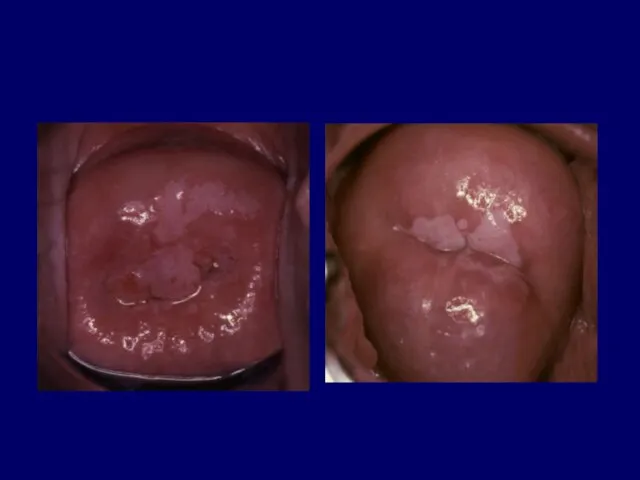
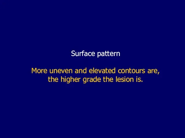
- 62. Surface pattern More uneven and elevated contours are, the higher grade the lesion is.
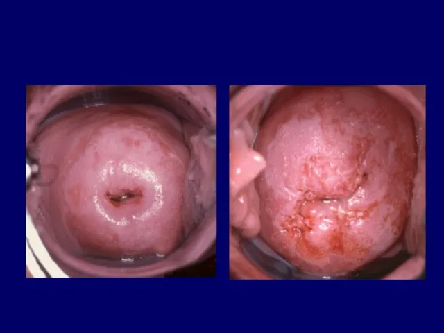
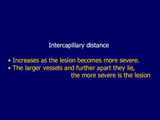
- 64. Intercapillary distance Increases as the lesion becomes more severe. The larger vessels and further apart they
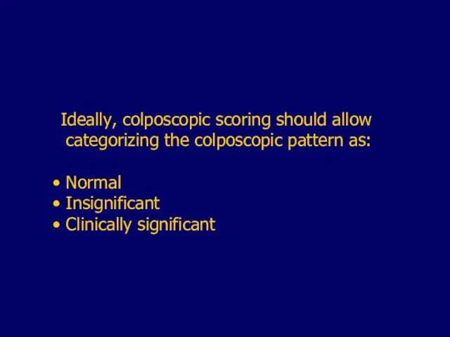
- 67. Ideally, colposcopic scoring should allow categorizing the colposcopic pattern as: Normal Insignificant Clinically significant
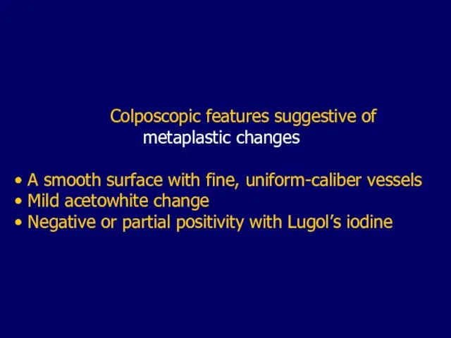
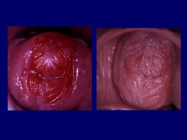
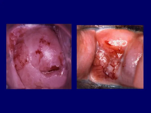
- 69. Colposcopic features suggestive of metaplastic changes A smooth surface with fine, uniform-caliber vessels Mild acetowhite change
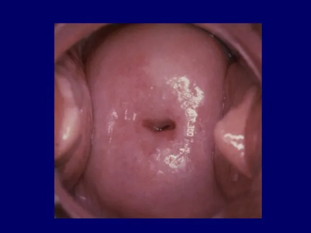
- 73. As the metaplastic cells transform into mature squamous cells, the coloration is indistinquisable from the mature
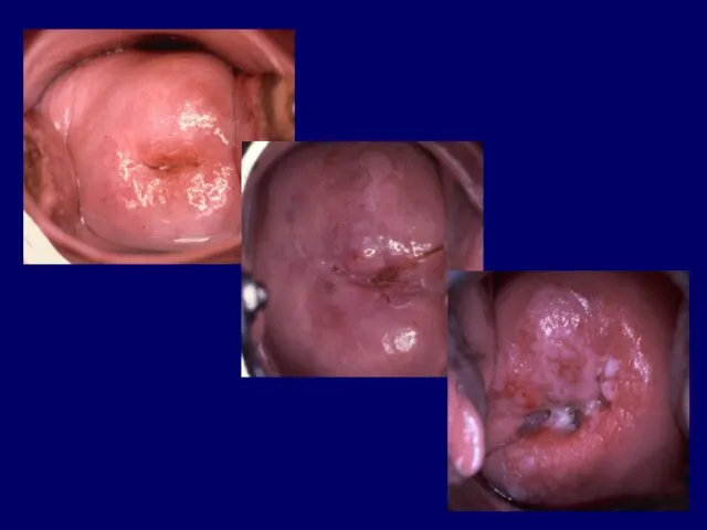
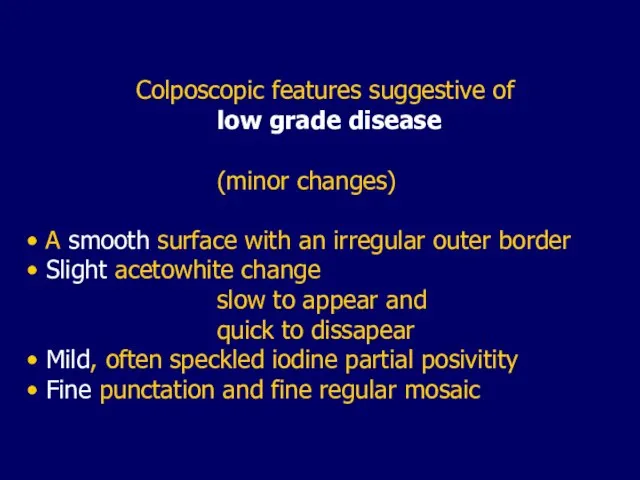
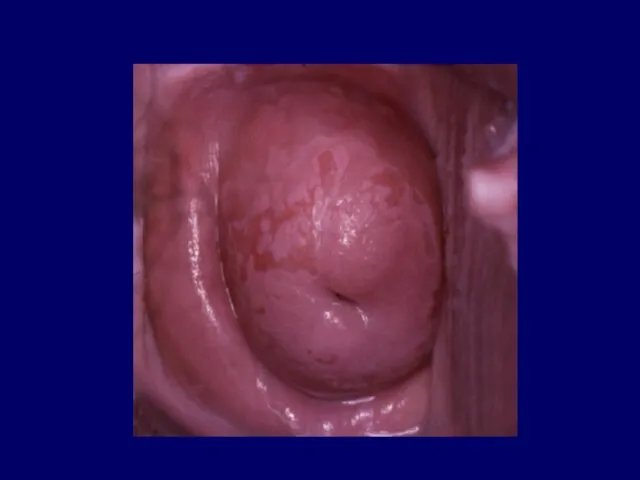
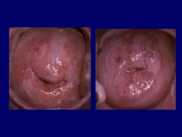
- 75. Colposcopic features suggestive of low grade disease (minor changes) A smooth surface with an irregular outer
- 78. The subtle differences between the features of squamous metaplasia and those of low-grade intraepithelial lesions make
- 80. It is easier to determine that a cervix is either normal or very abnormal, than it
- 81. Misinterpretation of trivial changes as atypical findings can lead to mismanagement and overtreatment of the patient
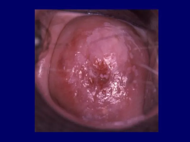
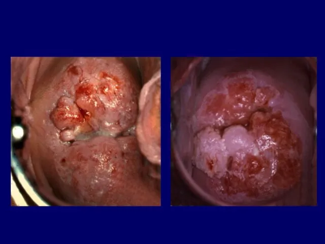
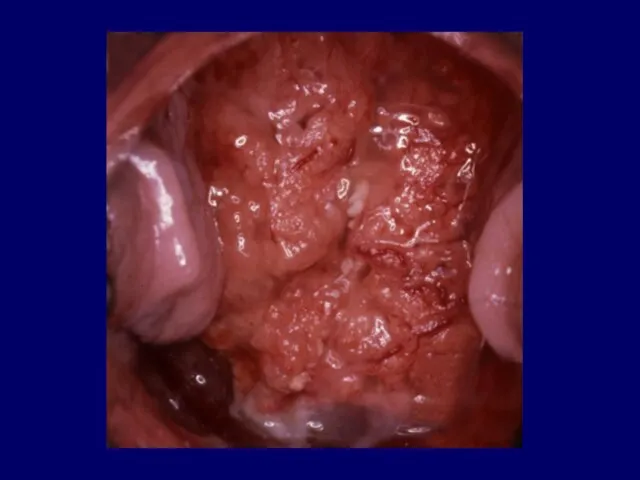
- 82. Colposcopic features suggestive of high- grade disease (major changes) A generally smooth surface with sharp outer
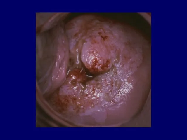
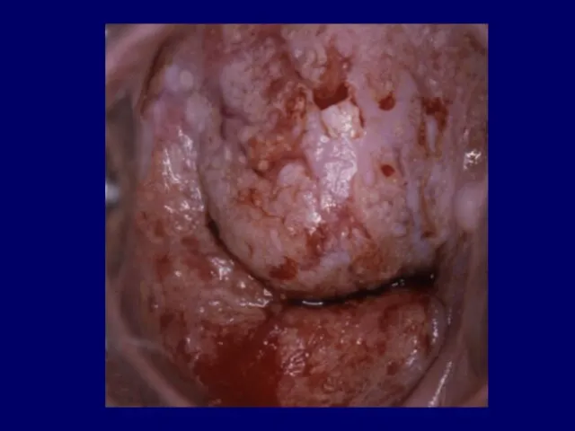
- 86. Signs of microinvasion Yellow discoloration Ulceration Thickened areas Nodularity Abnormal vascularity Rapid increase in size
- 89. There is a direct relationship between the size of a lesion and the likelihood of invasion
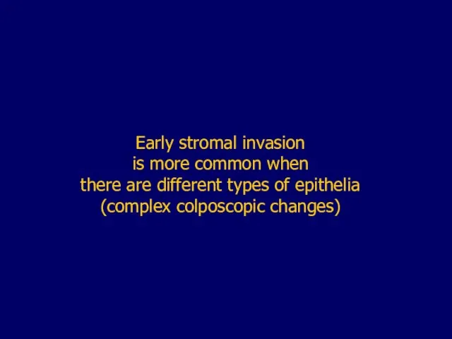
- 91. Early stromal invasion is more common when there are different types of epithelia (complex colposcopic changes)
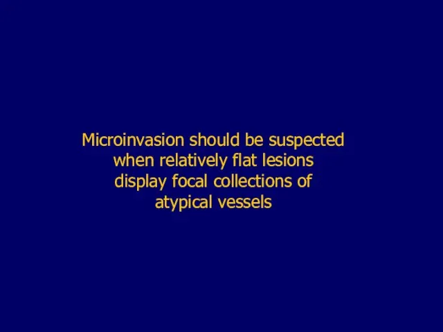
- 94. Microinvasion should be suspected when relatively flat lesions display focal collections of atypical vessels
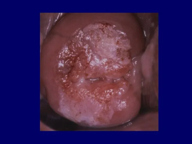
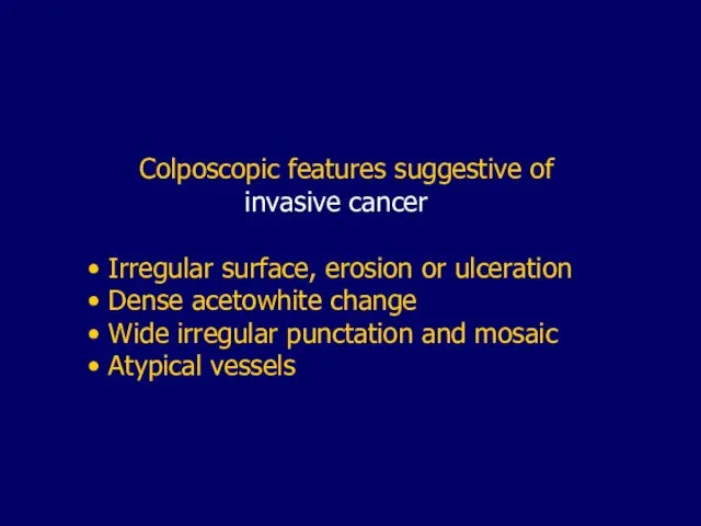
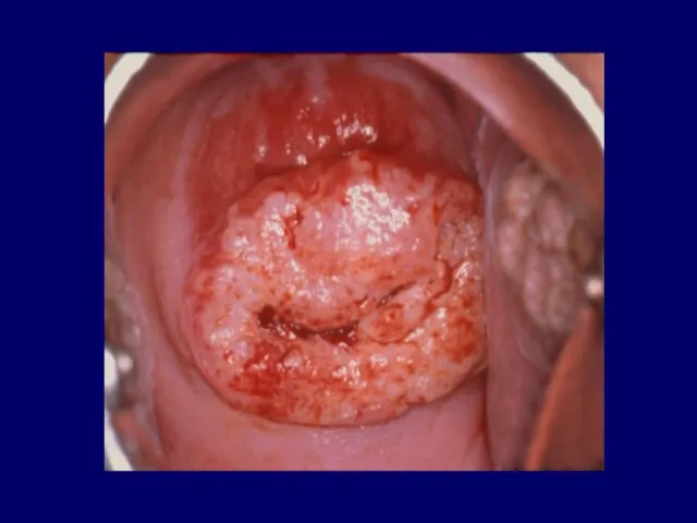
- 96. Colposcopic features suggestive of invasive cancer Irregular surface, erosion or ulceration Dense acetowhite change Wide irregular
- 105. In most cases biopsy is mandatory to establish the correct diagnosis
- 107. Скачать презентацию
Слайд 5 Squamous epithelium
Columnar epithelium
Squamo-columnar junction
Metaplasia
Transformation Zone
Squamous epithelium
Columnar epithelium
Squamo-columnar junction
Metaplasia
Transformation Zone
Слайд 6Squamous epithelium
Collumnar epithelium
Squamous epithelium
Collumnar epithelium
Слайд 7Squamo-collumnar
junction- SCJ
Squamo-collumnar
junction- SCJ

Слайд 11Metaplasia
a physiological and benign process
whereby the columnar epithelium is gradually
replaced by squamous
Metaplasia
a physiological and benign process
whereby the columnar epithelium is gradually
replaced by squamous
Слайд 13The result of normal metaplasia is
a normal Transformation zone
The result of normal metaplasia is
a normal Transformation zone

Слайд 15Immature metaplastic cells are
susceptible
to the development of
atypical cellular changes
Immature metaplastic cells are
susceptible
to the development of
atypical cellular changes
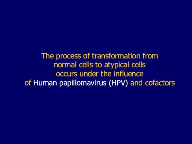
Слайд 16The process of transformation from
normal cells to atypical cells
occurs under
The process of transformation from
normal cells to atypical cells
occurs under
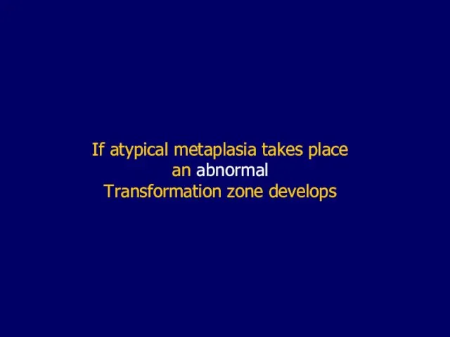
Слайд 17If atypical metaplasia takes place
an abnormal
Transformation zone develops
If atypical metaplasia takes place
an abnormal
Transformation zone develops
Слайд 19N
O
R
M
A
L
M
E
T
A
P
L
A
S
I
A
A
T
Y
P
I
C
A
L
N
O
R
M
A
L
M
E
T
A
P
L
A
S
I
A
A
T
Y
P
I
C
A
L
Слайд 20In colposcopy, it is essential to asses
whether Transformation zone is
normal
In colposcopy, it is essential to asses
whether Transformation zone is
normal
Слайд 22 International Federation for Cervical Pathology
and Colposcopy (IFCPC)
Colposcopic Classification
I Normal
International Federation for Cervical Pathology
and Colposcopy (IFCPC)
Colposcopic Classification
I Normal
Слайд 23Components of a normal Transformation zone
Islands of columnar epithelium
Cleft openings
Components of a normal Transformation zone
Islands of columnar epithelium
Cleft openings
Слайд 25The abnormal Transformation zone is manifested
as a wide spectrum of
epithelial and
The abnormal Transformation zone is manifested
as a wide spectrum of
epithelial and
Слайд 26Abnormal transformation zone is
presented by
abnormal (atypical) colposcopic findings
Abnormal transformation zone is
presented by
abnormal (atypical) colposcopic findings
Слайд 27Abnormal colposcopic findings
Leukoplakia
Acetowhite epithelium
Punctation
Mosaic
Iodine negativity
Atypical
Abnormal colposcopic findings
Leukoplakia
Acetowhite epithelium
Punctation
Mosaic
Iodine negativity
Atypical
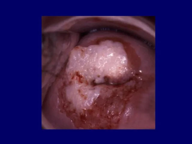
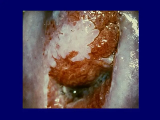
Слайд 28Leukoplakia
or white plaque
is visible grossly as a white often raised
area
Leukoplakia
or white plaque
is visible grossly as a white often raised
area
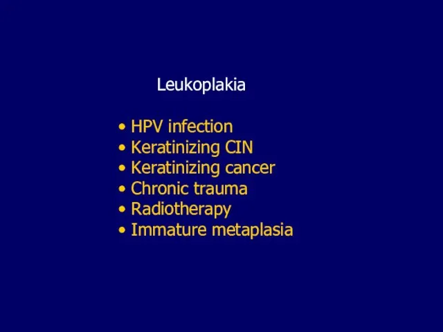
Слайд 33 Leukoplakia
HPV infection
Keratinizing CIN
Keratinizing cancer
Chronic trauma
Radiotherapy
Leukoplakia
HPV infection
Keratinizing CIN
Keratinizing cancer
Chronic trauma
Radiotherapy
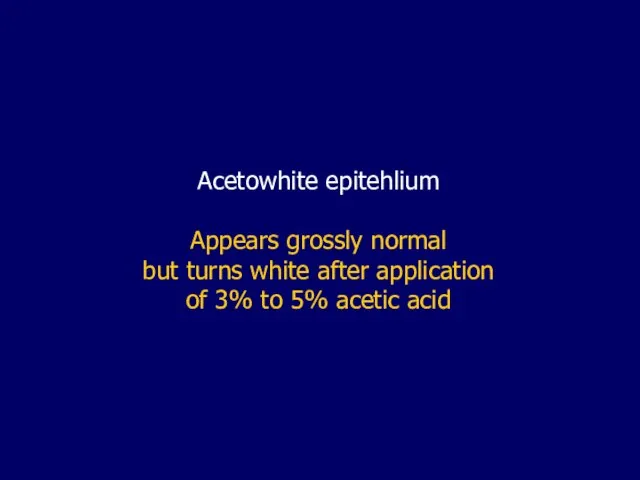
Слайд 34Acetowhite epitehlium
Appears grossly normal
but turns white after application
of 3% to 5%
Acetowhite epitehlium
Appears grossly normal
but turns white after application
of 3% to 5%
Слайд 37 Acetowhite epithelium
HPV infection
Immature squamous metaplasia
Regenerative or reparative changes
Acetowhite epithelium
HPV infection
Immature squamous metaplasia
Regenerative or reparative changes
Слайд 38Any cells with an enlarged nucleus
such as metaplatic cells or
cells
Any cells with an enlarged nucleus
such as metaplatic cells or
cells
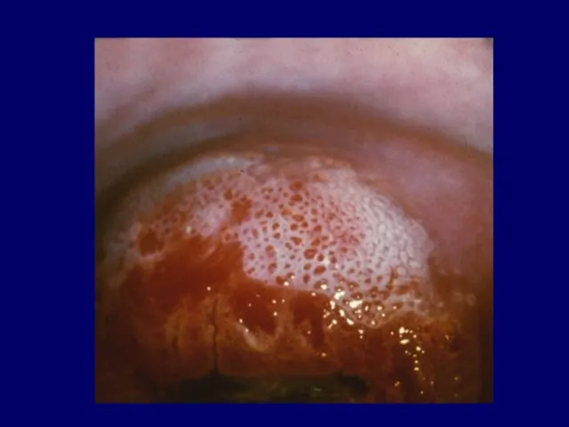
Слайд 39Punctation
a focal colposcopic pattern in which cappilaries
appear in stippled pattern.
Mozaik
a focal
Punctation
a focal colposcopic pattern in which cappilaries
appear in stippled pattern.
Mozaik
a focal
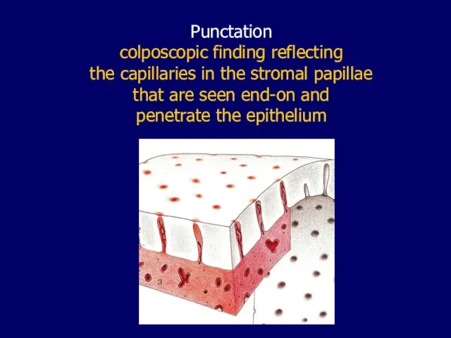
Слайд 40Punctation
colposcopic finding reflecting
the capillaries in the stromal papillae
that are
Punctation
colposcopic finding reflecting
the capillaries in the stromal papillae
that are
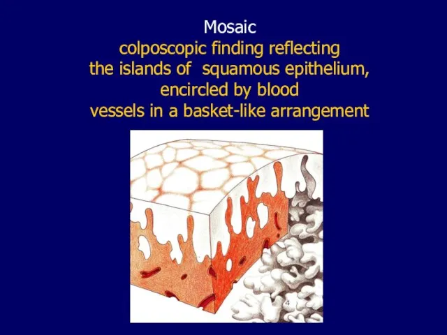
Слайд 43Mosaic
colposcopic finding reflecting
the islands of squamous epithelium,
encircled by blood
vessels
Mosaic
colposcopic finding reflecting
the islands of squamous epithelium,
encircled by blood
vessels
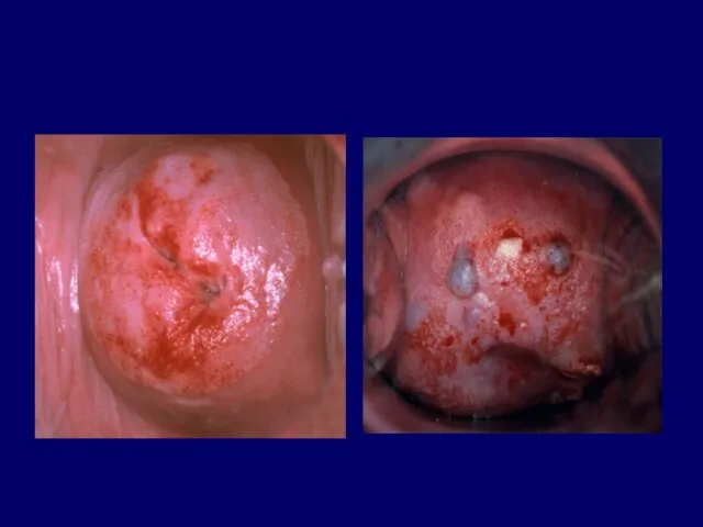
Слайд 46 Punctation and Mosaic
Inflammation
Rapidly growing metaplastic epithelium
CIN
Invasive squamous
Punctation and Mosaic
Inflammation
Rapidly growing metaplastic epithelium
CIN
Invasive squamous
Слайд 47If the punctation or mosaic
is not located
in a field of
If the punctation or mosaic
is not located
in a field of
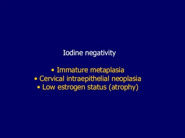
Слайд 48Iodine negativity
Immature metaplasia
Cervical intraepithelial neoplasia
Low estrogen status (atrophy)
Iodine negativity
Immature metaplasia
Cervical intraepithelial neoplasia
Low estrogen status (atrophy)
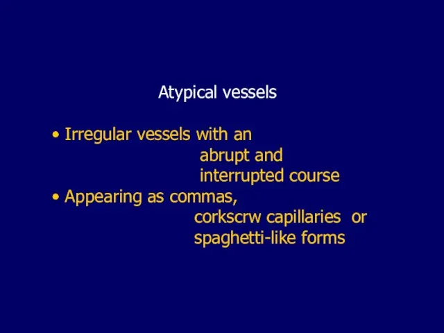
Слайд 49 Atypical vessels
Irregular vessels with an
abrupt and
interrupted
Atypical vessels
Irregular vessels with an
abrupt and
interrupted
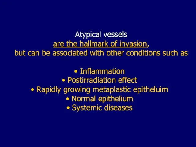
Слайд 50Atypical vessels
are the hallmark of invasion,
but can be associated with other
Atypical vessels
are the hallmark of invasion,
but can be associated with other
Слайд 54Development of abnormal
colposcopic features may be the
result of:
Immature physiologic
Development of abnormal
colposcopic features may be the
result of:
Immature physiologic
Слайд 55Colposcopic index (score)
a grading system used to evaluate the severity
of
Colposcopic index (score)
a grading system used to evaluate the severity
of
Слайд 56A number of scoring systems
have been introduced:
Coppleson & Pixley
A number of scoring systems
have been introduced:
Coppleson & Pixley

Слайд 57Grading of colposcopical findings
Vascular pattern
Intercapillary distance
Color tone
Grading of colposcopical findings
Vascular pattern
Intercapillary distance
Color tone
Слайд 58 Colour
Severe abnormalities become whiter
than minor lesions
They tend
Colour
Severe abnormalities become whiter
than minor lesions
They tend
Слайд 60Borders
A clear zone of demarcation exists between
the native squamous epithelium and
Borders
A clear zone of demarcation exists between
the native squamous epithelium and
Слайд 62Surface pattern
More uneven and elevated contours are,
the higher grade the lesion
Surface pattern
More uneven and elevated contours are,
the higher grade the lesion
Слайд 64 Intercapillary distance
Increases as the lesion becomes more severe.
The larger vessels
Intercapillary distance
Increases as the lesion becomes more severe.
The larger vessels
Слайд 67Ideally, colposcopic scoring should allow
categorizing the colposcopic pattern as:
Normal
Insignificant
Ideally, colposcopic scoring should allow
categorizing the colposcopic pattern as:
Normal
Insignificant
Слайд 69 Colposcopic features suggestive of
metaplastic changes
A smooth surface with
Colposcopic features suggestive of
metaplastic changes
A smooth surface with
Слайд 73As the metaplastic cells transform into
mature squamous cells,
the coloration is
As the metaplastic cells transform into
mature squamous cells,
the coloration is
Слайд 75 Colposcopic features suggestive of
low grade disease
(minor changes)
A
Colposcopic features suggestive of
low grade disease
(minor changes)
A
Слайд 78The subtle differences between the features of
squamous metaplasia and those of low-grade
The subtle differences between the features of
squamous metaplasia and those of low-grade
Слайд 80It is easier to determine that a cervix is
either normal or
It is easier to determine that a cervix is
either normal or

Слайд 81Misinterpretation of trivial changes
as atypical findings can lead
to mismanagement and
overtreatment
Misinterpretation of trivial changes
as atypical findings can lead
to mismanagement and
overtreatment

Слайд 82 Colposcopic features suggestive of
high- grade disease
(major changes)
A
Colposcopic features suggestive of
high- grade disease
(major changes)
A
Слайд 86Signs of microinvasion
Yellow discoloration
Ulceration
Thickened areas
Nodularity
Abnormal vascularity
Signs of microinvasion
Yellow discoloration
Ulceration
Thickened areas
Nodularity
Abnormal vascularity

Слайд 89There is a direct relationship
between the size of a lesion and
the
There is a direct relationship
between the size of a lesion and
the

Слайд 91Early stromal invasion
is more common when
there are different types of epithelia
(complex
Early stromal invasion
is more common when
there are different types of epithelia
(complex
Слайд 94Microinvasion should be suspected
when relatively flat lesions
display focal collections of
atypical vessels
Microinvasion should be suspected
when relatively flat lesions
display focal collections of
atypical vessels
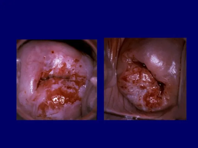
Слайд 96 Colposcopic features suggestive of
invasive cancer
Irregular surface, erosion or
Colposcopic features suggestive of
invasive cancer
Irregular surface, erosion or
Слайд 105In most cases
biopsy is mandatory to establish
the correct diagnosis
In most cases
biopsy is mandatory to establish
the correct diagnosis

 БАЗЫ ДАННЫХ
 Определение гидростатического давления
 НОУ ВПО «Волгоградский институт бизнеса» ЦЕНТР БЕСПЛАТНОЙ ЮРИДИЧЕСКОЙ ПОМОЩИ ЮРИДИЧЕСКАЯ КЛИНИКА ВОЛОНТЕРСКАЯ ОРГАНИЗАЦИЯ
 urok
urok ИНФОРМАЦИОННЫЕ ТЕХНОЛОГИИ
 Методы эффективного педагогического реагирования на факты проявления суицидального и деструктивного поведения обучающихся
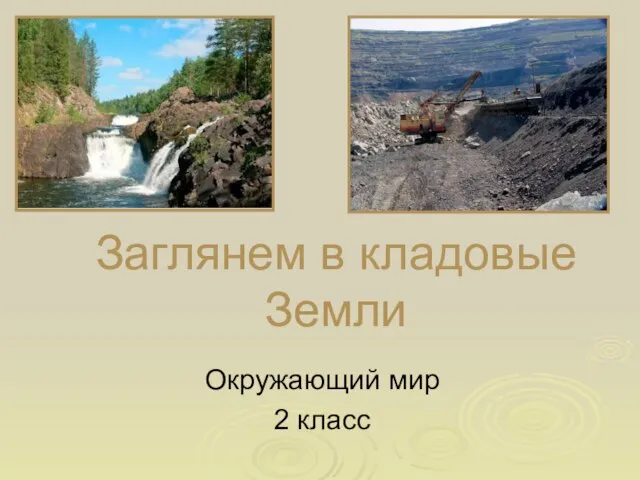 Заглянем в кладовые Земли
 Моя професія – різьбяр по дереву та бересті
 Кадастровая стоимость как база для расчета арендной платы за землю
 Директива Европейского Союза о потребительском кредитовании Правовое урегулирование в Европейском Союзе и его имплементация в
 Особенности культуры народов России
 Страховое право
 20141110_prezentatsiya5
 Графическое решение задач линейного программирования
 Презентация Microsoft PowerPoint
 Педагогический проект «Проектная деятельность как средство стимулирования познавательной активности младших школьников»
 Понятие о литературном портрете
  Прайс-лист на размещение рекламы
 Le corps humain
 Экономическая безопасность России Финансовая безопасность России
 Энергия связи. Дефект масс
 Николай Андреевич Римский-Корсаков (1844-1908)
 7. Монитор Хоара7.1. Общее описание
 Самопознание. Самооценка
 Значение птиц в природе и жизни человека
 Бессмертный взвод
 Ленивые голубцы
 Проект «Поклон ветеранам Великой Отечественной войны»