- Methods of Study of Nanosized Systems

Содержание
- 2. Objectives of lecture –presentation: Study of different methods of nanosystems investigations
- 3. Plan of lecture –presentation: Lecture 5 1. Method of Electron Microscopy 2. Sonde Microscopy 3. Diffraction
- 4. The basic methods of nanoparticles’ sizes and some properties in gaseous phase determination: ionization by photons
- 5. Methods of study of particles on the surface: X-ray and scanning electron microscopy (information about sizes/forms
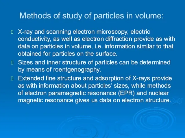
- 6. Methods of study of particles in volume: X-ray and scanning electron microscopy, electric conductivity, as well
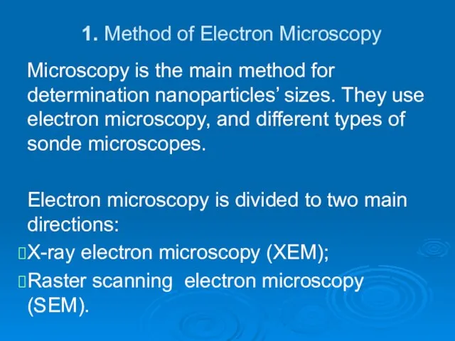
- 7. 1. Method of Electron Microscopy Microscopy is the main method for determination nanoparticles’ sizes. They use
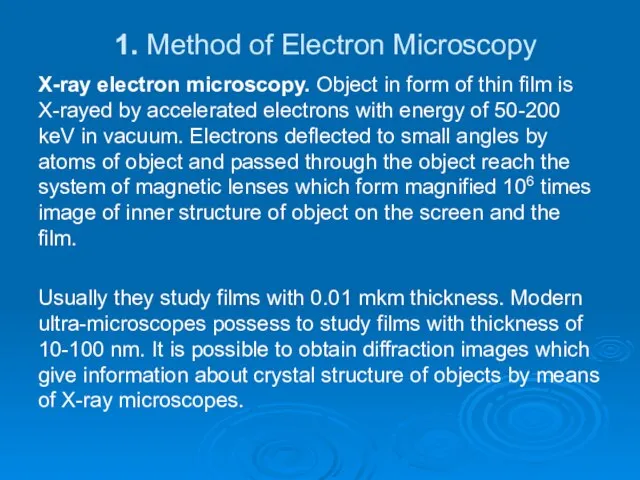
- 8. 1. Method of Electron Microscopy X-ray electron microscopy. Object in form of thin film is X-rayed
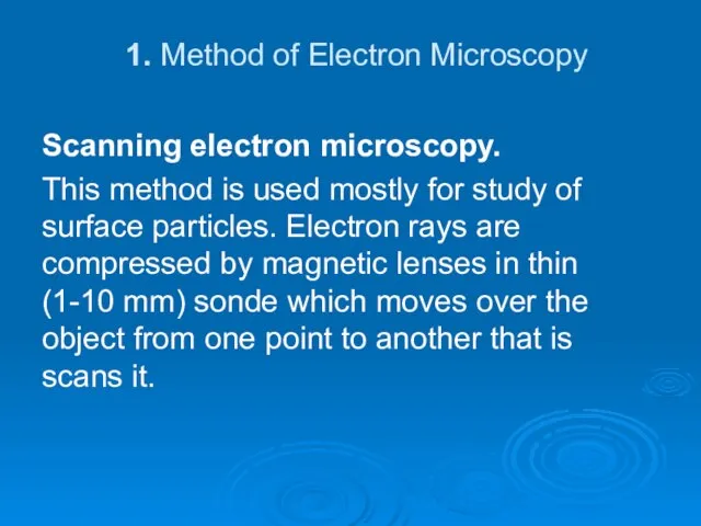
- 9. 1. Method of Electron Microscopy Scanning electron microscopy. This method is used mostly for study of
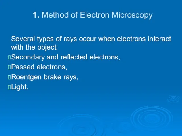
- 10. 1. Method of Electron Microscopy Several types of rays occur when electrons interact with the object:
- 11. 1. Method of Electron Microscopy The main value of this method – is that it is
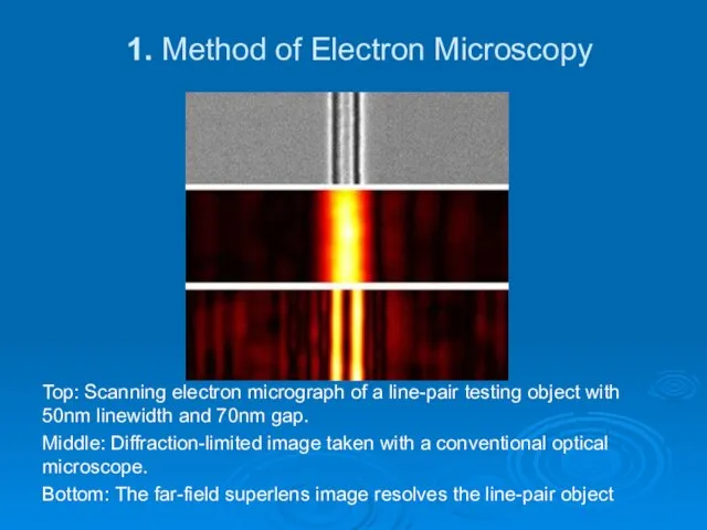
- 12. 1. Method of Electron Microscopy Top: Scanning electron micrograph of a line-pair testing object with 50nm
- 13. 2. Sonde Microscopy In 1981 Binnig and Rorer created scanning tunnel microscope (STM) and in 1986
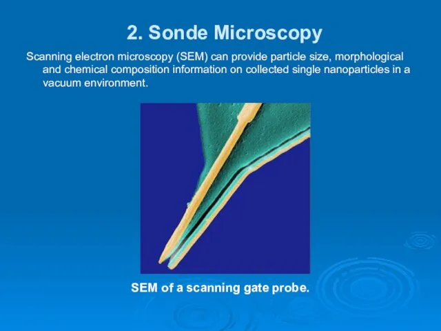
- 14. 2. Sonde Microscopy Scanning electron microscopy (SEM) can provide particle size, morphological and chemical composition information
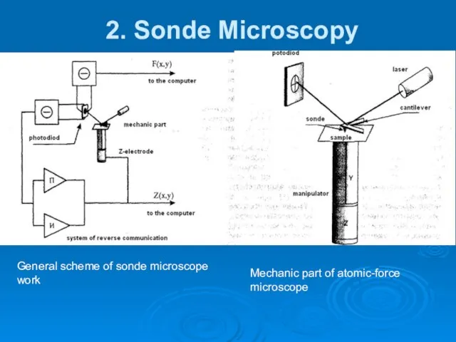
- 15. 2. Sonde Microscopy General scheme of sonde microscope work Mechanic part of atomic-force microscope
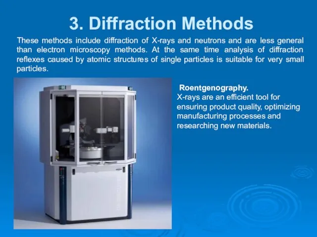
- 16. 3. Diffraction Methods . These methods include diffraction of X-rays and neutrons and are less general
- 17. 3. Diffraction Methods . Neutrons diffraction. Neutron is the particle which due to its properties is
- 18. Plan of lecture –presentation: Lecture 6 1. Mass-Spectrometry 2. Photoelectron Spectroscopy
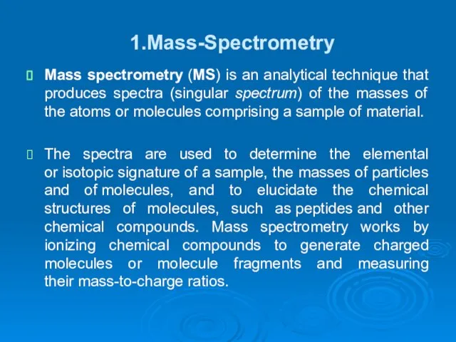
- 19. 1.Mass-Spectrometry Mass spectrometry (MS) is an analytical technique that produces spectra (singular spectrum) of the masses
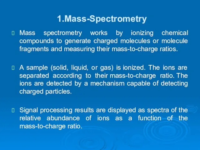
- 20. 1.Mass-Spectrometry Mass spectrometry works by ionizing chemical compounds to generate charged molecules or molecule fragments and
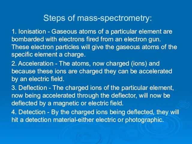
- 21. Steps of mass-spectrometry: 1. Ionisation - Gaseous atoms of a particular element are bombarded with electrons
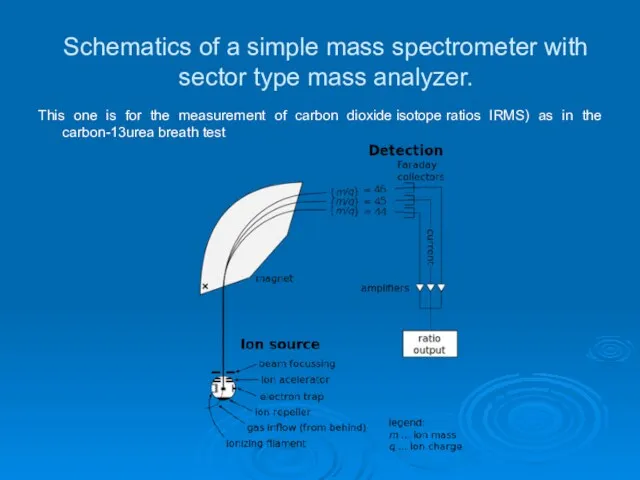
- 22. Schematics of a simple mass spectrometer with sector type mass analyzer. This one is for the
- 23. 2. Photoelectron Spectroscopy This method is based on the measurement of spectrum energies of electrons which
- 24. 2. Photoelectron Spectroscopy They determine energies of interactions of electrons and their energy levels in studied
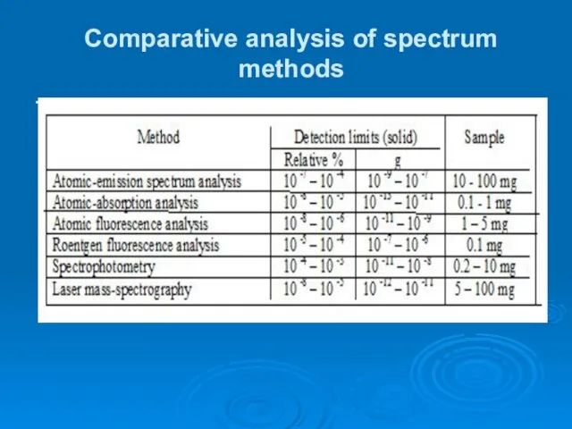
- 25. Comparative analysis of spectrum methods .
- 26. Check Yourself 1. What is the most significant for study of chemical interactions? 2. What are
- 27. Check Yourself 13. What restricts the resolution of conventional lenses? 14. Describe the method suggested by
- 28. Check Yourself 22. Which are the main requirements for crystals to be studied by roentgenography method?
- 30. Скачать презентацию
Слайд 3Plan of lecture –presentation:
Lecture 5
1. Method of Electron Microscopy
2. Sonde Microscopy
3. Diffraction
Plan of lecture –presentation:
Lecture 5
1. Method of Electron Microscopy
2. Sonde Microscopy
3. Diffraction
Слайд 4The basic methods of nanoparticles’ sizes and some properties in gaseous phase
The basic methods of nanoparticles’ sizes and some properties in gaseous phase
Слайд 5Methods of study of particles on the surface:
X-ray and scanning electron microscopy
Methods of study of particles on the surface:
X-ray and scanning electron microscopy
Слайд 6Methods of study of particles in volume:
X-ray and scanning electron microscopy, electric
Methods of study of particles in volume:
X-ray and scanning electron microscopy, electric
Слайд 71. Method of Electron Microscopy
Microscopy is the main method for determination nanoparticles’
1. Method of Electron Microscopy
Microscopy is the main method for determination nanoparticles’
Слайд 81. Method of Electron Microscopy
X-ray electron microscopy. Object in form of thin
1. Method of Electron Microscopy
X-ray electron microscopy. Object in form of thin
Слайд 91. Method of Electron Microscopy
Scanning electron microscopy.
This method is used mostly for
1. Method of Electron Microscopy
Scanning electron microscopy.
This method is used mostly for
Слайд 101. Method of Electron Microscopy
Several types of rays occur when electrons interact
1. Method of Electron Microscopy
Several types of rays occur when electrons interact
Слайд 111. Method of Electron Microscopy
The main value of this method – is
1. Method of Electron Microscopy
The main value of this method – is
Слайд 121. Method of Electron Microscopy
Top: Scanning electron micrograph of a line-pair testing
1. Method of Electron Microscopy
Top: Scanning electron micrograph of a line-pair testing
Слайд 132. Sonde Microscopy
In 1981 Binnig and Rorer created scanning tunnel microscope (STM)
2. Sonde Microscopy
In 1981 Binnig and Rorer created scanning tunnel microscope (STM)
Слайд 142. Sonde Microscopy
Scanning electron microscopy (SEM) can provide particle size, morphological and
2. Sonde Microscopy
Scanning electron microscopy (SEM) can provide particle size, morphological and
Слайд 152. Sonde Microscopy
General scheme of sonde microscope work
Mechanic part of atomic-force microscope
2. Sonde Microscopy
General scheme of sonde microscope work
Mechanic part of atomic-force microscope
Слайд 163. Diffraction Methods
.
These methods include diffraction of X-rays and neutrons and are
3. Diffraction Methods
.
These methods include diffraction of X-rays and neutrons and are
Слайд 173. Diffraction Methods
.
Neutrons diffraction.
Neutron is the particle which due to its properties
3. Diffraction Methods
.
Neutrons diffraction.
Neutron is the particle which due to its properties
Слайд 18Plan of lecture –presentation:
Lecture 6
1. Mass-Spectrometry
2. Photoelectron Spectroscopy
Plan of lecture –presentation:
Lecture 6
1. Mass-Spectrometry
2. Photoelectron Spectroscopy
Слайд 191.Mass-Spectrometry
Mass spectrometry (MS) is an analytical technique that produces spectra (singular spectrum) of
1.Mass-Spectrometry
Mass spectrometry (MS) is an analytical technique that produces spectra (singular spectrum) of
Слайд 201.Mass-Spectrometry
Mass spectrometry works by ionizing chemical compounds to generate charged molecules or
1.Mass-Spectrometry
Mass spectrometry works by ionizing chemical compounds to generate charged molecules or
Слайд 21Steps of mass-spectrometry:
1. Ionisation - Gaseous atoms of a particular element are bombarded
Steps of mass-spectrometry:
1. Ionisation - Gaseous atoms of a particular element are bombarded
Слайд 22Schematics of a simple mass spectrometer with sector type mass analyzer.
This
Schematics of a simple mass spectrometer with sector type mass analyzer.
This
Слайд 232. Photoelectron Spectroscopy
This method is based on the measurement of spectrum energies
2. Photoelectron Spectroscopy
This method is based on the measurement of spectrum energies
Слайд 242. Photoelectron Spectroscopy
They determine energies of interactions of electrons and their energy
2. Photoelectron Spectroscopy
They determine energies of interactions of electrons and their energy
Слайд 25Comparative analysis of spectrum methods
.
Comparative analysis of spectrum methods
.
Слайд 26Check Yourself
1. What is the most significant for study of chemical interactions?
2.
Check Yourself
1. What is the most significant for study of chemical interactions?
2.
Слайд 27Check Yourself
13. What restricts the resolution of conventional lenses?
14. Describe the method
Check Yourself
13. What restricts the resolution of conventional lenses?
14. Describe the method
Слайд 28Check Yourself
22. Which are the main requirements for crystals to be studied
Check Yourself
22. Which are the main requirements for crystals to be studied
 Товар и услуга в маркетинге
 Глобальные проблемы человечества
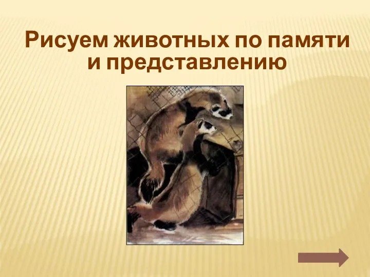 Урок 5. Рисуем животного по памяти и представлению
 Активные формы обучения студентов и слушателей кафедры Менеджмент НГТУ им. Р.Е. Алексеева
 Цветы в литературе
 История развития гистологии, цитологии и эмбриологии. Развитие гистологии в Республике Казахстан
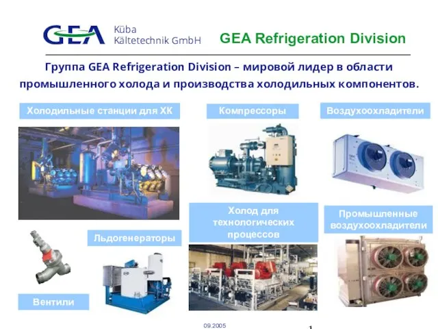 GEA Refrigeration Division
 Подготовка к ВПР
 Метод проектов. Что такое проект?
 13 Метод измерения v_biologii
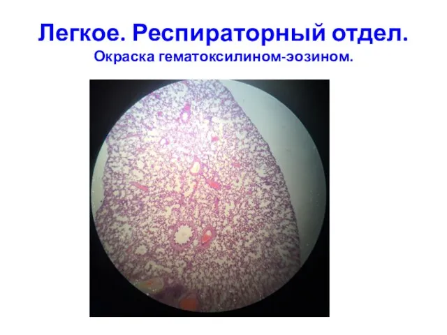 Легкое. Респираторный отдел. Окраска гематоксилином-эозином
  АО НК «КазМунайГаз»: Развитие казахстанского содержания Астана, апрель 2012 года
 Украина - переход к новой модели рынка электроэнергии
 Роль системы развития персонала организации
 Технологии в системе экономических отношений
 Мастер-класс по армянской кухне
 Презентация%20по%20теме%20правописание%20приставок%20и%20предлогов
 Большое агентство – бутиковый подход. Смотрим на маркетинг шире
 TEMPUS IV- ПЯТЫЙ КОНКУРС ЗАЯВОК Как подготовить конкурентоспособное проектное предложение Исполнительное агентство по образованию, а
 Шитьё фартука
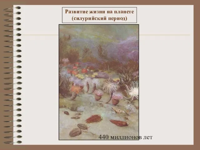 Презентация на тему Развитие жизни на планете
 The British Monarchy
  Второстепенные члены предложения. Обстоятельство
 Pyaterochka. Пятерочка
 Декабристы В Сибири
 How to teach children to understand mass media ?
 Изучение методов педагогической диагностики в соответствии с новым ФГОС Н. В. Нилова учитель начальных классов, руководитель МО
  Юридическая ответственность