- Microbiology

Содержание
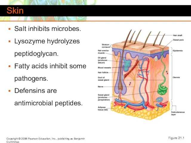
- 2. Skin Salt inhibits microbes. Lysozyme hydrolyzes peptidoglycan. Fatty acids inhibit some pathogens. Defensins are antimicrobial peptides.
- 3. Mucous Membranes Line body cavities. The epithelial cells are attached to an extracellular matrix. Cells secrete
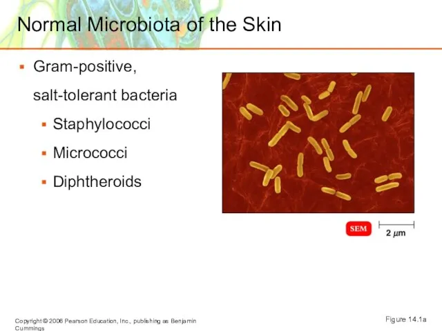
- 4. Normal Microbiota of the Skin Gram-positive, salt-tolerant bacteria Staphylococci Micrococci Diphtheroids Figure 14.1a
- 5. Microbial Diseases of the Skin Exanthem: Skin rash arising from another focus of the infection. Enanthem:
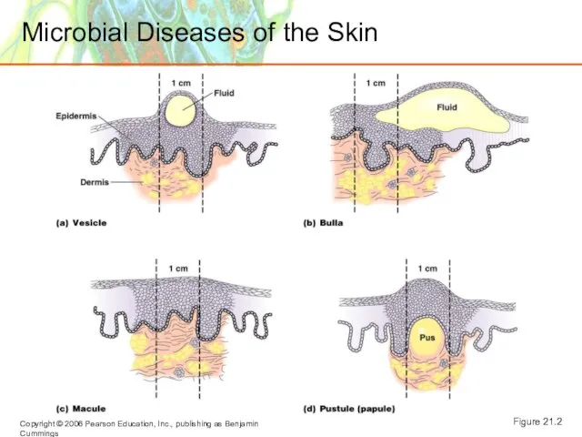
- 6. Microbial Diseases of the Skin Figure 21.2
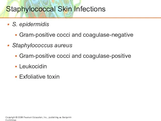
- 7. Staphylococcal Skin Infections S. epidermidis Gram-positive cocci and coagulase-negative Staphylococcus aureus Gram-positive cocci and coagulase-positive Leukocidin
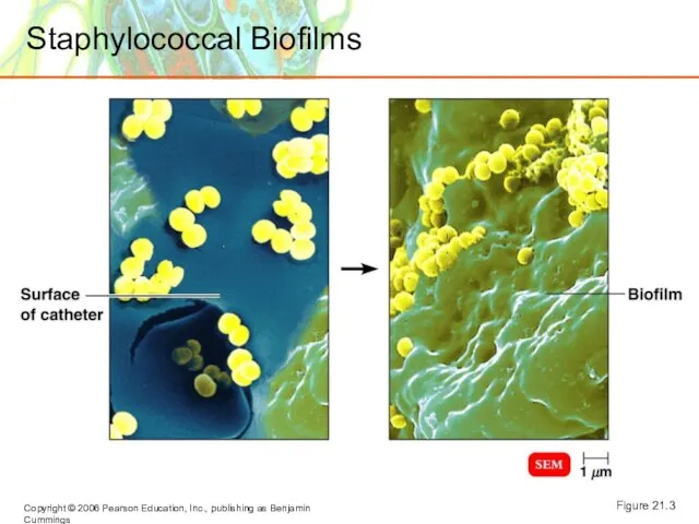
- 8. Staphylococcal Biofilms Figure 21.3
- 9. Staphylococcal Skin Infections Folliculitis: Infections of the hair follicles. Sty: Folliculitis of an eyelash. Furuncle: Abscess;
- 10. Staphylococcal Skin Infections Impetigo of the newborn Toxemia Scalded skin syndrome Toxic shock syndrome Figure 21.4
- 11. Streptococcal Skin Infections Streptococcus pyogenes Group A beta-hemolytic streptococci M proteins Figure 21.5
- 12. Streptococcal Skin Infections Erysipelas Impetigo Figures 21.6, 21.7
- 13. Invasive Group A Streptococcal Infections Streptokinases Hyaluronidase Exotoxin A, superantigen Cellulitis Necrotizing fasciitis Figure 21.8
- 14. Infections by Pseudomonads Pseudomonas aeruginosa Gram-negative, aerobic rod Pyocyanin produces a blue-green pus Pseudomonas dermatitis Otitis
- 15. Acne Comedonal acne occurs when sebum channels are blocked with shed cells. Inflammatory acne Propionibacterium acnes
- 16. Acne Inflammatory acne (continued) Nodular cystic acne Treatment: isotretinoin
- 17. Warts Papillomaviruses Treatment Removal Imiquimod (stimulates interferon production) Interferon
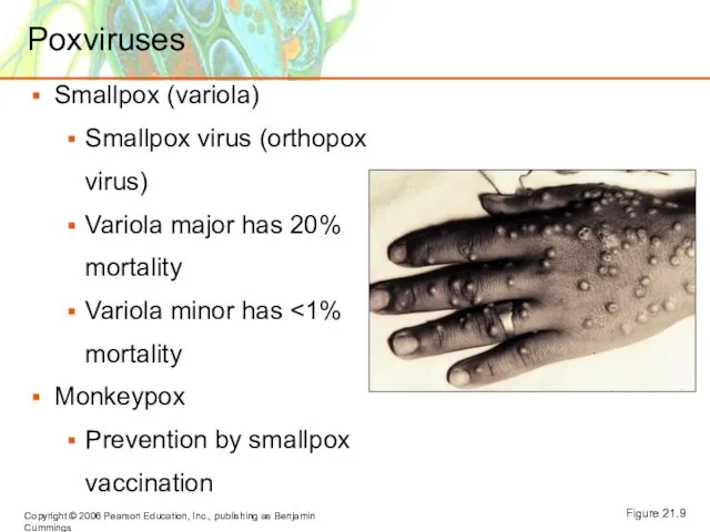
- 18. Poxviruses Smallpox (variola) Smallpox virus (orthopox virus) Variola major has 20% mortality Variola minor has Monkeypox
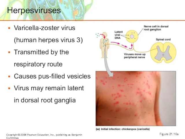
- 19. Herpesviruses Varicella-zoster virus (human herpes virus 3) Transmitted by the respiratory route Causes pus-filled vesicles Virus
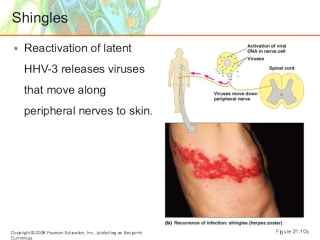
- 20. Shingles Reactivation of latent HHV-3 releases viruses that move along peripheral nerves to skin. Figure 21.10b
- 21. Herpes Simplex 1 and Herpes Simplex 2 Human herpes virus 1 and HHV-2 Cold sores or
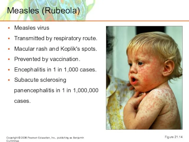
- 22. Measles (Rubeola) Measles virus Transmitted by respiratory route. Macular rash and Koplik's spots. Prevented by vaccination.
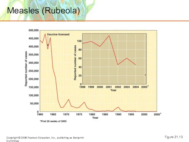
- 23. Measles (Rubeola) Figure 21.13
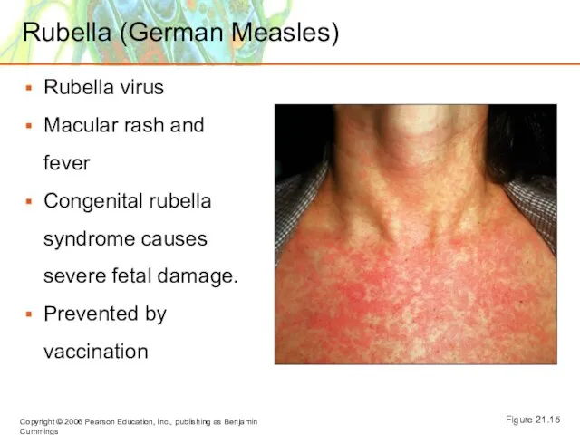
- 24. Rubella (German Measles) Rubella virus Macular rash and fever Congenital rubella syndrome causes severe fetal damage.
- 25. A 1905 list of skin rashes included (1)measles, (2)scarlet fever, (3)rubella, (4)Filatow-Dukes (mild scarlet fever), and
- 26. Cutaneous Mycoses Dermatomycoses: Tineas or ringworm Metabolize keratin Trichophyton: Infects hair, skin, and nails Epidermophyton: Infects
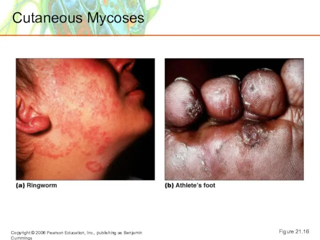
- 27. Cutaneous Mycoses Figure 21.16
- 28. Subcutaneous Mycoses Sporotrichosis Sporothrix schenckii enters puncture wound Treated with KI
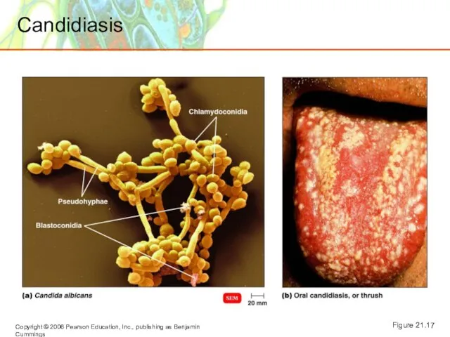
- 29. Candidiasis Candida albicans (yeast) Candidiasis may result from suppression of competing bacteria by antibiotics. Occurs in
- 30. Candidiasis Figure 21.17
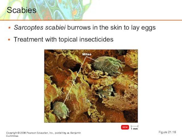
- 31. Scabies Sarcoptes scabiei burrows in the skin to lay eggs Treatment with topical insecticides Figure 21.18
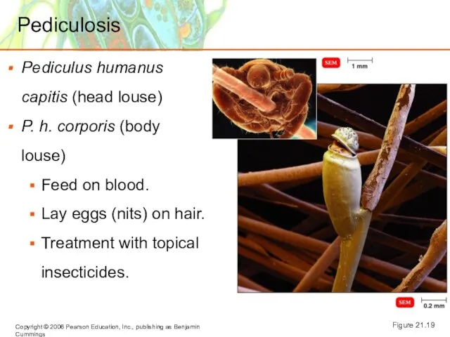
- 32. Pediculosis Pediculus humanus capitis (head louse) P. h. corporis (body louse) Feed on blood. Lay eggs
- 33. Macular Rashes A 9-year-old girl with a history of cough, conjunctivitis, and fever (38°C) has a
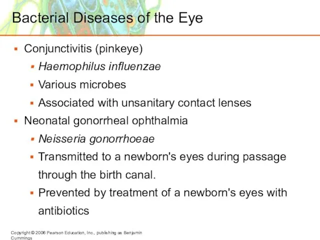
- 34. Bacterial Diseases of the Eye Conjunctivitis (pinkeye) Haemophilus influenzae Various microbes Associated with unsanitary contact lenses
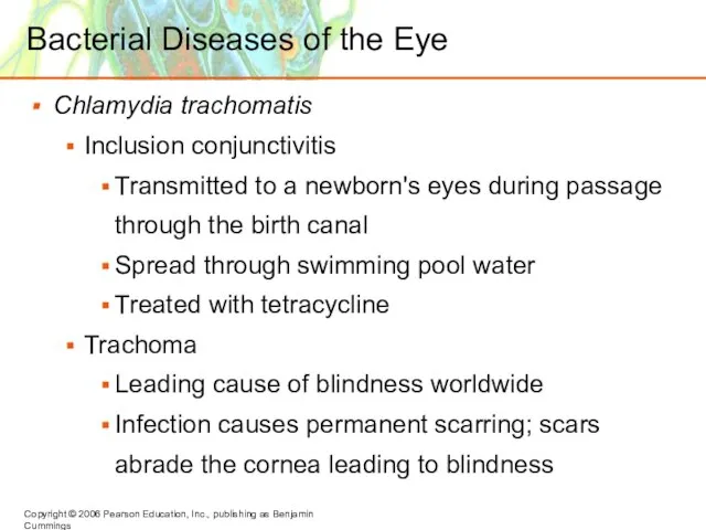
- 35. Bacterial Diseases of the Eye Chlamydia trachomatis Inclusion conjunctivitis Transmitted to a newborn's eyes during passage
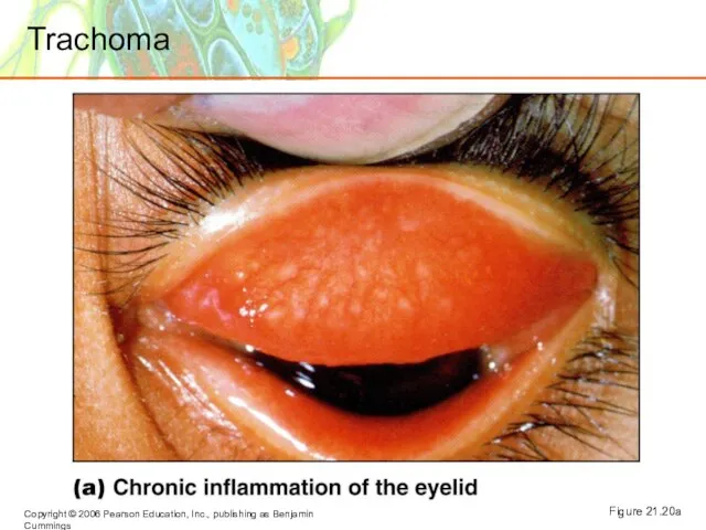
- 36. Figure 21.20a Trachoma
- 37. Viral Diseases of the Eye Conjunctivitis Adenoviruses Herpetic keratitis Herpes simplex virus 1 (HHV-1). Infects cornea
- 39. Скачать презентацию
Слайд 2Skin
Salt inhibits microbes.
Lysozyme hydrolyzes peptidoglycan.
Fatty acids inhibit some pathogens.
Defensins are antimicrobial peptides.
Figure
Skin
Salt inhibits microbes.
Lysozyme hydrolyzes peptidoglycan.
Fatty acids inhibit some pathogens.
Defensins are antimicrobial peptides.
Figure
Слайд 3Mucous Membranes
Line body cavities.
The epithelial cells are attached to an extracellular matrix.
Cells
Mucous Membranes
Line body cavities.
The epithelial cells are attached to an extracellular matrix.
Cells
Слайд 4Normal Microbiota of the Skin
Gram-positive, salt-tolerant bacteria
Staphylococci
Micrococci
Diphtheroids
Figure 14.1a
Normal Microbiota of the Skin
Gram-positive, salt-tolerant bacteria
Staphylococci
Micrococci
Diphtheroids
Figure 14.1a
Слайд 5Microbial Diseases of the Skin
Exanthem: Skin rash arising from another focus
of
Microbial Diseases of the Skin
Exanthem: Skin rash arising from another focus of
Слайд 6Microbial Diseases of the Skin
Figure 21.2
Microbial Diseases of the Skin
Figure 21.2
Слайд 7Staphylococcal Skin Infections
S. epidermidis
Gram-positive cocci and coagulase-negative
Staphylococcus aureus
Gram-positive cocci and coagulase-positive
Leukocidin
Exfoliative toxin
Staphylococcal Skin Infections
S. epidermidis
Gram-positive cocci and coagulase-negative
Staphylococcus aureus
Gram-positive cocci and coagulase-positive
Leukocidin
Exfoliative toxin
Слайд 8Staphylococcal Biofilms
Figure 21.3
Staphylococcal Biofilms
Figure 21.3
Слайд 9Staphylococcal Skin Infections
Folliculitis: Infections of the hair follicles.
Sty: Folliculitis of an eyelash.
Furuncle:
Staphylococcal Skin Infections
Folliculitis: Infections of the hair follicles.
Sty: Folliculitis of an eyelash.
Furuncle:
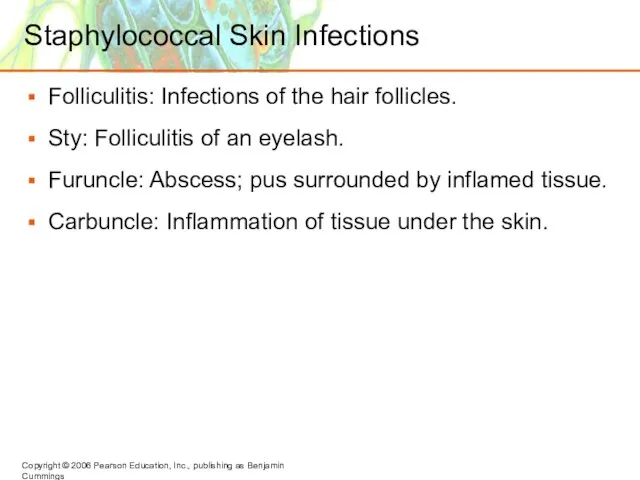
Слайд 10Staphylococcal Skin Infections
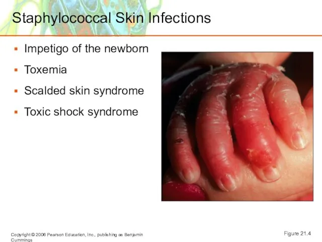
Impetigo of the newborn
Toxemia
Scalded skin syndrome
Toxic shock syndrome
Figure 21.4
Staphylococcal Skin Infections
Impetigo of the newborn
Toxemia
Scalded skin syndrome
Toxic shock syndrome
Figure 21.4
Слайд 11Streptococcal Skin Infections
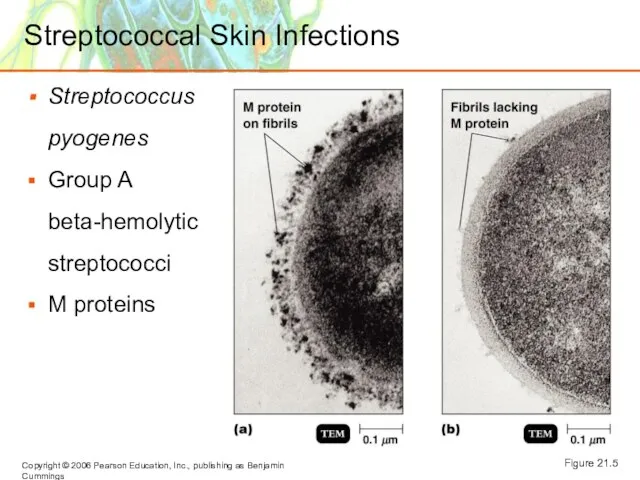
Streptococcus pyogenes
Group A beta-hemolytic streptococci
M proteins
Figure 21.5
Streptococcal Skin Infections
Streptococcus pyogenes
Group A beta-hemolytic streptococci
M proteins
Figure 21.5
Слайд 12Streptococcal Skin Infections
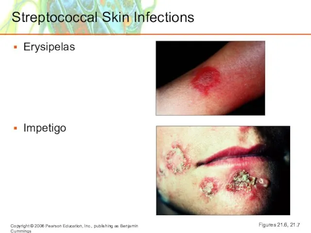
Erysipelas
Impetigo
Figures 21.6, 21.7
Streptococcal Skin Infections
Erysipelas
Impetigo
Figures 21.6, 21.7
Слайд 13Invasive Group A Streptococcal Infections
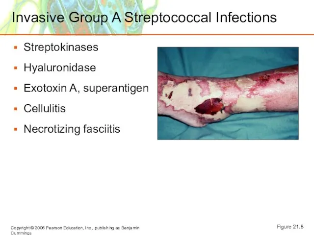
Streptokinases
Hyaluronidase
Exotoxin A, superantigen
Cellulitis
Necrotizing fasciitis
Figure 21.8
Invasive Group A Streptococcal Infections
Streptokinases
Hyaluronidase
Exotoxin A, superantigen
Cellulitis
Necrotizing fasciitis
Figure 21.8
Слайд 14Infections by Pseudomonads
Pseudomonas aeruginosa
Gram-negative, aerobic rod
Pyocyanin produces a blue-green pus
Pseudomonas dermatitis
Otitis externa
Post-burn
Infections by Pseudomonads
Pseudomonas aeruginosa
Gram-negative, aerobic rod
Pyocyanin produces a blue-green pus
Pseudomonas dermatitis
Otitis externa
Post-burn
Слайд 15Acne
Comedonal acne occurs when sebum channels are blocked with shed cells.
Inflammatory acne
Propionibacterium
Acne
Comedonal acne occurs when sebum channels are blocked with shed cells.
Inflammatory acne
Propionibacterium
Слайд 16Acne
Inflammatory acne (continued)
Nodular cystic acne
Treatment: isotretinoin
Acne
Inflammatory acne (continued)
Nodular cystic acne
Treatment: isotretinoin
Слайд 17Warts
Papillomaviruses
Treatment
Removal
Imiquimod (stimulates interferon production)
Interferon
Warts
Papillomaviruses
Treatment
Removal
Imiquimod (stimulates interferon production)
Interferon
Слайд 18Poxviruses
Smallpox (variola)
Smallpox virus (orthopox virus)
Variola major has 20% mortality
Variola minor has <1%
Poxviruses
Smallpox (variola)
Smallpox virus (orthopox virus)
Variola major has 20% mortality
Variola minor has <1%
Слайд 19Herpesviruses
Varicella-zoster virus (human herpes virus 3)
Transmitted by the respiratory route
Causes pus-filled vesicles
Virus
Herpesviruses
Varicella-zoster virus (human herpes virus 3)
Transmitted by the respiratory route
Causes pus-filled vesicles
Virus
Слайд 20Shingles
Reactivation of latent HHV-3 releases viruses that move along peripheral nerves to
Shingles
Reactivation of latent HHV-3 releases viruses that move along peripheral nerves to
Слайд 21Herpes Simplex 1 and Herpes Simplex 2
Human herpes virus 1 and HHV-2
Cold
Herpes Simplex 1 and Herpes Simplex 2
Human herpes virus 1 and HHV-2
Cold
Слайд 22Measles (Rubeola)
Measles virus
Transmitted by respiratory route.
Macular rash and Koplik's spots.
Prevented by vaccination.
Encephalitis
Measles (Rubeola)
Measles virus
Transmitted by respiratory route.
Macular rash and Koplik's spots.
Prevented by vaccination.
Encephalitis
Слайд 23Measles (Rubeola)
Figure 21.13
Measles (Rubeola)
Figure 21.13
Слайд 24Rubella (German Measles)
Rubella virus
Macular rash and fever
Congenital rubella syndrome causes severe fetal
Rubella (German Measles)
Rubella virus
Macular rash and fever
Congenital rubella syndrome causes severe fetal
Слайд 25A 1905 list of skin rashes included (1)measles, (2)scarlet fever, (3)rubella, (4)Filatow-Dukes
A 1905 list of skin rashes included (1)measles, (2)scarlet fever, (3)rubella, (4)Filatow-Dukes
Слайд 26Cutaneous Mycoses
Dermatomycoses: Tineas or ringworm
Metabolize keratin
Trichophyton: Infects hair, skin, and nails
Epidermophyton:
Cutaneous Mycoses
Dermatomycoses: Tineas or ringworm
Metabolize keratin
Trichophyton: Infects hair, skin, and nails
Epidermophyton:
Слайд 27Cutaneous Mycoses
Figure 21.16
Cutaneous Mycoses
Figure 21.16
Слайд 28Subcutaneous Mycoses
Sporotrichosis
Sporothrix schenckii enters puncture wound
Treated with KI
Subcutaneous Mycoses
Sporotrichosis
Sporothrix schenckii enters puncture wound
Treated with KI
Слайд 29Candidiasis
Candida albicans (yeast)
Candidiasis may result from suppression of competing bacteria by antibiotics.
Occurs
Candidiasis
Candida albicans (yeast)
Candidiasis may result from suppression of competing bacteria by antibiotics.
Occurs
Слайд 30Candidiasis
Figure 21.17
Candidiasis
Figure 21.17
Слайд 31Scabies
Sarcoptes scabiei burrows in the skin to lay eggs
Treatment with topical insecticides
Figure
Scabies
Sarcoptes scabiei burrows in the skin to lay eggs
Treatment with topical insecticides
Figure
Слайд 32Pediculosis
Pediculus humanus capitis (head louse)
P. h. corporis (body louse)
Feed on blood.
Lay eggs
Pediculosis
Pediculus humanus capitis (head louse)
P. h. corporis (body louse)
Feed on blood.
Lay eggs
Слайд 33Macular Rashes
A 9-year-old girl with a history of cough, conjunctivitis, and fever
Macular Rashes
A 9-year-old girl with a history of cough, conjunctivitis, and fever
Слайд 34Bacterial Diseases of the Eye
Conjunctivitis (pinkeye)
Haemophilus influenzae
Various microbes
Associated with unsanitary contact lenses
Neonatal
Bacterial Diseases of the Eye
Conjunctivitis (pinkeye)
Haemophilus influenzae
Various microbes
Associated with unsanitary contact lenses
Neonatal
Слайд 35Bacterial Diseases of the Eye
Chlamydia trachomatis
Inclusion conjunctivitis
Transmitted to a newborn's eyes during
Bacterial Diseases of the Eye
Chlamydia trachomatis
Inclusion conjunctivitis
Transmitted to a newborn's eyes during
Слайд 36Figure 21.20a
Trachoma
Figure 21.20a
Trachoma
Слайд 37Viral Diseases of the Eye
Conjunctivitis
Adenoviruses
Herpetic keratitis
Herpes simplex virus 1 (HHV-1).
Infects cornea and
Viral Diseases of the Eye
Conjunctivitis
Adenoviruses
Herpetic keratitis
Herpes simplex virus 1 (HHV-1).
Infects cornea and
 Я-концепция по Р. Бернсу
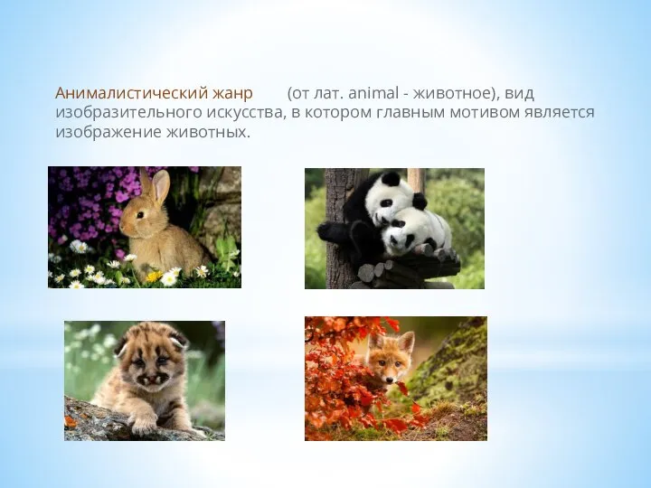 Анималистический жанр
 Приём в первые классы
 Анализ лирического стихотворения
 Форми держави
 Системы управления базами данных
 ПРОЕКТ ПРОФЕССИЯ (1)
 Жостово. Роспись подносов
 Нужные профессии
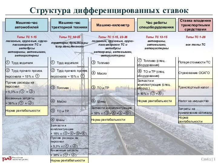 Структура дифференцированных ставок
 Как вести себя во время теракта
 Проект стикеры
 Автотранспорт
 Школа Дионисия и Симон Ушаков
 Куб. Создание изображений в графическом редакторе Paint
 Влияние среды на развитие растения из семени
 Презентация на тему КОМПЬЮТЕР
 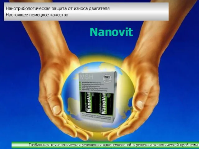 Nanovit
 Прикметники І-ІІ відміни
 Методика бенчмаркинга. Финансовая информация и принятие решений
 Масштаб
 Храмов Сергей МихайловичЛАТЕНТНАЯ ПРЕСТУПНОСТЬ:МЕТОДОЛОГИЯ ПОЗНАНИЯ И ОСНОВНЫЕ НАПРАВЛЕНИЯ ПРОТИВОДЕЙСТВИЯ
 Проект
 Презентация о библиотеке
 Беременность и пролапс митрального клапана
 «Салон Дона и Баса»
 Рубительные машины
 The Greatest Writers of the World