- The Autonomic Nervous System

Содержание
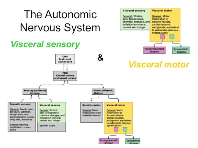
- 2. The Autonomic Nervous System Visceral sensory Visceral motor &
- 3. Autonomic nervous system The autonomic nervous system is the subdivision of the peripheral nervous system that
- 4. ANS is the subdivision of the peripheral nervous system that regulates body activities that are generally
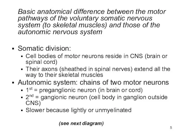
- 5. Basic anatomical difference between the motor pathways of the voluntary somatic nervous system (to skeletal muscles)
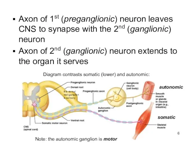
- 6. Axon of 1st (preganglionic) neuron leaves CNS to synapse with the 2nd (ganglionic) neuron Axon of
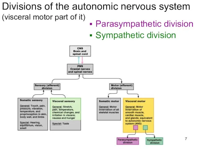
- 7. Divisions of the autonomic nervous system (visceral motor part of it) Parasympathetic division Sympathetic division
- 8. Divisions of the autonomic nervous system Parasympathetic division Sympathetic division Serve most of the same organs
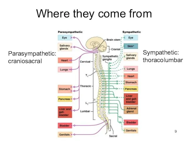
- 9. Where they come from Parasympathetic: craniosacral Sympathetic: thoracolumbar
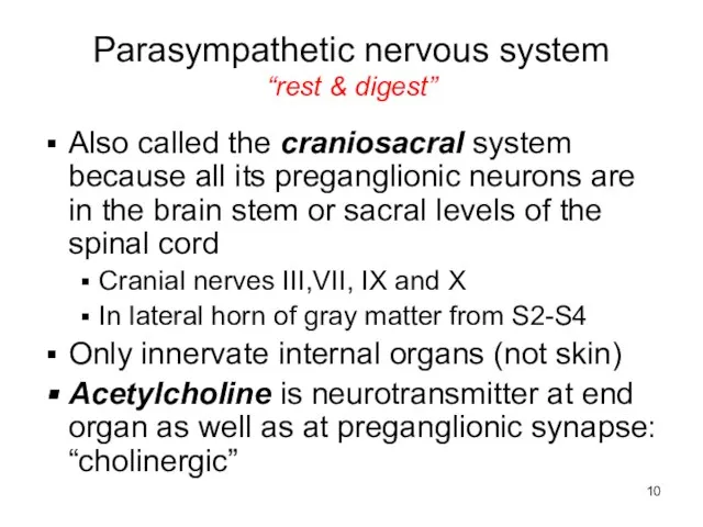
- 10. Parasympathetic nervous system “rest & digest” Also called the craniosacral system because all its preganglionic neurons
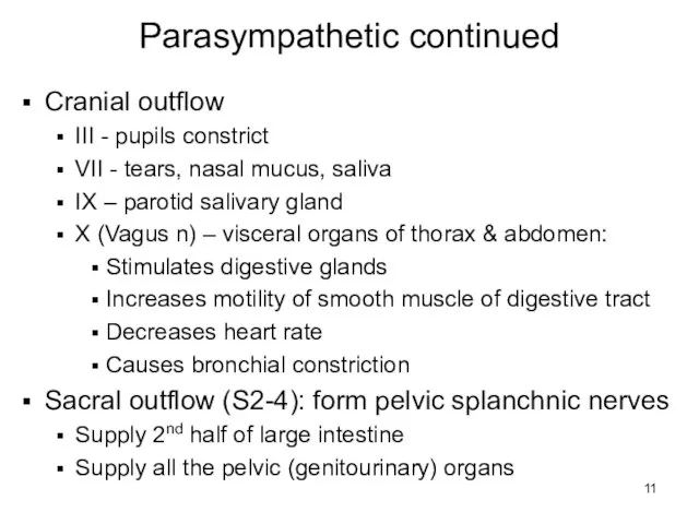
- 11. Parasympathetic continued Cranial outflow III - pupils constrict VII - tears, nasal mucus, saliva IX –
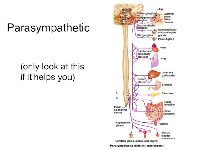
- 12. Parasympathetic (only look at this if it helps you)
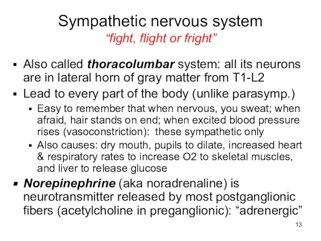
- 13. Sympathetic nervous system “fight, flight or fright” Also called thoracolumbar system: all its neurons are in
- 14. Sympathetic nervous system continued Regardless of target, all begin same Preganglionic axons exit spinal cord through
- 15. Options of preganglionic axons in sympathetic trunk Synapse on postganglionic neuron in chain ganglion then return
- 16. Synapse in chain ganglia at same level or different level
- 17. Pass through ganglia and synapse in prevertebral ganglion
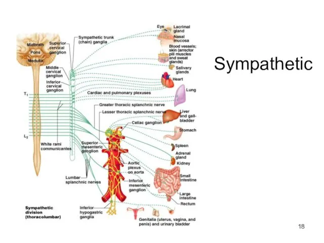
- 18. Sympathetic
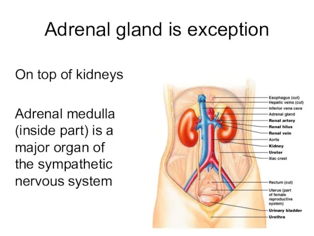
- 19. Adrenal gland is exception On top of kidneys Adrenal medulla (inside part) is a major organ
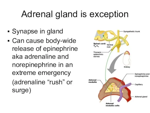
- 20. Adrenal gland is exception Synapse in gland Can cause body-wide release of epinephrine aka adrenaline and
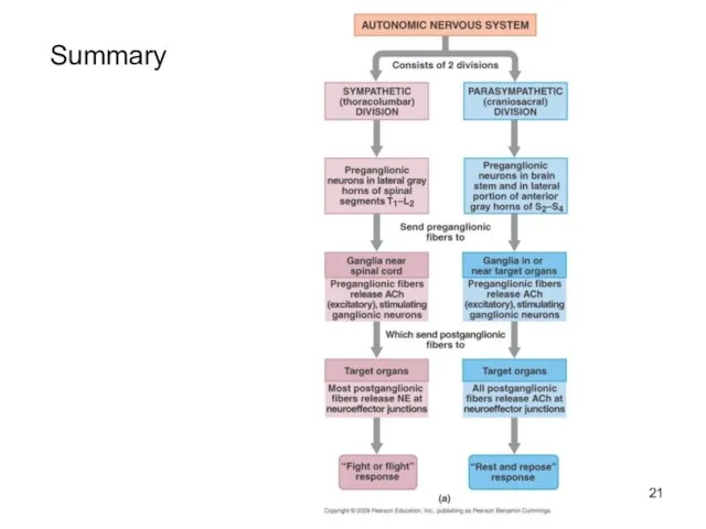
- 21. Summary
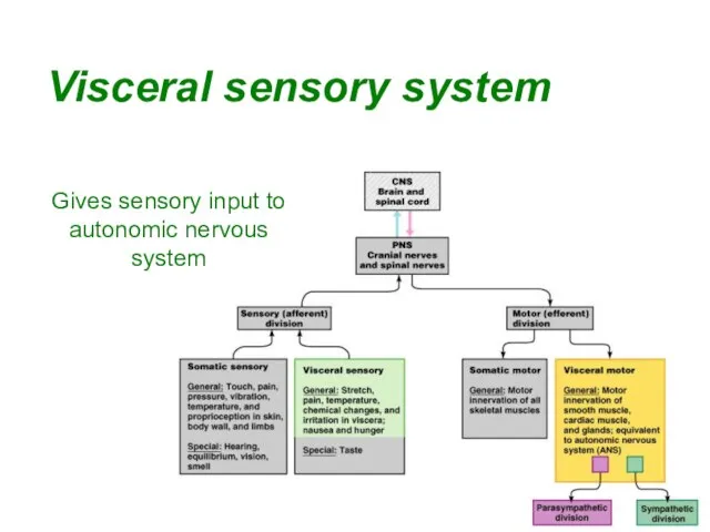
- 22. Visceral sensory system Gives sensory input to autonomic nervous system
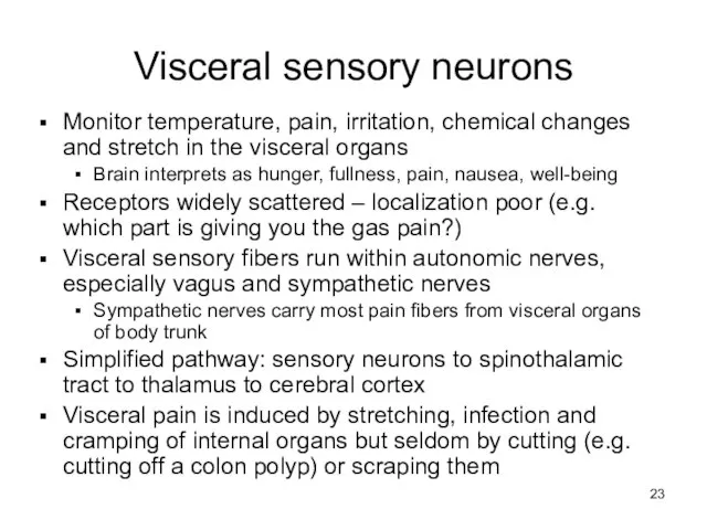
- 23. Visceral sensory neurons Monitor temperature, pain, irritation, chemical changes and stretch in the visceral organs Brain
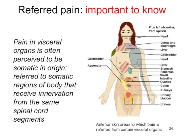
- 24. Referred pain: important to know Pain in visceral organs is often perceived to be somatic in
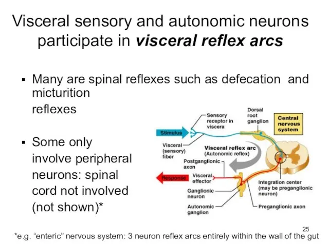
- 25. Visceral sensory and autonomic neurons participate in visceral reflex arcs Many are spinal reflexes such as
- 27. Скачать презентацию
Слайд 3Autonomic nervous system
The autonomic nervous system is the subdivision of the peripheral
Autonomic nervous system
The autonomic nervous system is the subdivision of the peripheral
Слайд 4ANS is the subdivision of the peripheral nervous system that regulates body
ANS is the subdivision of the peripheral nervous system that regulates body
Слайд 5 Basic anatomical difference between the motor pathways of the voluntary somatic nervous
Basic anatomical difference between the motor pathways of the voluntary somatic nervous
Слайд 6Axon of 1st (preganglionic) neuron leaves CNS to synapse with the 2nd
Axon of 1st (preganglionic) neuron leaves CNS to synapse with the 2nd
Слайд 7Divisions of the autonomic nervous system (visceral motor part of it)
Parasympathetic division
Sympathetic
Divisions of the autonomic nervous system (visceral motor part of it)
Parasympathetic division
Sympathetic
Слайд 8Divisions of the autonomic nervous system
Parasympathetic division
Sympathetic division
Serve most of the same
Divisions of the autonomic nervous system
Parasympathetic division
Sympathetic division
Serve most of the same
Слайд 9Where they come from
Parasympathetic:
craniosacral
Sympathetic:
thoracolumbar
Where they come from
Parasympathetic:
craniosacral
Sympathetic:
thoracolumbar
Слайд 10Parasympathetic nervous system
“rest & digest”
Also called the craniosacral system because all its
Parasympathetic nervous system
“rest & digest”
Also called the craniosacral system because all its
Слайд 11Parasympathetic continued
Cranial outflow
III - pupils constrict
VII - tears, nasal mucus, saliva
IX –
Parasympathetic continued
Cranial outflow
III - pupils constrict
VII - tears, nasal mucus, saliva
IX –
Слайд 12Parasympathetic
(only look at this if it helps you)
Parasympathetic
(only look at this if it helps you)
Слайд 13Sympathetic nervous system
“fight, flight or fright”
Also called thoracolumbar system: all its neurons
Sympathetic nervous system
“fight, flight or fright”
Also called thoracolumbar system: all its neurons
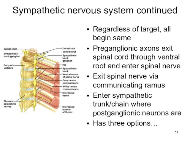
Слайд 14Sympathetic nervous system continued
Regardless of target, all begin same
Preganglionic axons exit spinal
Sympathetic nervous system continued
Regardless of target, all begin same
Preganglionic axons exit spinal
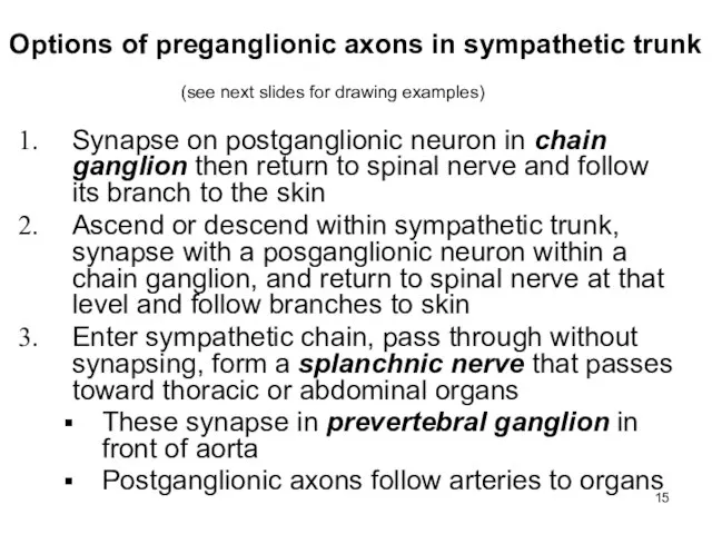
Слайд 15Options of preganglionic axons in sympathetic trunk
Synapse on postganglionic neuron in chain
Options of preganglionic axons in sympathetic trunk
Synapse on postganglionic neuron in chain
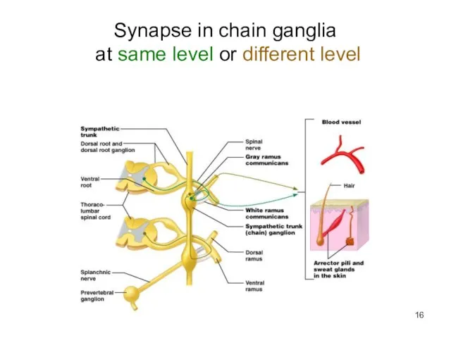
Слайд 16Synapse in chain ganglia
at same level or different level
Synapse in chain ganglia
at same level or different level
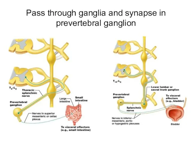
Слайд 17Pass through ganglia and synapse in prevertebral ganglion
Pass through ganglia and synapse in prevertebral ganglion
Слайд 18Sympathetic
Sympathetic
Слайд 19Adrenal gland is exception
On top of kidneys
Adrenal medulla (inside part) is a
Adrenal gland is exception
On top of kidneys
Adrenal medulla (inside part) is a
Слайд 20Adrenal gland is exception
Synapse in gland
Can cause body-wide release of epinephrine aka
Adrenal gland is exception
Synapse in gland
Can cause body-wide release of epinephrine aka
Слайд 21Summary
Summary
Слайд 22Visceral sensory system
Gives sensory input to autonomic nervous system
Visceral sensory system
Gives sensory input to autonomic nervous system
Слайд 23Visceral sensory neurons
Monitor temperature, pain, irritation, chemical changes and stretch in the
Visceral sensory neurons
Monitor temperature, pain, irritation, chemical changes and stretch in the
Слайд 24Referred pain: important to know
Pain in visceral organs is often perceived to
Referred pain: important to know
Pain in visceral organs is often perceived to
Слайд 25Visceral sensory and autonomic neurons participate in visceral reflex arcs
Many are spinal
Visceral sensory and autonomic neurons participate in visceral reflex arcs
Many are spinal
 ПРОЕКТ : Модифицирование стали Гадфильда (110Г13) при производстве крупных отливок (стрелочные переводы, зубья и передние стенки ков
 Разумная и обоснованная стоимость ремонта – страхование КАСКО морских судов.
 Методологический аппарат исследования
 Презентация на тему Безопасность детей в интернете
 Bazovye_ponyatia_informatiki
 Исследование статической устойчивости асинхронной нагрузки при питании их от шин бесконечной мощности
 Учебные курсы Microsoft для средней школы
 Здоровьесберегающие технологии на уроках русского языка
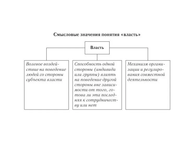 Власть. Типы (системы) политического господства
 Prezentatsia1
 Как Достичь Успеха Когда ты уже Успешен или Был УспешнымСессия №4
 Москва 4 июня 2008 г.
 Особенности крупного бизнеса
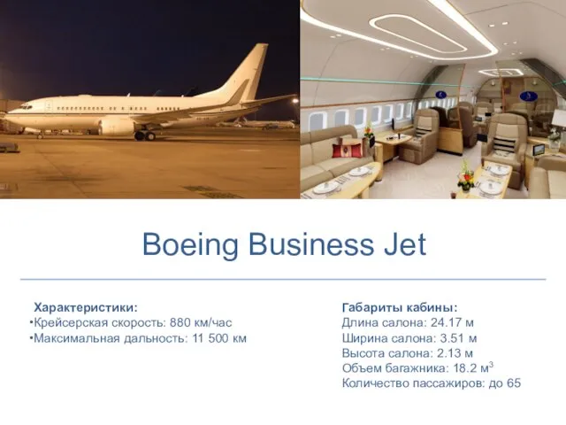 Boeing Business Jet
 0006d06c-2b7ab0fb
 Роль рекламы в обществе
 Буквица
 Ищем достойного хозяина!
 Мужская половая система
 контент-план
 Реализация Закона Краснодарского края № 1539 от 21 июля 2008 года «О мерах по профилактике безнадзорности и правонарушений среди несо
 Календарно-тематическое планирование
 Гражданин РФ
 Производство магния
 Формулы квадрата суммы и квадрата разности двух выражений
 право
право Автоматизация звука Р в словах
 Networking в стиле Бутерброд