- The nervous system

Содержание
- 2. Peripheral Nervous System 3 kinds of neurons connect CNS to the body sensory motor interneurons Motor
- 3. Peripheral Nervous System
- 4. Somatic System Nerves to/from spinal cord control muscle movements somatosensory inputs Both Voluntary and reflex movements
- 5. Autonomic System Two divisions: sympathetic Parasympatheitic Control involuntary functions heartbeat blood pressure respiration perspiration digestion Can
- 6. Sympathetic “ Fight or flight” response Release adrenaline and noradrenaline Increases heart rate and blood pressure
- 7. Parasympathetic “ Rest and digest ” system Calms body to conserve and maintain energy Lowers heartbeat,
- 8. Summary of autonomic differences
- 9. Central Nervous System Brain and Spinal Cord
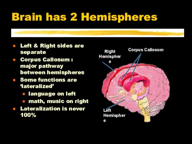
- 10. Left & Right sides are separate Corpus Callosum : major pathway between hemispheres Some functions are
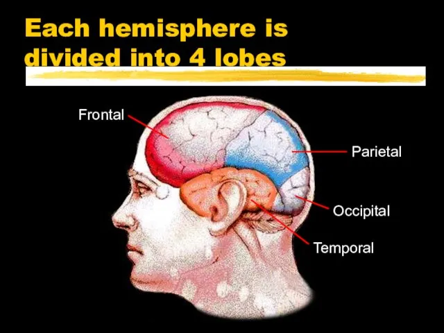
- 11. Each hemisphere is divided into 4 lobes
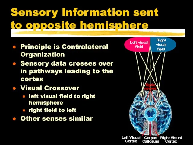
- 12. Sensory Information sent to opposite hemisphere Principle is Contralateral Organization Sensory data crosses over in pathways
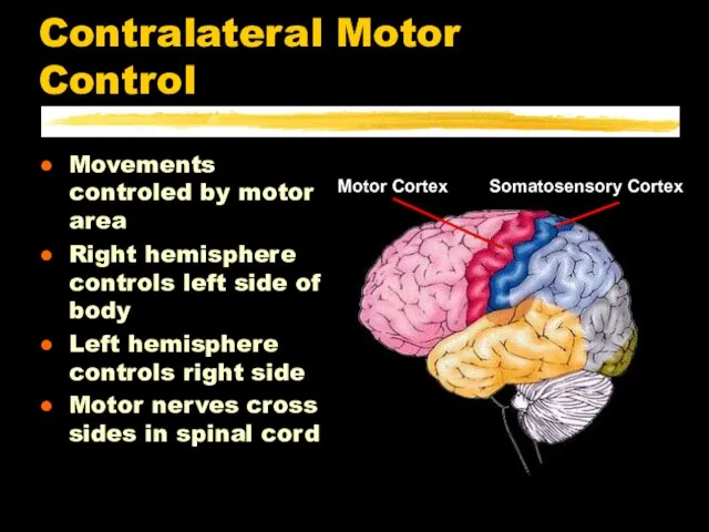
- 13. Contralateral Motor Control Movements controled by motor area Right hemisphere controls left side of body Left
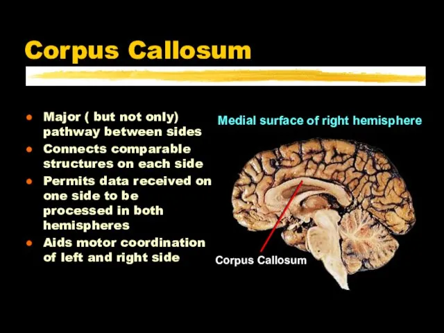
- 14. Corpus Callosum Major ( but not only) pathway between sides Connects comparable structures on each side
- 15. Corpus Callosum What happens when the corpus callosum is cut? Sensory inputs are still crossed Motor
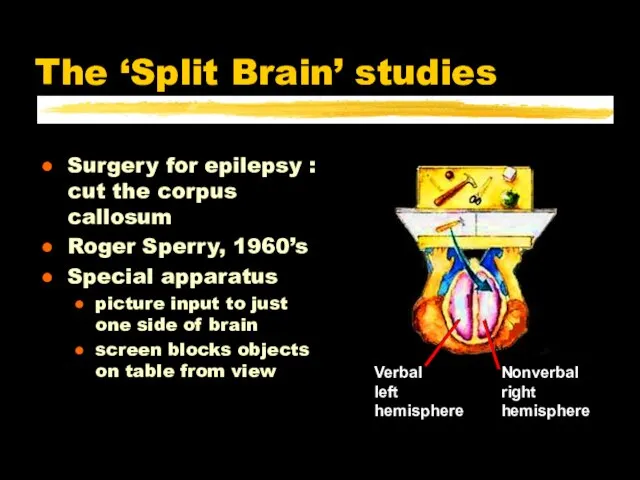
- 16. The ‘Split Brain’ studies Surgery for epilepsy : cut the corpus callosum Roger Sperry, 1960’s Special
- 17. Picture to left brain can name the object left hand cannot identify by touch Picture to
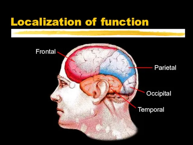
- 18. Localization of function
- 19. Occipital Lobe Input from Optic nerve Contains primary visual cortex most is on surface inside central
- 20. Temporal Lobe Inputs are auditory, visual patterns speech recognition face recognition word recognition memory formation Outputs
- 21. Parietal Lobe Inputs from multiple senses contains primary somatosensory cortex borders visual & auditory cortex Outputs
- 22. Frontal Lobe Contains primary motor cortex No direct sensory input Important planning and sequencing areas Broca’s
- 23. Frontal Lobe Disorders Broca’s area productive aphasia Prefrontal area lose track of ongoing context fail to
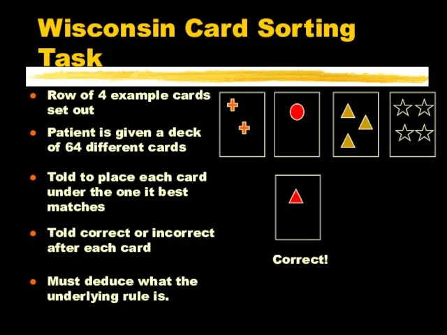
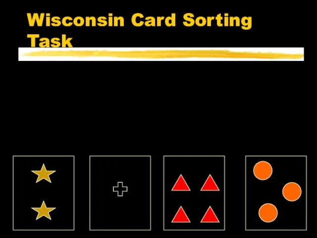
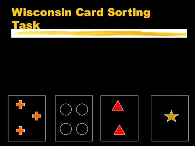
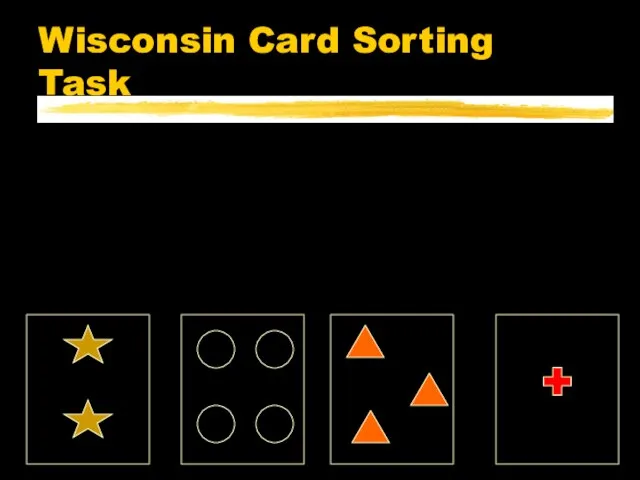
- 24. Wisconsin Card Sorting Task Patient is given a deck of 64 different cards Told to place
- 25. Wisconsin Card Sorting Task
- 26. Wisconsin Card Sorting Task
- 27. Wisconsin Card Sorting Task
- 28. Wisconsin Card Sorting Task
- 29. Wisconsin Card Sorting Task
- 30. Wisconsin Card Sorting Task
- 32. Скачать презентацию
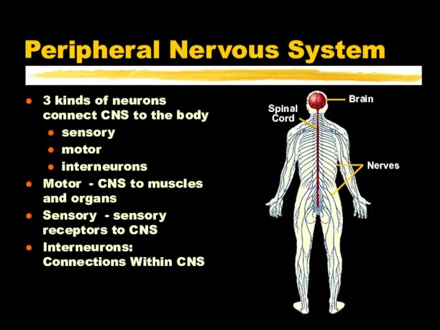
Слайд 2Peripheral Nervous System
3 kinds of neurons connect CNS to the body
sensory
motor
interneurons
Motor -
Peripheral Nervous System
3 kinds of neurons connect CNS to the body
sensory
motor
interneurons
Motor -
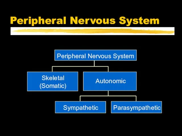
Слайд 3Peripheral Nervous System
Peripheral Nervous System
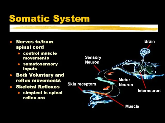
Слайд 4Somatic System
Nerves to/from spinal cord
control muscle movements
somatosensory inputs
Both Voluntary and reflex movements
Skeletal
Somatic System
Nerves to/from spinal cord
control muscle movements
somatosensory inputs
Both Voluntary and reflex movements
Skeletal
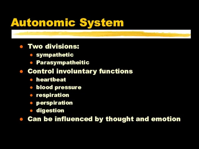
Слайд 5Autonomic System
Two divisions:
sympathetic
Parasympatheitic
Control involuntary functions
heartbeat
blood pressure
respiration
perspiration
digestion
Can be influenced by thought and
Autonomic System
Two divisions:
sympathetic
Parasympatheitic
Control involuntary functions
heartbeat
blood pressure
respiration
perspiration
digestion
Can be influenced by thought and
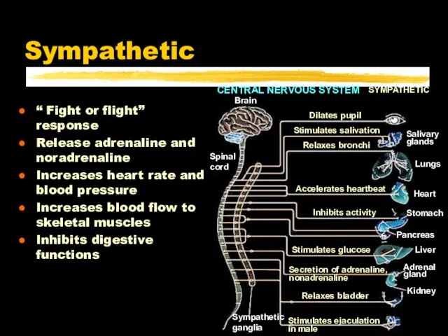
Слайд 6Sympathetic
“ Fight or flight” response
Release adrenaline and noradrenaline
Increases heart rate and
Sympathetic
“ Fight or flight” response
Release adrenaline and noradrenaline
Increases heart rate and
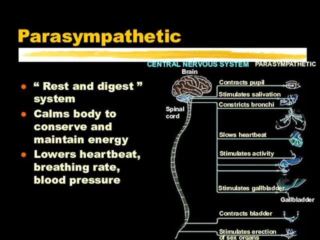
Слайд 7Parasympathetic
“ Rest and digest ” system
Calms body to conserve and maintain energy
Lowers
Parasympathetic
“ Rest and digest ” system
Calms body to conserve and maintain energy
Lowers
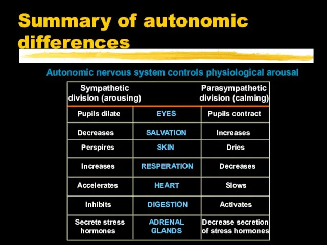
Слайд 8Summary of autonomic differences
Summary of autonomic differences
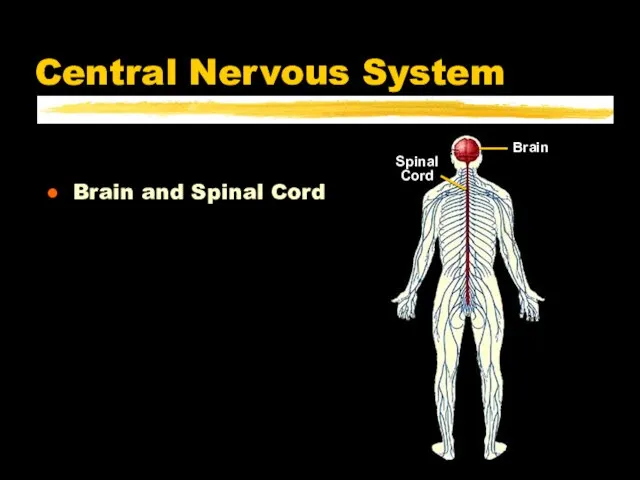
Слайд 9Central Nervous System
Brain and Spinal Cord
Central Nervous System
Brain and Spinal Cord
Слайд 10Left & Right sides are separate
Corpus Callosum : major pathway between hemispheres
Some
Left & Right sides are separate
Corpus Callosum : major pathway between hemispheres
Some
Слайд 11Each hemisphere is
divided into 4 lobes
Each hemisphere is
divided into 4 lobes
Слайд 12Sensory Information sent to opposite hemisphere
Principle is Contralateral Organization
Sensory data crosses over
Sensory Information sent to opposite hemisphere
Principle is Contralateral Organization
Sensory data crosses over
Слайд 13Contralateral Motor Control
Movements controled by motor area
Right hemisphere controls left side of
Contralateral Motor Control
Movements controled by motor area
Right hemisphere controls left side of
Слайд 14Corpus Callosum
Major ( but not only) pathway between sides
Connects comparable structures on
Corpus Callosum
Major ( but not only) pathway between sides
Connects comparable structures on
Слайд 15Corpus Callosum
What happens when the corpus callosum is cut?
Sensory inputs are still
Corpus Callosum
What happens when the corpus callosum is cut?
Sensory inputs are still
Слайд 16The ‘Split Brain’ studies
Surgery for epilepsy : cut the corpus callosum
Roger Sperry,
The ‘Split Brain’ studies
Surgery for epilepsy : cut the corpus callosum
Roger Sperry,
Слайд 17Picture to left brain
can name the object
left hand cannot identify by touch
Picture
Picture to left brain
can name the object
left hand cannot identify by touch
Picture

Слайд 18Localization of function
Localization of function
Слайд 19Occipital Lobe
Input from Optic nerve
Contains primary visual cortex
most is on surface inside
Occipital Lobe
Input from Optic nerve
Contains primary visual cortex
most is on surface inside

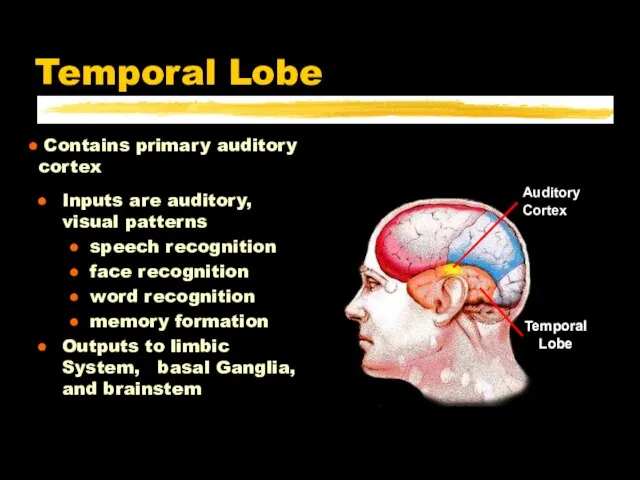
Слайд 20Temporal Lobe
Inputs are auditory, visual patterns
speech recognition
face recognition
word recognition
memory formation
Outputs to limbic
Temporal Lobe
Inputs are auditory, visual patterns
speech recognition
face recognition
word recognition
memory formation
Outputs to limbic
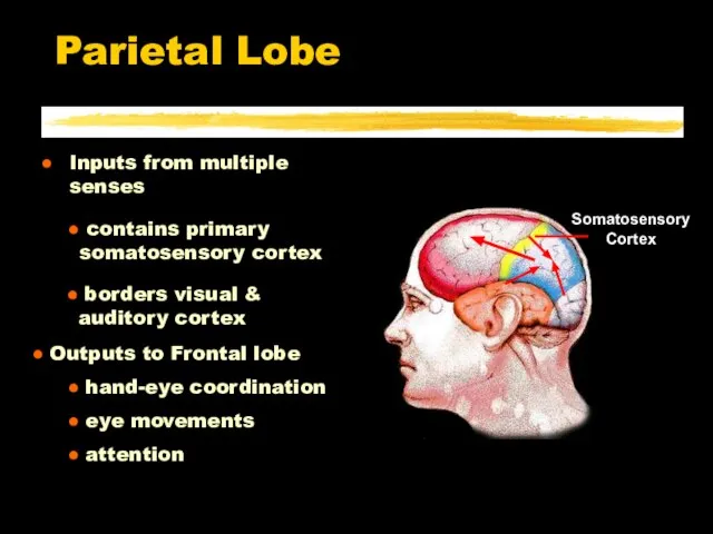
Слайд 21Parietal Lobe
Inputs from multiple senses
contains primary somatosensory cortex
borders visual &
Parietal Lobe
Inputs from multiple senses
contains primary somatosensory cortex
borders visual &
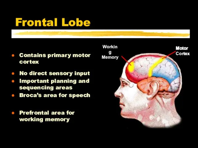
Слайд 22Frontal Lobe
Contains primary motor cortex
No direct sensory input
Important planning and sequencing areas
Broca’s
Frontal Lobe
Contains primary motor cortex
No direct sensory input
Important planning and sequencing areas
Broca’s
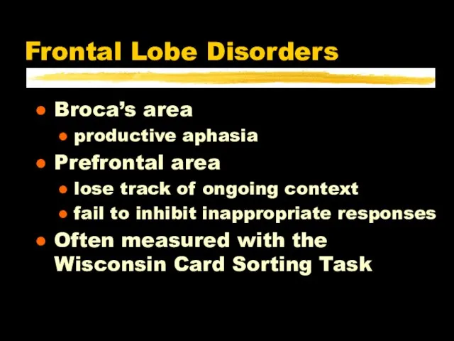
Слайд 23Frontal Lobe Disorders
Broca’s area
productive aphasia
Prefrontal area
lose track of ongoing context
fail to inhibit
Frontal Lobe Disorders
Broca’s area
productive aphasia
Prefrontal area
lose track of ongoing context
fail to inhibit
Слайд 24Wisconsin Card Sorting Task
Patient is given a deck of 64 different cards
Wisconsin Card Sorting Task
Patient is given a deck of 64 different cards
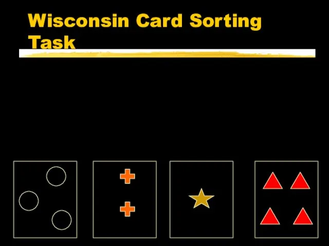
Слайд 25Wisconsin Card Sorting Task
Wisconsin Card Sorting Task
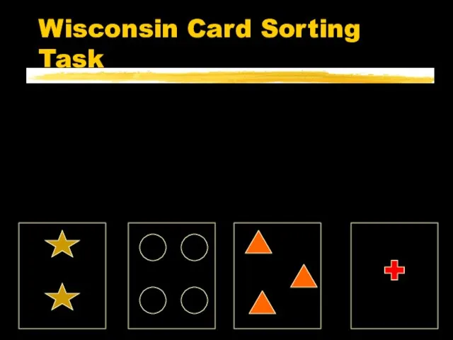
Слайд 26Wisconsin Card Sorting Task
Wisconsin Card Sorting Task
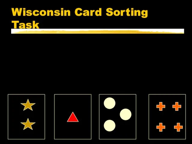
Слайд 27Wisconsin Card Sorting Task
Wisconsin Card Sorting Task
Слайд 28Wisconsin Card Sorting Task
Wisconsin Card Sorting Task
Слайд 29Wisconsin Card Sorting Task
Wisconsin Card Sorting Task
Слайд 30Wisconsin Card Sorting Task
Wisconsin Card Sorting Task
 Презентация на тему Социальный портрет молодежи 8/24/16
  Презентация-ООО-ХимСталь
 Грамоты благодарности родителям за хорошее воспитание детей
 Языковой центр Top Way Language Center
 Портфолио Карпенко Анастасии
 Рисуночный тест Н.Г. Хитровой
 Аэродинамика тепловых установок
 Цифровое телевидение и инновации
 Дом как отражение личности
 Великие изобретения человечества
 Глаз. Особенности зрения
 Презентация на тему Создание надежной системы безопасности, сбалансированной по критерию безопасность-стоимость
  Кафедра таможенного дела Магистерская диссертация Эффективность таможенно-тарифной политики в создании безбарьерной внешнетор
 Пенсионная реформа 2015 года
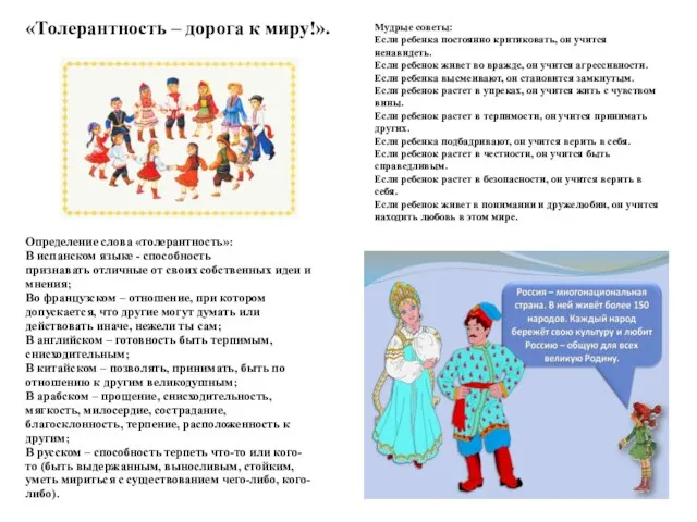 Толерантность – дорога к миру
 Технология приготовления сложных горячих соусов
 Звонят колокола в память о растерзанной душе Убеженского храма (вечер-экскурс в историю Михайло-Архангельской церкви станицы Убе
 Презентация на тему Демографическая проблема
 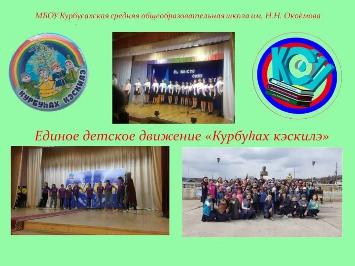 Лидер ЕДД: Прядезникова Варя
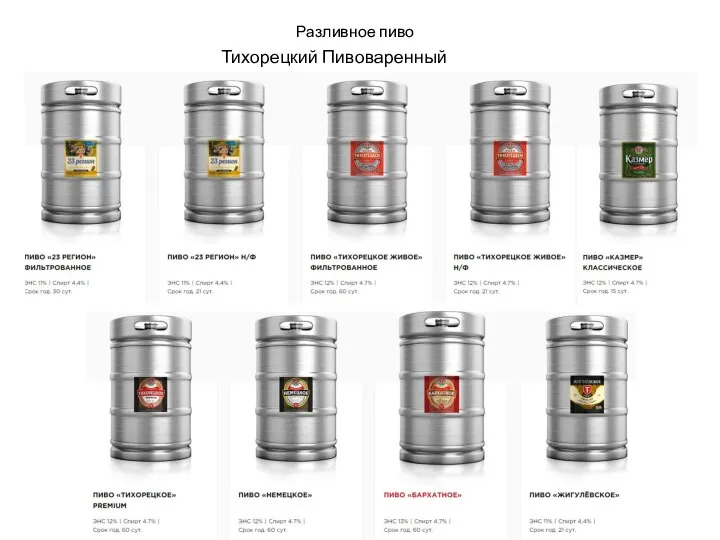 Пиво, напитки, энергетические коктейли
 Характеристика отдельных видов транспорта
 Лого заголовок
 Восточные походы Александра Македонского
 Групповая динамика в команде разработчиков Игорь Лужанский
 Сложение чисел с помощью координатной прямой
 Панно Корзина с цветами
 Межрегиональный гуманитарный научный комитет им. К.П. Победоносцева
 "Обучение жизненно важным навыкам"