- The human heart

Содержание
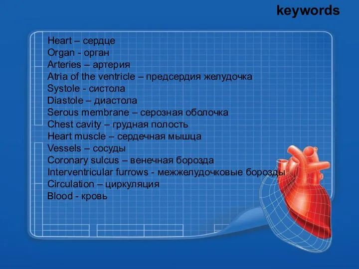
- 2. keywords Heart – сердце Organ - орган Arteries – артерия Atria of the ventricle – предсердия
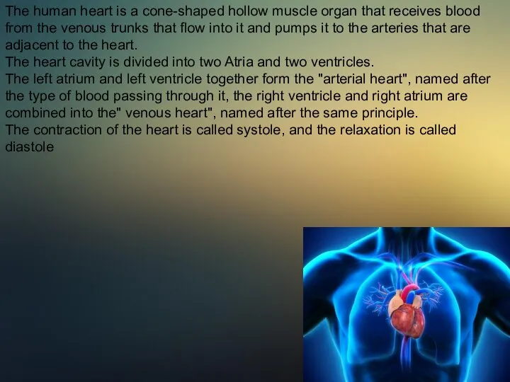
- 3. The human heart is a cone-shaped hollow muscle organ that receives blood from the venous trunks
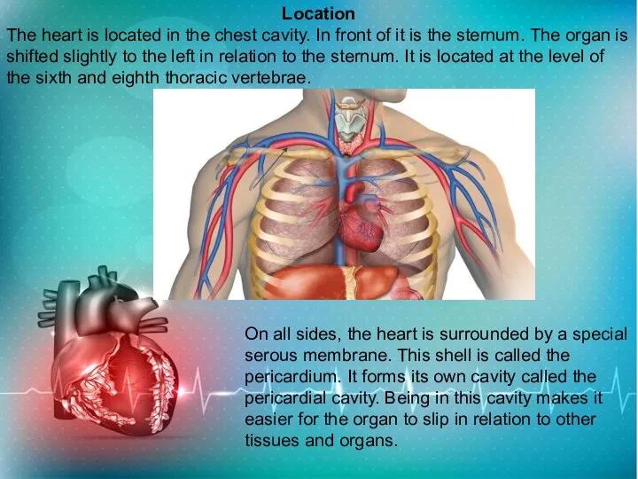
- 4. Location The heart is located in the chest cavity. In front of it is the sternum.
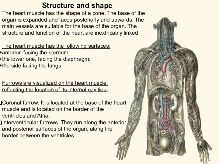
- 5. Structure and shape The heart muscle has the shape of a cone. The base of the
- 6. Anatomical features of the human heart Since the normal functioning of the heart directly depends on
- 7. Phases of the heart To continuously move blood through the blood vessels, the heart must contract.
- 8. Heart muscle The Uniqueness of the heart muscle is its ability to rhythmically automatic contractions, alternating
- 9. Circulation The heart conducts two circulations: Small-begins in the right ventricle and ends in the left
- 10. How does the blood flow in the heart work: Blood from veins with a high content
- 11. Heart disease It is not surprising that the number of cardiovascular diseases is increasing in the
- 12. Lifestyle and heart health The full functioning of the heart directly affects the state of the
- 14. Скачать презентацию
Слайд 3The human heart is a cone-shaped hollow muscle organ that receives blood
The human heart is a cone-shaped hollow muscle organ that receives blood
Слайд 4Location
The heart is located in the chest cavity. In front of it
Location
The heart is located in the chest cavity. In front of it
Слайд 5Structure and shape
The heart muscle has the shape of a cone. The
Structure and shape
The heart muscle has the shape of a cone. The
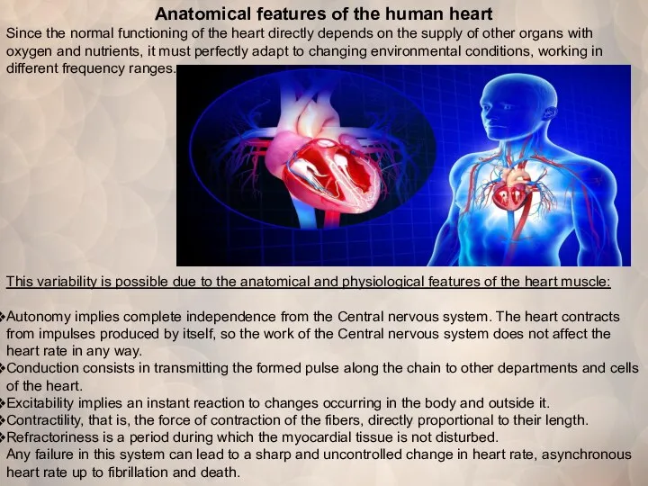
Слайд 6Anatomical features of the human heart
Since the normal functioning of the heart
Anatomical features of the human heart
Since the normal functioning of the heart
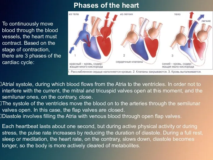
Слайд 7Phases of the heart
To continuously move blood through the blood vessels, the
Phases of the heart
To continuously move blood through the blood vessels, the
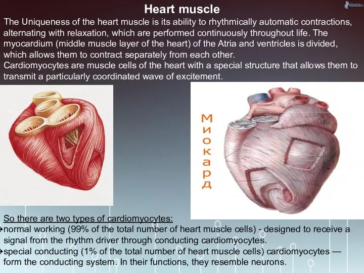
Слайд 8Heart muscle
The Uniqueness of the heart muscle is its ability to
Heart muscle
The Uniqueness of the heart muscle is its ability to
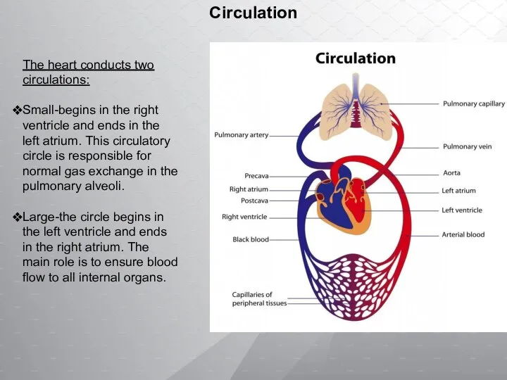
Слайд 9Circulation
The heart conducts two circulations:
Small-begins in the right ventricle and ends in
Circulation
The heart conducts two circulations:
Small-begins in the right ventricle and ends in
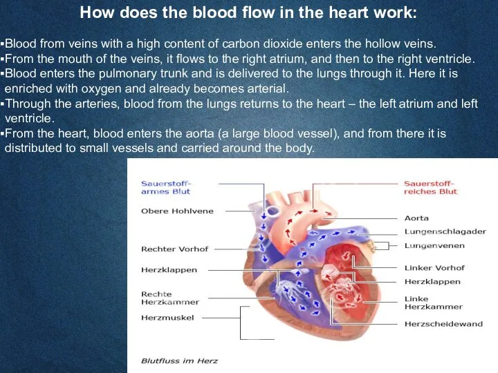
Слайд 10How does the blood flow in the heart work:
Blood from veins with
How does the blood flow in the heart work:
Blood from veins with
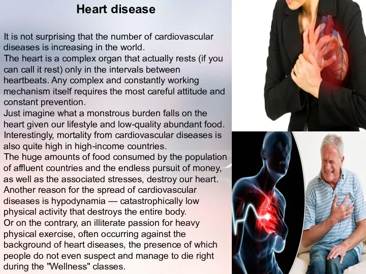
Слайд 11Heart disease
It is not surprising that the number of cardiovascular diseases is
Heart disease
It is not surprising that the number of cardiovascular diseases is
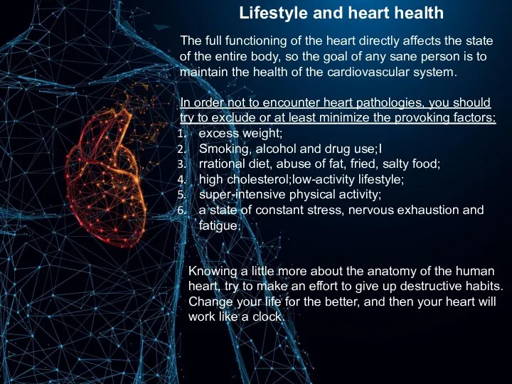
Слайд 12Lifestyle and heart health
The full functioning of the heart directly affects the
Lifestyle and heart health
The full functioning of the heart directly affects the
 С-парадокс. Размер генома
 Manchots royaux
 Физиология микроорганизмов
 Взаимоотношения в системе паразит-хозяин
 Rosen
Rosen Тест Животные
 Презентация на тему Инфекционные заболевания людей
  Секретный мир природы. Мир птиц
 Углеводы. Важнейшие моносахариды
 Зрение. Зрительный анализатор
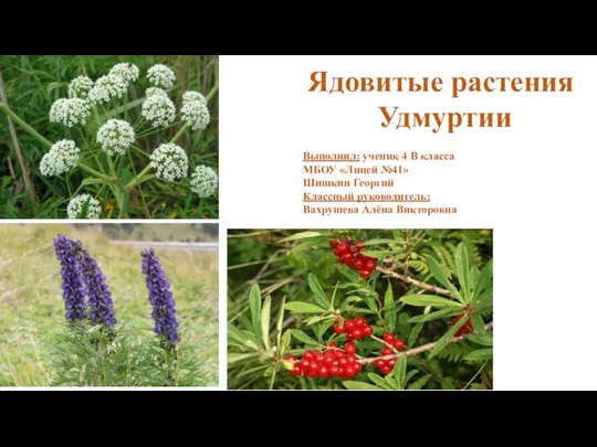 Ядовитые растения Удмуртии
 Тип простейшие. Класс споровики
 Клетка и клеточная теория Шлейдона и Шванна
 Презентация на тему "Какие бывают жуки" - презентации по Биологии
 Презентация на тему Есть ли корень у гриба?
  Нижняя челюсть, её форма и значение при рисовании головы человека
 Ладирование. По курсу Юговой
 Сцепленное наследование генов и кроссинговер
 Мох и его роль в жизни человека
 Регуляция метаболизма у бактерий
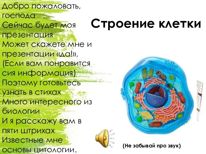 Строение клетки
 Имена в биологии. 10 класс
 Диаграмма цветка
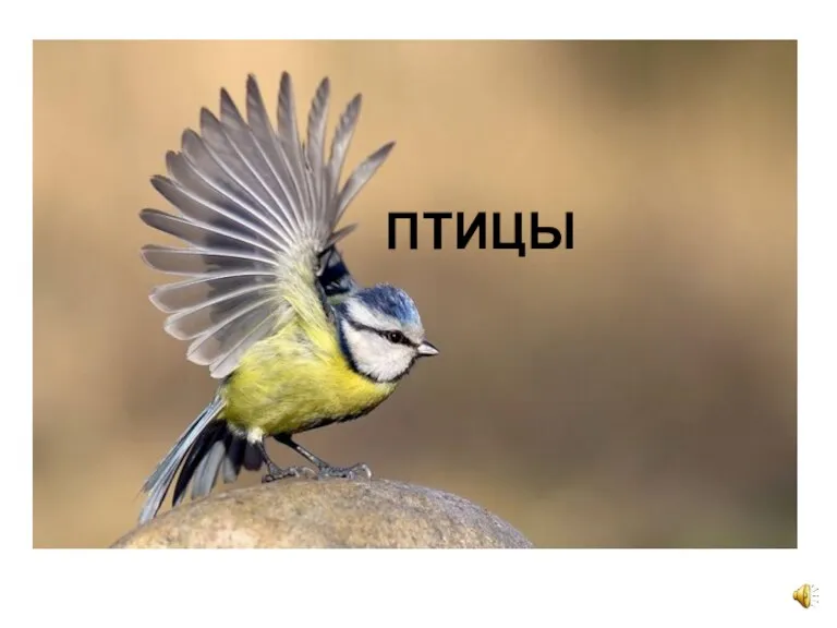 Класс Птицы
 Прямое и непрямое развитие насекомых
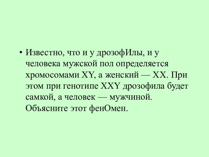 Хромосомная и балансовая теория пола
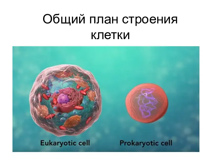 Общий план строения клетки
 Возрастное развитие систем пищеварения, выделения, опорно-двигательного аппарата, обмена веществ