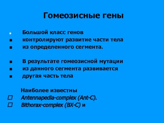
- Молекулярная генетика развития

Содержание
- 3. Молекулярная генетика развития Вельков В В 2013 Gene Regulation during Development
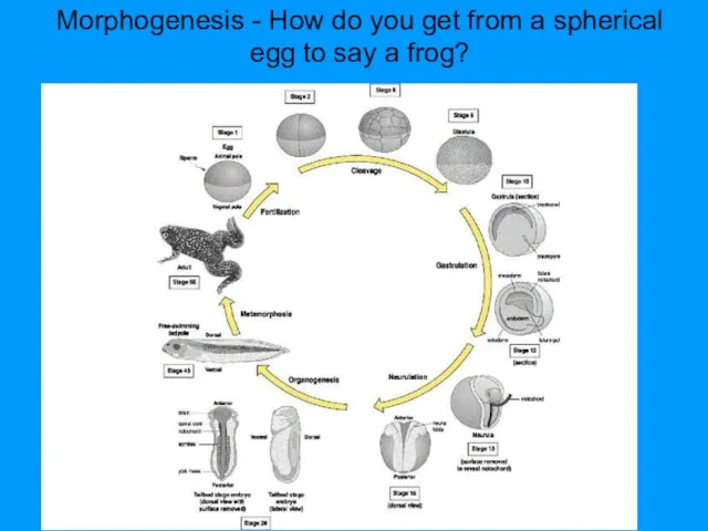
- 4. Morphogenesis - How do you get from a spherical egg to say a frog?
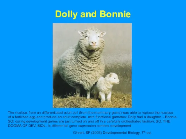
- 5. Dolly and Bonnie Gilbert, SF (2003) Developmental Biology, 7th ed. The nucleus from an differentiated adult
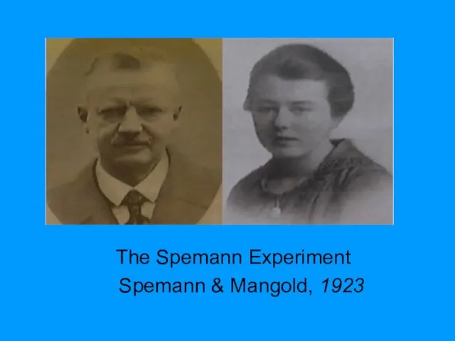
- 6. The Spemann Experiment Spemann & Mangold, 1923
- 7. Spemann Experiment Outline: Graft a tissue from one embryo into another embryo, and see what happens
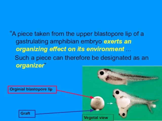
- 8. “A piece taken from the upper blastopore lip of a gastrulating amphibian embryo exerts an organizing
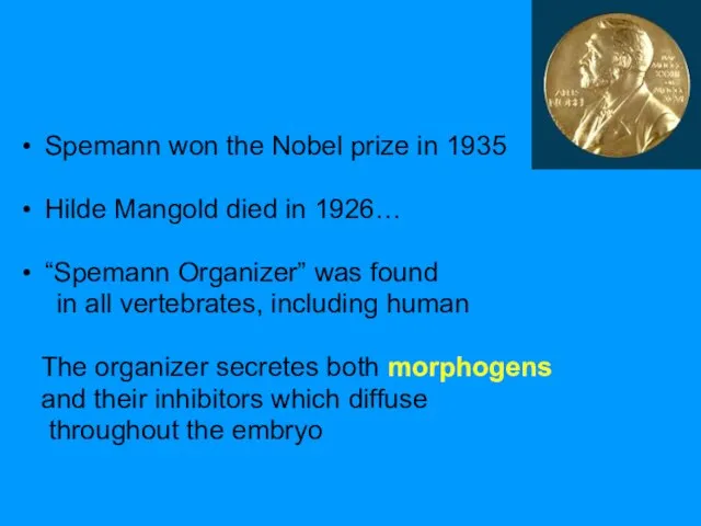
- 9. Spemann won the Nobel prize in 1935 Hilde Mangold died in 1926… “Spemann Organizer” was found
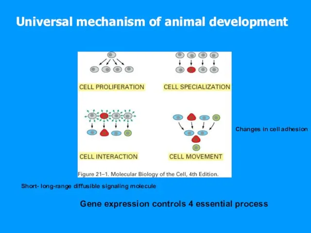
- 10. Universal mechanism of animal development Gene expression controls 4 essential process Short- long-range diffusible signaling molecule
- 11. Development is progressive Specification of cell fate: determination All cells still ‘look the same’ Can be
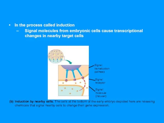
- 12. In the process called induction Signal molecules from embryonic cells cause transcriptional changes in nearby target
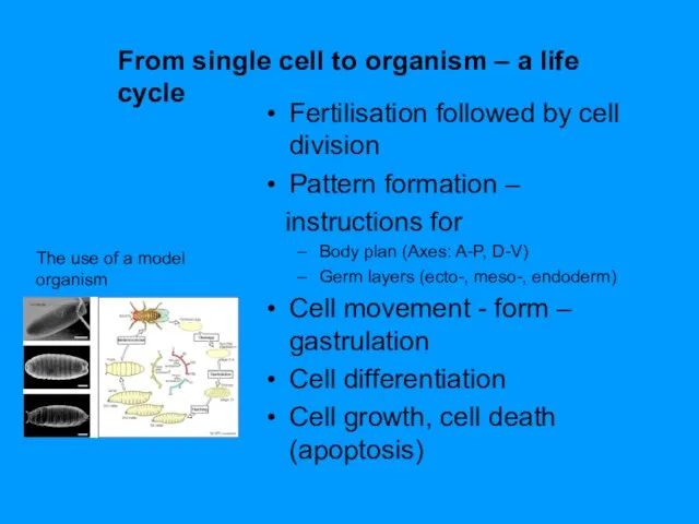
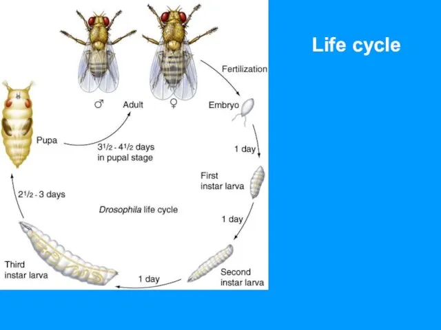
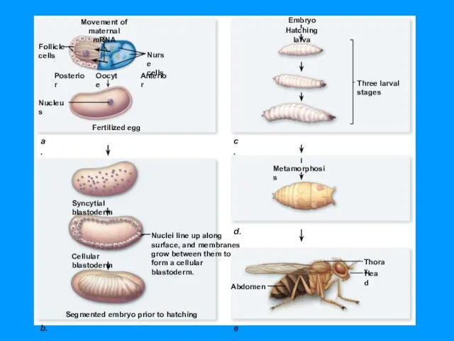
- 13. From single cell to organism – a life cycle The use of a model organism Fertilisation
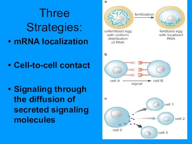
- 15. Three Strategies: mRNA localization Cell-to-cell contact Signaling through the diffusion of secreted signaling molecules
- 17. Морфогены и рецепторы морфогенов
- 18. Morphogen – substances that define different cell fates in a concentration-dependent manner Interaction of two signaling
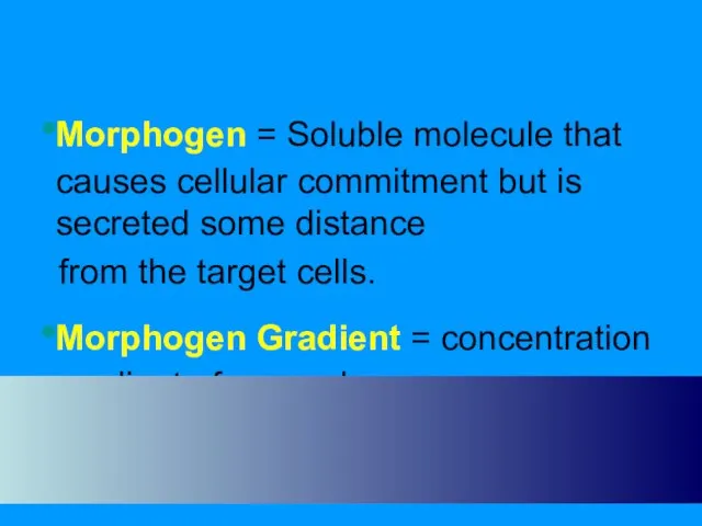
- 19. Morphogen = Soluble molecule that causes cellular commitment but is secreted some distance from the target
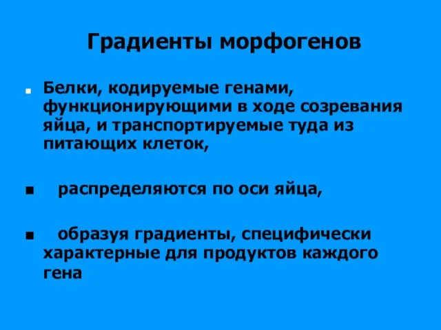
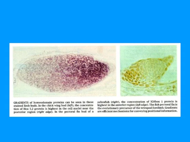
- 20. Градиенты морфогенов Белки, кодируемые генами, функционирующими в ходе созревания яйца, и транспортируемые туда из питающих клеток,
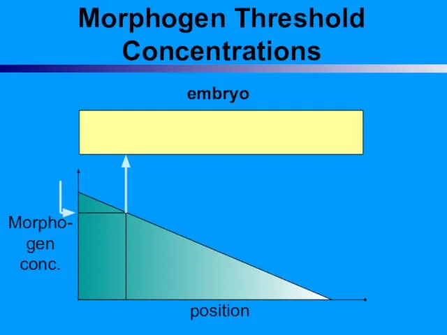
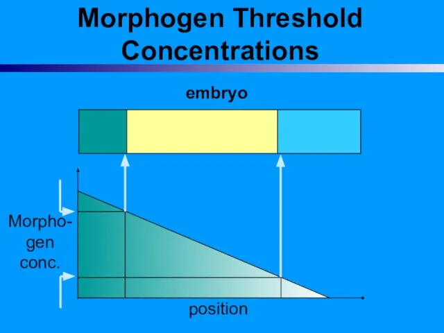
- 21. Morphogen Threshold Concentrations embryo
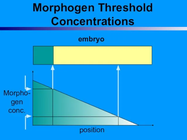
- 22. Morphogen Threshold Concentrations embryo Morpho- gen conc. position
- 23. Morphogen Threshold Concentrations embryo Morpho- gen conc. position
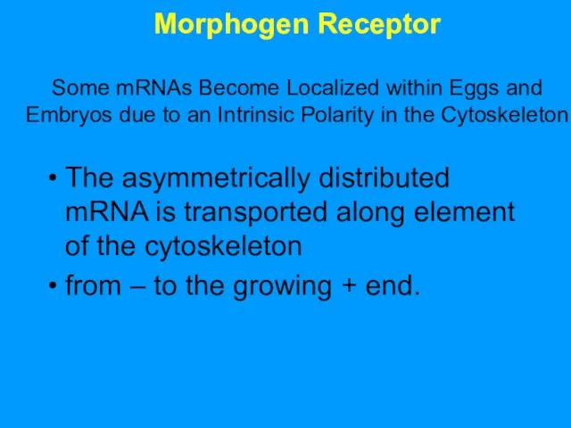
- 24. Morphogen Receptor Some mRNAs Become Localized within Eggs and Embryos due to an Intrinsic Polarity in
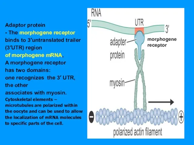
- 25. Adaptor protein - The morphogene receptor binds to 3’untranslated trailer (3’UTR) region of morphogene mRNA, A
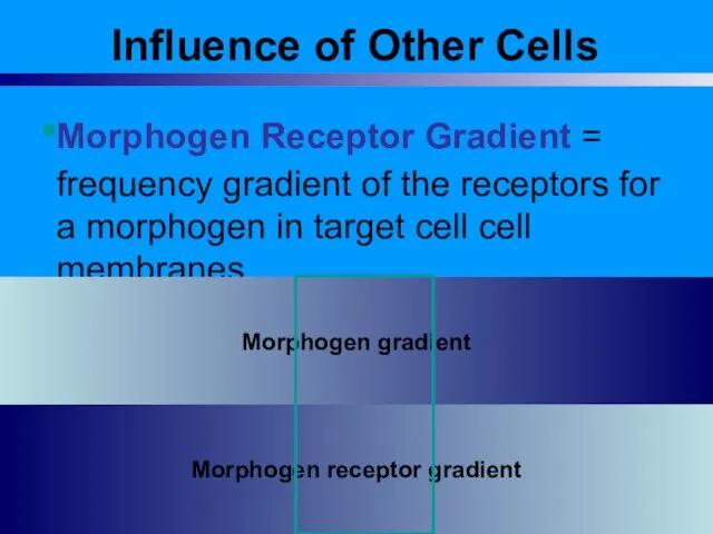
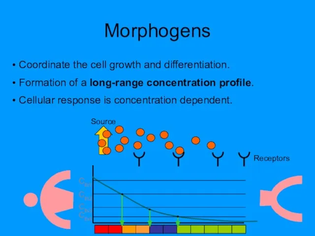
- 26. Influence of Other Cells Morphogen Receptor Gradient = frequency gradient of the receptors for a morphogen
- 27. Morphogens Coordinate the cell growth and differentiation. Formation of a long-range concentration profile. Cellular response is
- 28. Copyright © The McGraw-Hill Companies, Inc. Permission required to reproduce or display
- 29. Life cycle
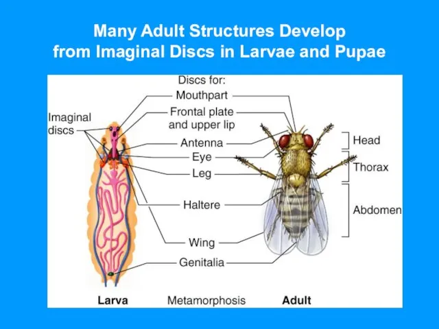
- 30. Many Adult Structures Develop from Imaginal Discs in Larvae and Pupae
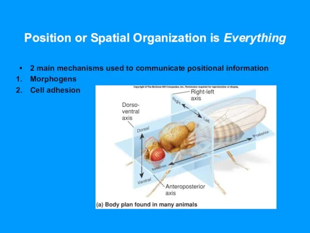
- 31. Position or Spatial Organization is Everything 2 main mechanisms used to communicate positional information Morphogens Cell
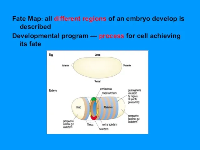
- 32. Fate Map: all different regions of an embryo develop is described Developmental program — process for
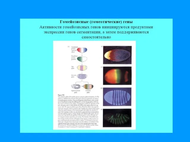
- 35. Морфогены активируют Гены сегментации активируют Гомеозисные гены активируют…
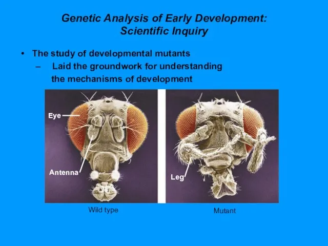
- 36. Genetic Analysis of Early Development: Scientific Inquiry The study of developmental mutants Laid the groundwork for
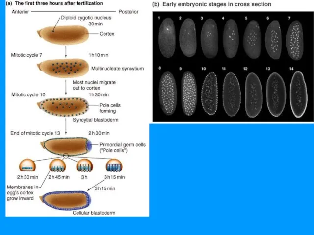
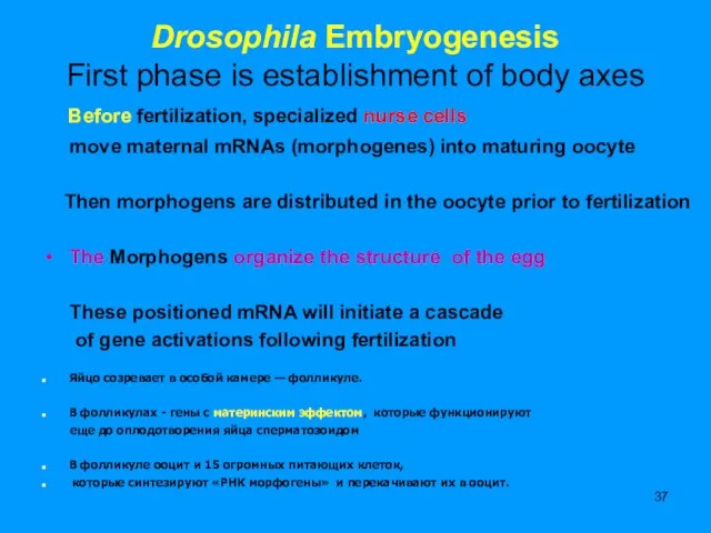
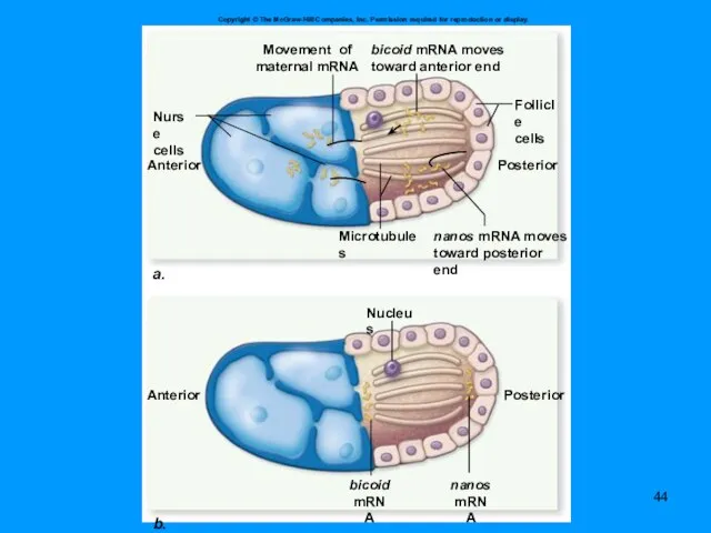
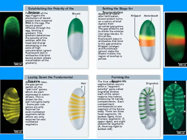
- 37. Drosophila Embryogenesis First phase is establishment of body axes Before fertilization, specialized nurse cells move maternal
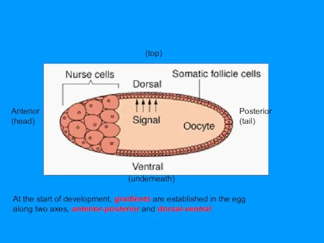
- 38. At the start of development, gradients are established in the egg along two axes, anterior-posterior and
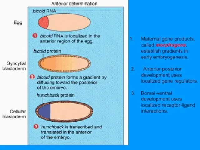
- 40. Maternal gene products, called morphogens, establish gradients in early embryogenesis. 2. Anterior-posterior development uses localized gene
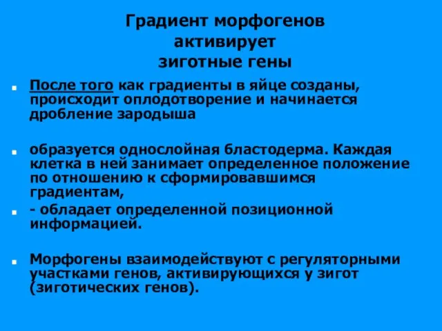
- 41. Градиент морфогенов активирует зиготные гены После того как градиенты в яйце созданы, происходит оплодотворение и начинается
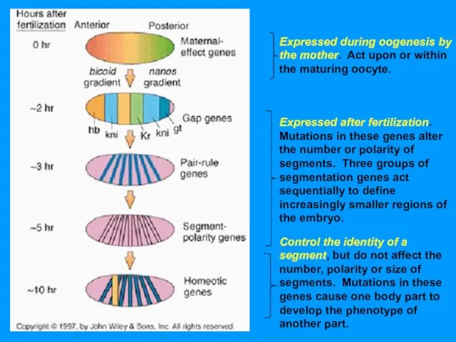
- 42. Expressed during oogenesis by the mother. Act upon or within the maturing oocyte. Expressed after fertilization.
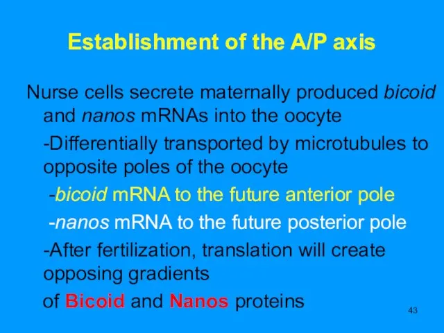
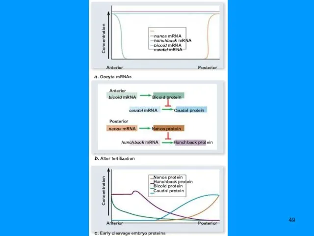
- 43. Establishment of the A/P axis Nurse cells secrete maternally produced bicoid and nanos mRNAs into the
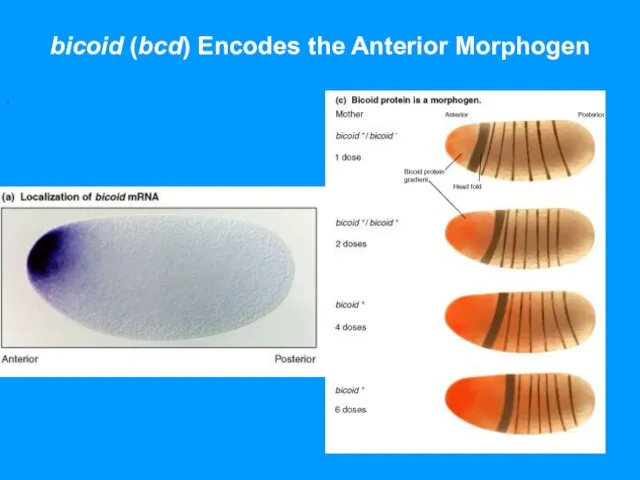
- 45. bicoid (bcd) Encodes the Anterior Morphogen .
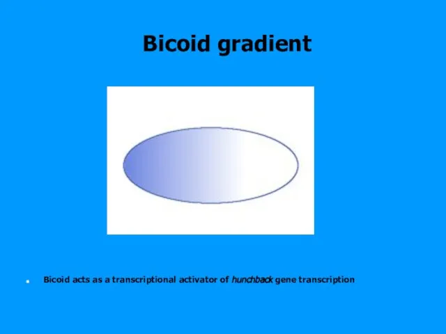
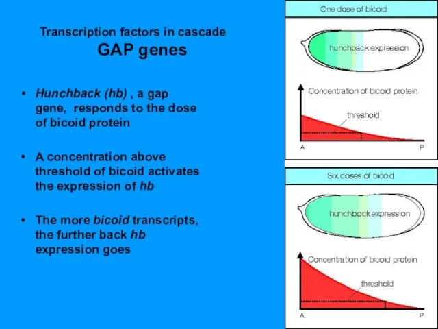
- 46. Bicoid gradient Bicoid acts as a transcriptional activator of hunchback gene transcription
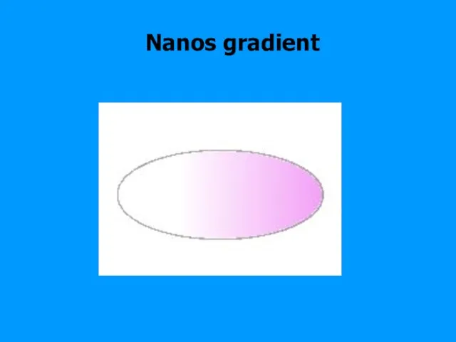
- 47. Nanos gradient
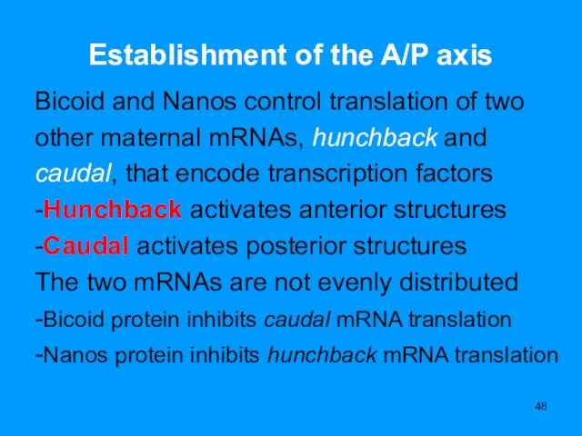
- 48. Establishment of the A/P axis Bicoid and Nanos control translation of two other maternal mRNAs, hunchback
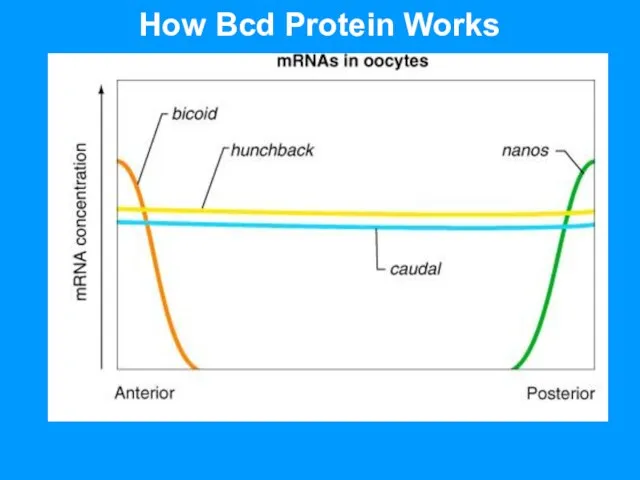
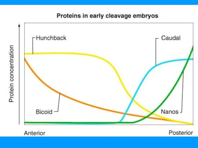
- 50. How Bcd Protein Works
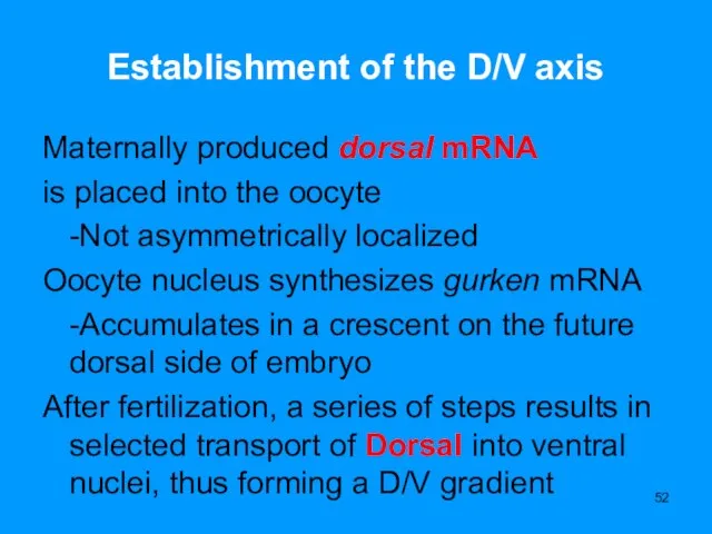
- 52. Establishment of the D/V axis Maternally produced dorsal mRNA is placed into the oocyte -Not asymmetrically
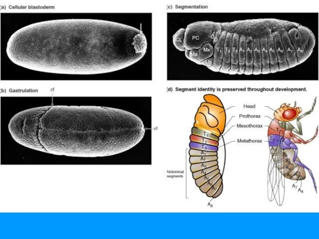
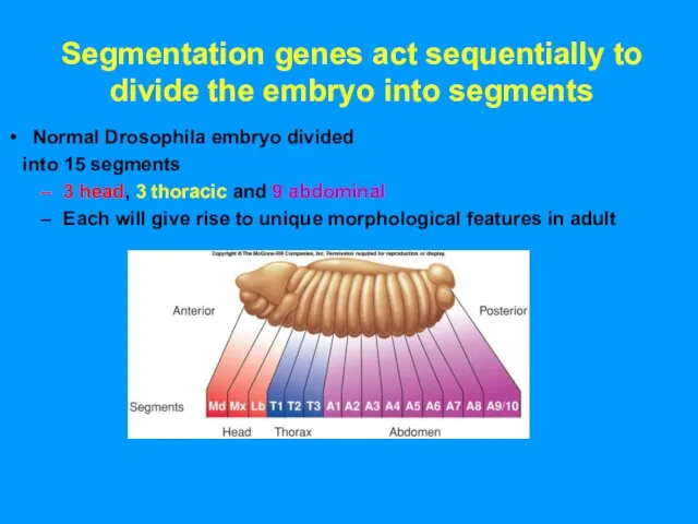
- 53. Segmentation genes act sequentially to divide the embryo into segments Normal Drosophila embryo divided into 15
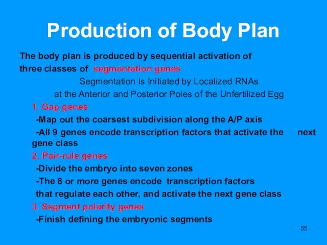
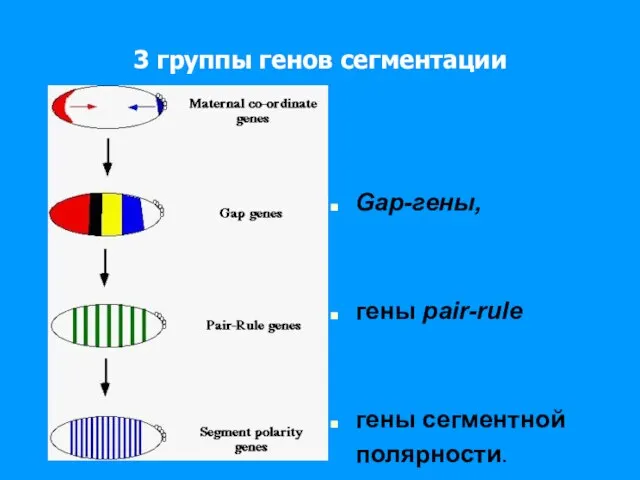
- 55. Production of Body Plan The body plan is produced by sequential activation of three classes of
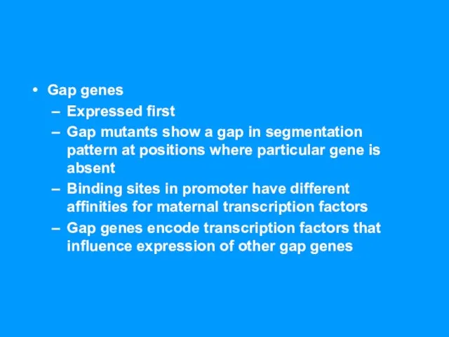
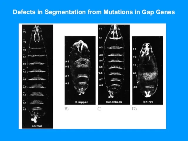
- 57. Gap genes Expressed first Gap mutants show a gap in segmentation pattern at positions where particular
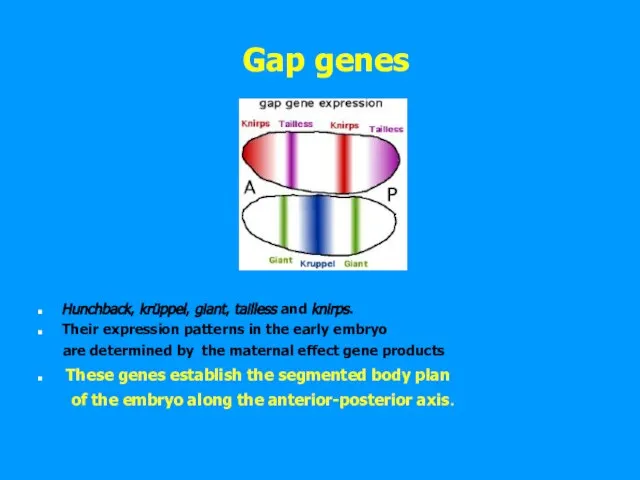
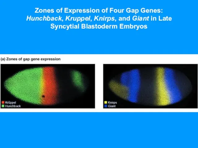
- 58. Gap genes Hunchback, krüppel, giant, tailless and knirps. Their expression patterns in the early embryo are
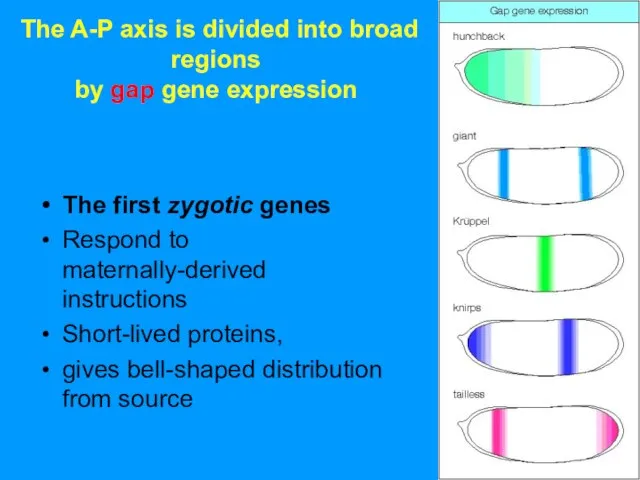
- 59. The A-P axis is divided into broad regions by gap gene expression The first zygotic genes
- 60. Zones of Expression of Four Gap Genes: Hunchback, Kruppel, Knirps, and Giant in Late Syncytial Blastoderm
- 61. Defects in Segmentation from Mutations in Gap Genes A) B) C) D)
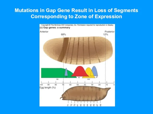
- 62. Mutations in Gap Gene Result in Loss of Segments Corresponding to Zone of Expression
- 63. Transcription factors in cascade GAP genes Hunchback (hb) , a gap gene, responds to the dose
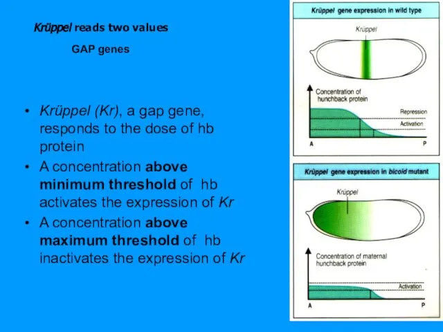
- 64. Krüppel reads two values Krüppel (Kr), a gap gene, responds to the dose of hb protein
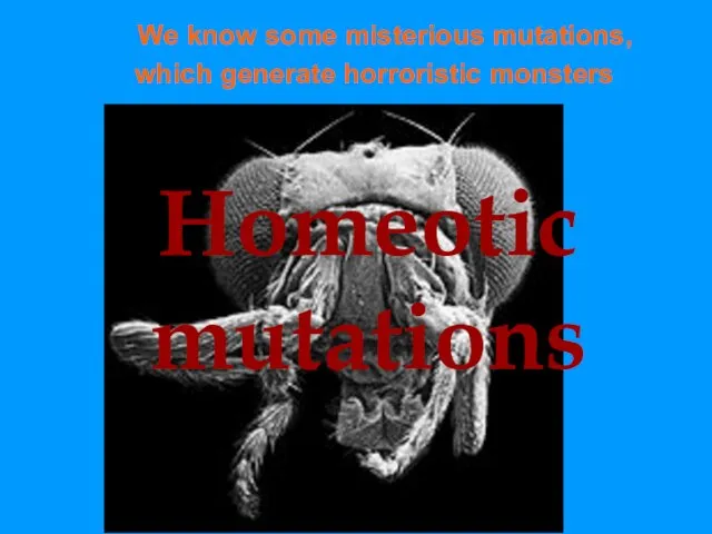
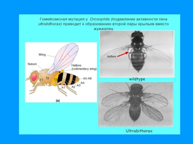
- 66. Homeotic mutations We know some misterious mutations, which generate horroristic monsters
- 67. Homeotic-Selector/ HOX Genes
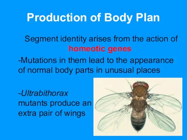
- 68. Production of Body Plan Segment identity arises from the action of homeotic genes -Mutations in them
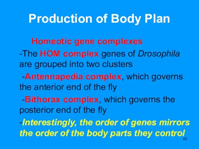
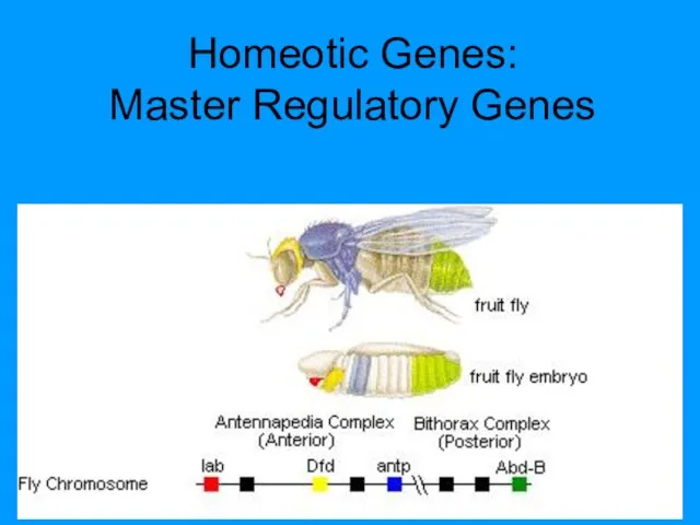
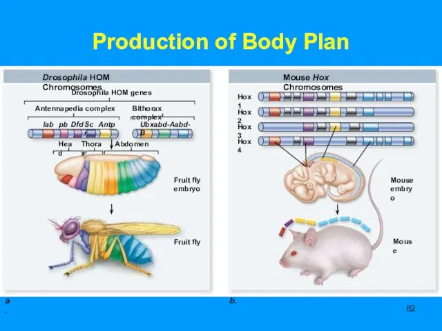
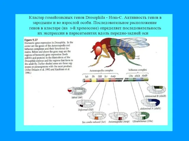
- 69. Production of Body Plan Homeotic gene complexes -The HOM complex genes of Drosophila are grouped into
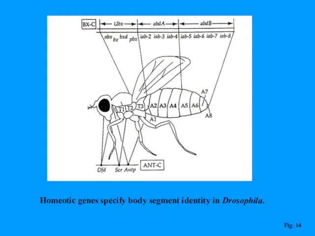
- 70. Homeotic genes specify body segment identity in Drosophila. Fig. 14
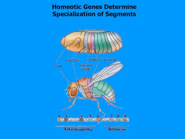
- 71. Homeotic Genes Determine Specialization of Segments
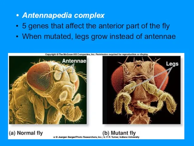
- 73. Antennapedia complex 5 genes that affect the anterior part of the fly When mutated, legs grow
- 74. Homeotic Genes: Master Regulatory Genes
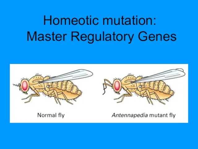
- 75. Homeotic mutation: Master Regulatory Genes
- 77. Halteres into wings Gilbert, SF (2003) Developmental Biology, 7th ed. One structure is placed in the
- 81. Production of Body Plan Homeotic gene complexes -All of these genes contain a conserved 180-base sequence,
- 82. Production of Body Plan
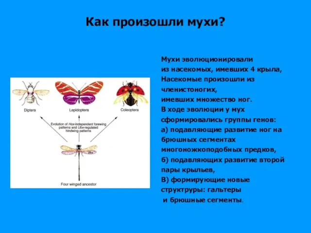
- 83. Как произошли мухи? Мухи эволюционировали из насекомых, имевших 4 крыла, Насекомые произошли из членистоногих, имевших множество
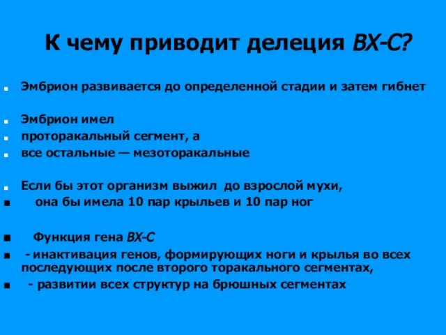
- 84. К чему приводит делеция ВХ-С? Эмбрион развивается до определенной стадии и затем гибнет Эмбрион имел проторакальный
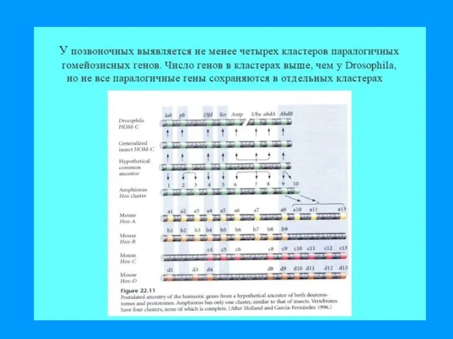
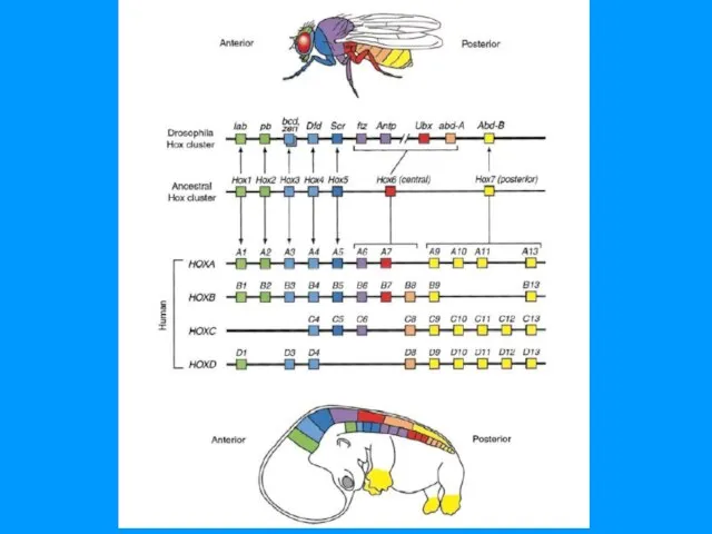
- 87. Vertebrates have four Hox complexes, with about 10 genes each. They can be aligned in 13
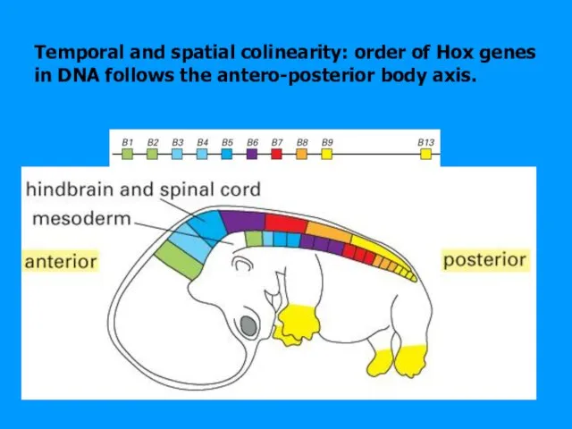
- 89. Temporal and spatial colinearity: order of Hox genes in DNA follows the antero-posterior body axis.
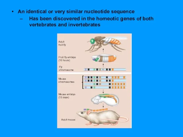
- 90. An identical or very similar nucleotide sequence Has been discovered in the homeotic genes of both
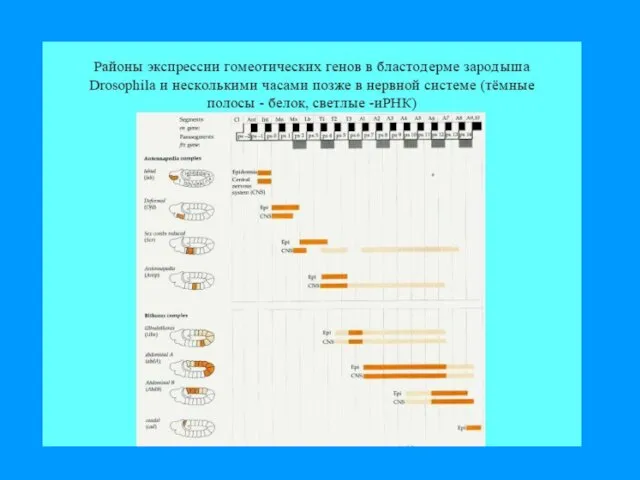
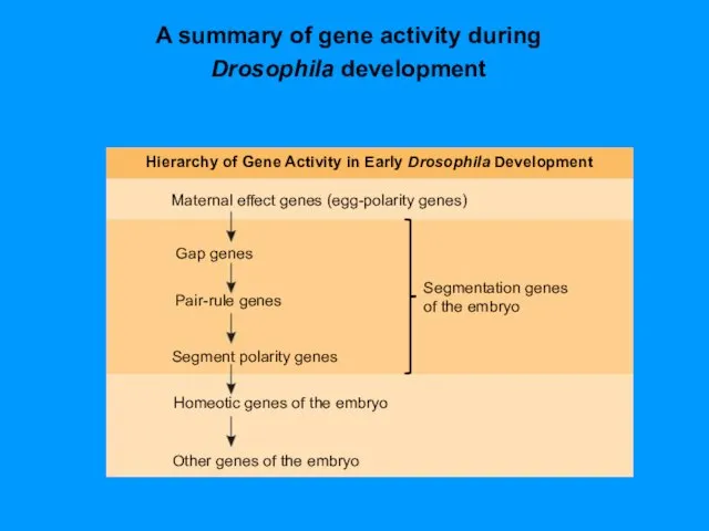
- 95. A summary of gene activity during Drosophila development
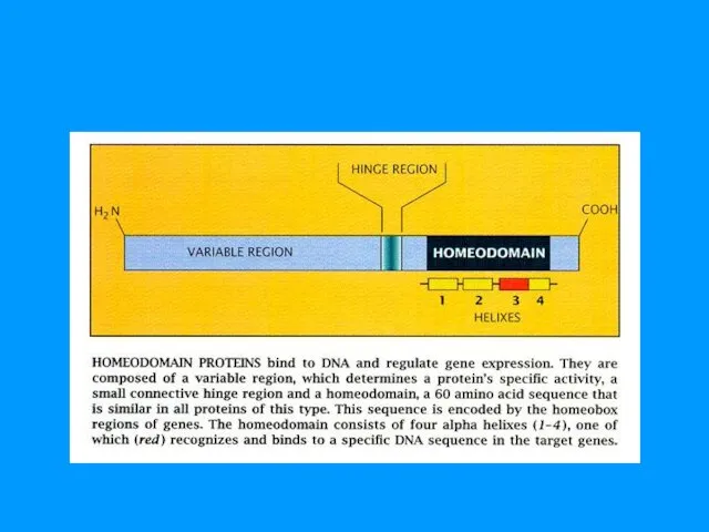
- 96. Homeobox & Homeodomain ДНК связывающий модуль факторов транскрипции генов дифференцировки 180 пн 60 аминокислотных остатков
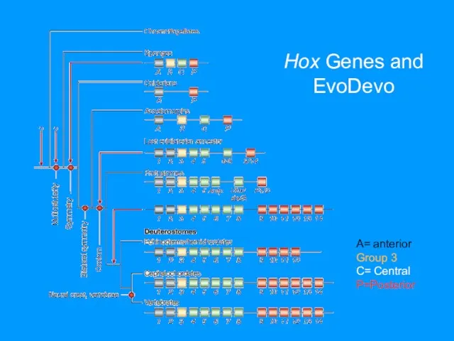
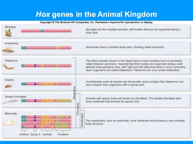
- 102. Hox Genes and EvoDevo A= anterior Group 3 C= Central P=Posterior
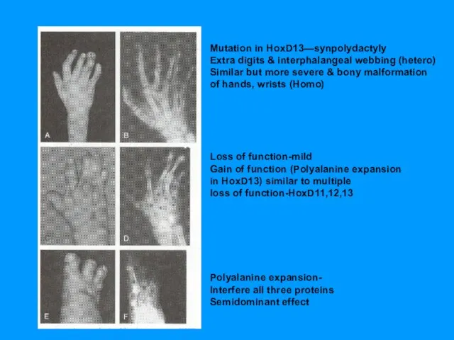
- 103. Mutation in HoxD13—synpolydactyly Extra digits & interphalangeal webbing (hetero) Similar but more severe & bony malformation
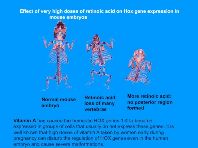
- 104. Normal mouse embryo Retinoic acid: loss of many vertebrae More retinoic acid: no posterior region formed
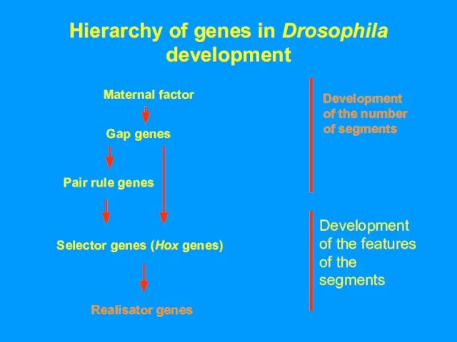
- 105. Hierarchy of genes in Drosophila development Maternal factor Development of the number of segments
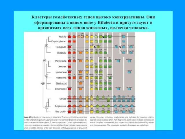
- 106. Hox genes in the Animal Kingdom
- 108. Скачать презентацию

Слайд 3Молекулярная генетика развития
Вельков В В 2013
Gene Regulation during Development
Молекулярная генетика развития
Вельков В В 2013
Gene Regulation during Development

Слайд 4Morphogenesis - How do you get from a spherical egg to say
Morphogenesis - How do you get from a spherical egg to say
Слайд 5Dolly and Bonnie
Gilbert, SF (2003) Developmental Biology, 7th ed.
The nucleus from an
Dolly and Bonnie
Gilbert, SF (2003) Developmental Biology, 7th ed.
The nucleus from an
Слайд 6The Spemann Experiment
Spemann & Mangold, 1923
The Spemann Experiment
Spemann & Mangold, 1923
Слайд 7Spemann Experiment
Outline: Graft a tissue from one embryo into another embryo, and
Spemann Experiment
Outline: Graft a tissue from one embryo into another embryo, and
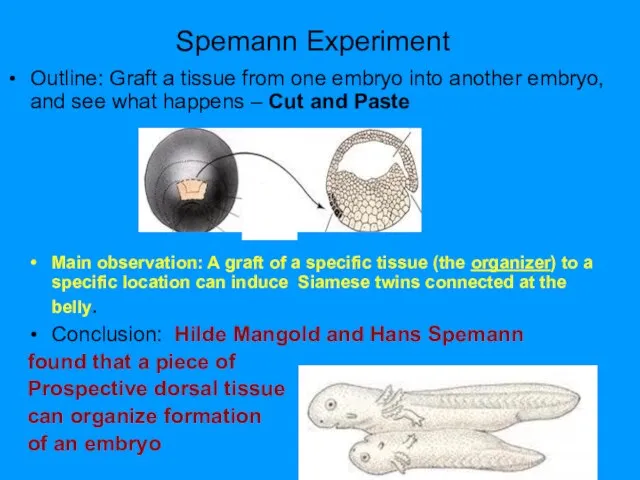
Слайд 8“A piece taken from the upper blastopore lip of a gastrulating amphibian
“A piece taken from the upper blastopore lip of a gastrulating amphibian
Слайд 9Spemann won the Nobel prize in 1935
Hilde Mangold died in 1926…
“Spemann
Hilde Mangold died in 1926…
“Spemann
Слайд 10Universal mechanism of animal development
Gene expression controls 4 essential process
Short- long-range diffusible
Universal mechanism of animal development
Gene expression controls 4 essential process
Short- long-range diffusible
Слайд 11Development is progressive
Specification of cell fate: determination
All cells still ‘look the same’
Can
Development is progressive
Specification of cell fate: determination
All cells still ‘look the same’
Can
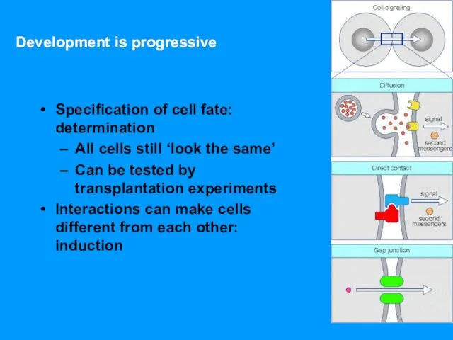
Слайд 12In the process called induction
Signal molecules from embryonic cells cause transcriptional changes
In the process called induction
Signal molecules from embryonic cells cause transcriptional changes
Слайд 13From single cell to organism – a life cycle
The use of a
From single cell to organism – a life cycle
The use of a

Слайд 15Three Strategies:
mRNA localization
Cell-to-cell contact
Signaling through the diffusion of secreted signaling molecules
Three Strategies:
mRNA localization
Cell-to-cell contact
Signaling through the diffusion of secreted signaling molecules

Слайд 17
Морфогены
и рецепторы
морфогенов
Морфогены
и рецепторы
морфогенов

Слайд 18Morphogen – substances that define different cell fates in a concentration-dependent manner
Interaction
Morphogen – substances that define different cell fates in a concentration-dependent manner
Interaction
Слайд 19Morphogen = Soluble molecule that causes cellular commitment but is secreted some
Morphogen = Soluble molecule that causes cellular commitment but is secreted some
Слайд 20Градиенты морфогенов
Белки, кодируемые генами, функционирующими в ходе созревания яйца, и транспортируемые туда
Градиенты морфогенов
Белки, кодируемые генами, функционирующими в ходе созревания яйца, и транспортируемые туда
Слайд 21Morphogen Threshold Concentrations
embryo
Morphogen Threshold Concentrations
embryo
Слайд 22Morphogen Threshold Concentrations
embryo
Morpho-
gen
conc.
position
Morphogen Threshold Concentrations
embryo
Morpho-
gen
conc.
position
Слайд 23Morphogen Threshold Concentrations
embryo
Morpho-
gen
conc.
position
Morphogen Threshold Concentrations
embryo
Morpho-
gen
conc.
position
Слайд 24Morphogen Receptor
Some mRNAs Become Localized within Eggs and Embryos due to
Morphogen Receptor Some mRNAs Become Localized within Eggs and Embryos due to
Слайд 25Adaptor protein
- The morphogene receptor
binds to 3’untranslated trailer
(3’UTR) region
of morphogene
Adaptor protein
- The morphogene receptor
binds to 3’untranslated trailer
(3’UTR) region
of morphogene
Слайд 26Influence of Other Cells
Morphogen Receptor Gradient = frequency gradient of the receptors
Influence of Other Cells
Morphogen Receptor Gradient = frequency gradient of the receptors
Слайд 27Morphogens
Coordinate the cell growth and differentiation.
Formation of a long-range concentration
Morphogens
Coordinate the cell growth and differentiation.
Formation of a long-range concentration
Слайд 28Copyright © The McGraw-Hill Companies, Inc. Permission required to reproduce or display
Copyright © The McGraw-Hill Companies, Inc. Permission required to reproduce or display
Слайд 29Life cycle
Life cycle
Слайд 30Many Adult Structures Develop
from Imaginal Discs in Larvae and Pupae
Many Adult Structures Develop
from Imaginal Discs in Larvae and Pupae
Слайд 31Position or Spatial Organization is Everything
2 main mechanisms used to communicate positional
Position or Spatial Organization is Everything
2 main mechanisms used to communicate positional
Слайд 32Fate Map: all different regions of an embryo develop is described
Developmental program
Fate Map: all different regions of an embryo develop is described
Developmental program
Слайд 35Морфогены
активируют
Гены сегментации
активируют
Гомеозисные гены
активируют…
активируют
Гены сегментации
активируют
Гомеозисные гены
активируют…

Слайд 36Genetic Analysis of Early Development:
Scientific Inquiry
The study of developmental mutants
Laid the
Genetic Analysis of Early Development:
Scientific Inquiry
The study of developmental mutants
Laid the
Слайд 37Drosophila Embryogenesis
First phase is establishment of body axes
Before fertilization, specialized nurse
Drosophila Embryogenesis
First phase is establishment of body axes
Before fertilization, specialized nurse
Слайд 38At the start of development, gradients are established in the egg
along
At the start of development, gradients are established in the egg
along
Слайд 40Maternal gene products, called morphogens, establish gradients in early embryogenesis.
2. Anterior-posterior development
Maternal gene products, called morphogens, establish gradients in early embryogenesis.
2. Anterior-posterior development
Слайд 41Градиент морфогенов
активирует
зиготные гены
После того как градиенты в яйце созданы, происходит
Градиент морфогенов
активирует
зиготные гены
После того как градиенты в яйце созданы, происходит
Слайд 42Expressed during oogenesis by the mother. Act upon or within the maturing
Expressed during oogenesis by the mother. Act upon or within the maturing
Слайд 43Establishment of the A/P axis
Nurse cells secrete maternally produced bicoid and nanos
Establishment of the A/P axis
Nurse cells secrete maternally produced bicoid and nanos
Слайд 45bicoid (bcd) Encodes the Anterior Morphogen
.
bicoid (bcd) Encodes the Anterior Morphogen
.
Слайд 46Bicoid gradient
Bicoid acts as a transcriptional activator of hunchback gene transcription
Bicoid gradient
Bicoid acts as a transcriptional activator of hunchback gene transcription
Слайд 47Nanos gradient
Nanos gradient
Слайд 48Establishment of the A/P axis
Bicoid and Nanos control translation of two
other maternal
Establishment of the A/P axis
Bicoid and Nanos control translation of two
other maternal
Слайд 50How Bcd Protein Works
How Bcd Protein Works
Слайд 52Establishment of the D/V axis
Maternally produced dorsal mRNA
is placed into the
Establishment of the D/V axis
Maternally produced dorsal mRNA
is placed into the
Слайд 53Segmentation genes act sequentially to divide the embryo into segments
Normal Drosophila embryo
Segmentation genes act sequentially to divide the embryo into segments
Normal Drosophila embryo
Слайд 55Production of Body Plan
The body plan is produced by sequential activation of
Production of Body Plan
The body plan is produced by sequential activation of
Слайд 57Gap genes
Expressed first
Gap mutants show a gap in segmentation pattern at positions
Gap genes
Expressed first
Gap mutants show a gap in segmentation pattern at positions
Слайд 58Gap genes
Hunchback, krüppel, giant, tailless and knirps.
Their expression patterns in the
Gap genes
Hunchback, krüppel, giant, tailless and knirps.
Their expression patterns in the
Слайд 59The A-P axis is divided into broad regions
by gap gene expression
The
The A-P axis is divided into broad regions
by gap gene expression
The
Слайд 60Zones of Expression of Four Gap Genes:
Hunchback, Kruppel, Knirps, and Giant
Zones of Expression of Four Gap Genes: Hunchback, Kruppel, Knirps, and Giant
Слайд 61Defects in Segmentation from Mutations in Gap Genes
A)
B)
C)
D)
Defects in Segmentation from Mutations in Gap Genes
A)
B)
C)
D)
Слайд 62Mutations in Gap Gene Result in Loss of Segments Corresponding to Zone
Mutations in Gap Gene Result in Loss of Segments Corresponding to Zone
Слайд 63 Transcription factors in cascade
GAP genes
Hunchback (hb) , a gap gene, responds
Transcription factors in cascade
GAP genes
Hunchback (hb) , a gap gene, responds
Слайд 64Krüppel reads two values
Krüppel (Kr), a gap gene, responds to the dose
Krüppel reads two values
Krüppel (Kr), a gap gene, responds to the dose
Слайд 66Homeotic mutations
We know some misterious mutations,
which generate horroristic monsters
Homeotic mutations
We know some misterious mutations,
which generate horroristic monsters
Слайд 67Homeotic-Selector/ HOX Genes
Homeotic-Selector/ HOX Genes
Слайд 68Production of Body Plan
Segment identity arises from the action of homeotic
Production of Body Plan
Segment identity arises from the action of homeotic
Слайд 69Production of Body Plan
Homeotic gene complexes
-The HOM complex genes of Drosophila
Production of Body Plan
Homeotic gene complexes
-The HOM complex genes of Drosophila
Слайд 70Homeotic genes specify body segment identity in Drosophila.
Fig. 14
Homeotic genes specify body segment identity in Drosophila.
Fig. 14
Слайд 71Homeotic Genes Determine
Specialization of Segments
Homeotic Genes Determine
Specialization of Segments
Слайд 73Antennapedia complex
5 genes that affect the anterior part of the fly
When mutated,
Antennapedia complex
5 genes that affect the anterior part of the fly
When mutated,
Слайд 74Homeotic Genes:
Master Regulatory Genes
Homeotic Genes:
Master Regulatory Genes
Слайд 75Homeotic mutation:
Master Regulatory Genes
Homeotic mutation:
Master Regulatory Genes
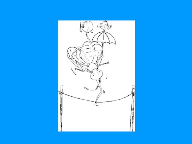
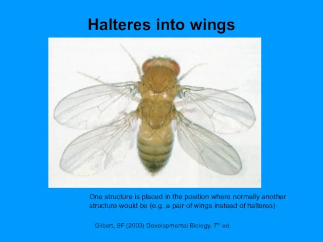
Слайд 77Halteres into wings
Gilbert, SF (2003) Developmental Biology, 7th ed.
One structure is placed
Halteres into wings
Gilbert, SF (2003) Developmental Biology, 7th ed.
One structure is placed
Слайд 81Production of Body Plan
Homeotic gene complexes
-All of these genes contain a conserved
Production of Body Plan
Homeotic gene complexes
-All of these genes contain a conserved
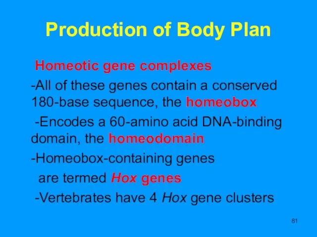
Слайд 82Production of Body Plan
Production of Body Plan
Слайд 83Как произошли мухи?
Мухи эволюционировали
из насекомых, имевших 4 крыла,
Насекомые произошли из
членистоногих,
Как произошли мухи?
Мухи эволюционировали
из насекомых, имевших 4 крыла,
Насекомые произошли из
членистоногих,
Слайд 84К чему приводит делеция ВХ-С?
Эмбрион развивается до определенной стадии и затем
К чему приводит делеция ВХ-С?
Эмбрион развивается до определенной стадии и затем
Слайд 87Vertebrates have four Hox complexes,
with about 10 genes each.
They can
Vertebrates have four Hox complexes,
with about 10 genes each.
They can
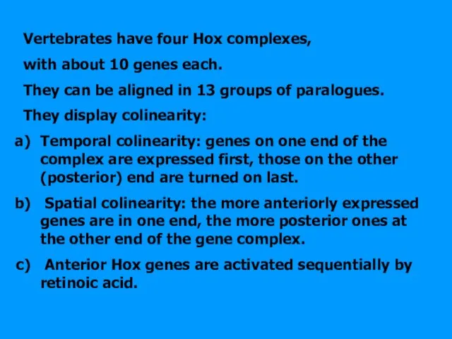
Слайд 89Temporal and spatial colinearity: order of Hox genes in DNA follows the
Temporal and spatial colinearity: order of Hox genes in DNA follows the
Слайд 90An identical or very similar nucleotide sequence
Has been discovered in the homeotic
An identical or very similar nucleotide sequence
Has been discovered in the homeotic
Слайд 95A summary of gene activity during
Drosophila development
A summary of gene activity during
Drosophila development
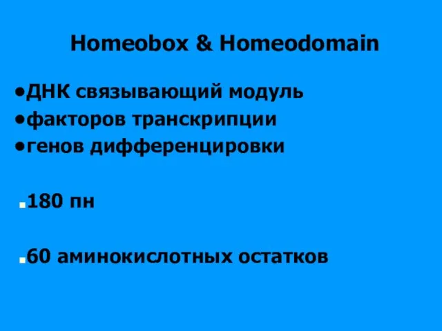
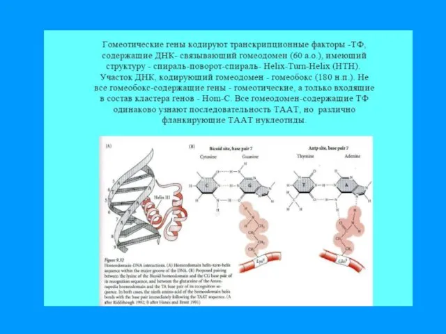
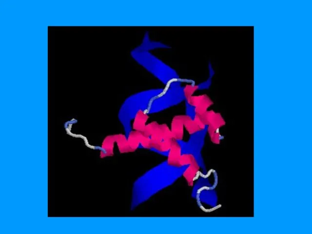
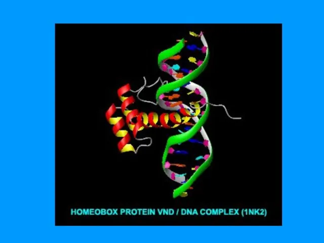
Слайд 96Homeobox & Homeodomain
ДНК связывающий модуль
факторов транскрипции
генов дифференцировки
180 пн
60
Homeobox & Homeodomain
ДНК связывающий модуль
факторов транскрипции
генов дифференцировки
180 пн
60
Слайд 102Hox Genes and EvoDevo
A= anterior
Group 3
C= Central
P=Posterior
Hox Genes and EvoDevo
A= anterior
Group 3
C= Central
P=Posterior
Слайд 103Mutation in HoxD13—synpolydactyly
Extra digits & interphalangeal webbing (hetero)
Similar but more severe &
Mutation in HoxD13—synpolydactyly
Extra digits & interphalangeal webbing (hetero)
Similar but more severe &
Слайд 104Normal mouse
embryo
Retinoic acid:
loss of many
vertebrae
More retinoic acid:
no posterior
Normal mouse
embryo
Retinoic acid:
loss of many
vertebrae
More retinoic acid: no posterior
Слайд 105Hierarchy of genes in Drosophila development
Maternal factor
Development of the number of segments
Hierarchy of genes in Drosophila development
Maternal factor
Development of the number of segments
Слайд 106Hox genes in the Animal Kingdom
Hox genes in the Animal Kingdom
 Судебный этикет как составляющая культуры уголовно-процессуальной деятельности. Нормативные основы судебного этикета
 Оружие массового поражения
 Модельный ряд грузовых автомобилей Mercedes-Benz
 Энергетика: вчера, сегодня, завтра
 ГИИС ЭБ. Запрос на аннулирование
 Элементы интернет-маркетинга и их взаимодействие
 1. ОСНОВНЫЕ ПОНЯТИЯ Компьютерный исполнитель – это виртуальный объект, действующий в виртуальной среде обитания. Примеры: –Чертеж
 Beerfest
 Красота осени
 Социально-психологическая служба в школе
 Как принимать управленческие решения
 Денежные переводы в Республике Таджикистан
 Темы в Drupal 6 Что нового, и чем оно грозит
 Память в камне
 Помещение для открытия магазина Белорусская косметика
 Огневая подготовка. Версия 3
 Горный Дагестан
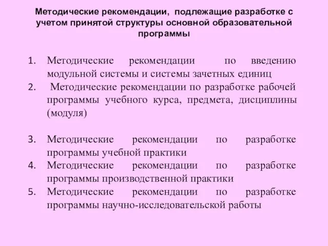 Методические рекомендации по введению модульной системы и системы зачетных единиц Методические рекомендации по разработке рабо
 Внутренний мир болезни
 Техническое обслуживание и ремонт трансформатора ТДТНГ 40500\115\38,5\6,6 кВ Смоленск – 1
 Кинестетик
 Илья Ефимович Репин –великий русский художник
 Václavské náměstí
 Презентация на тему Иван третий (4 класс)
 11 причин инвестировать в Свердловскую область
 Технология установки врезного замка
 Религия и мораль
 Караевское сельское поселение. Золотые руки мастера по бисероплетению