- Regulation of the Respiration

Содержание
- 2. Respiratory Center and Formation of the Respiratory Rhythm 1 Respiratory Center
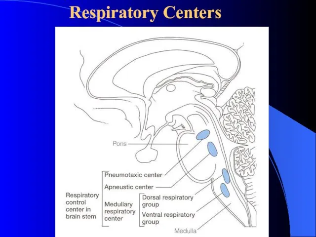
- 5. Respiratory Centers
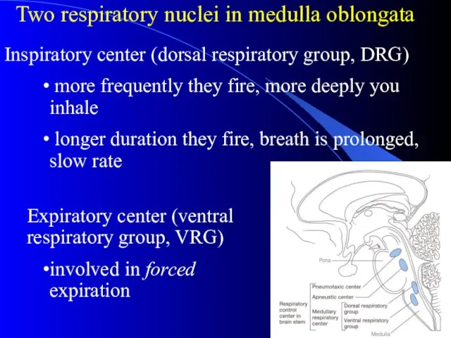
- 6. Two respiratory nuclei in medulla oblongata Expiratory center (ventral respiratory group, VRG) involved in forced expiration
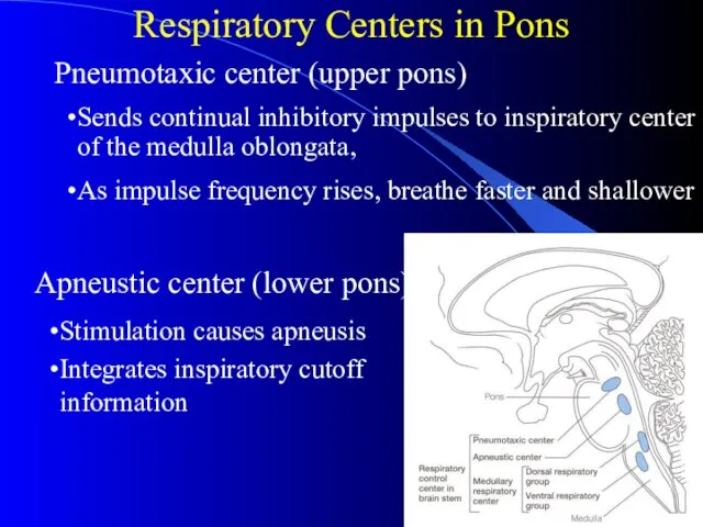
- 7. Respiratory Centers in Pons Apneustic center (lower pons) Sends continual inhibitory impulses to inspiratory center of
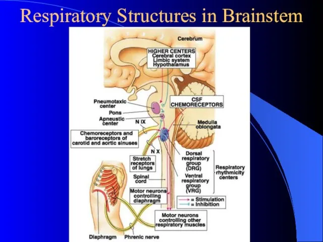
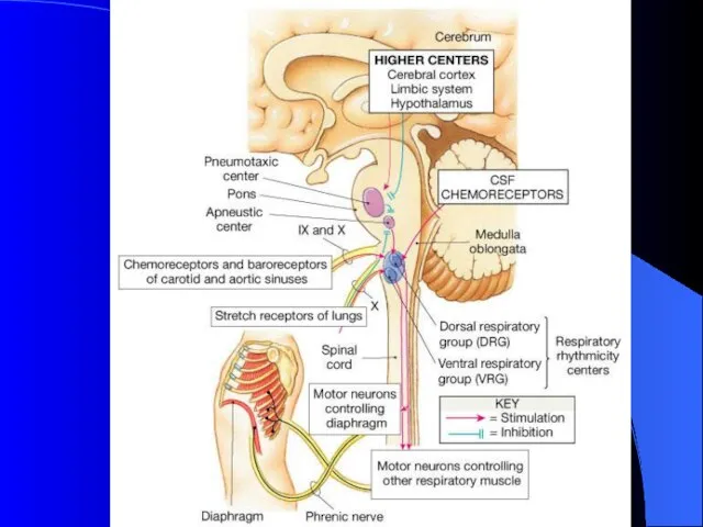
- 8. Respiratory Structures in Brainstem
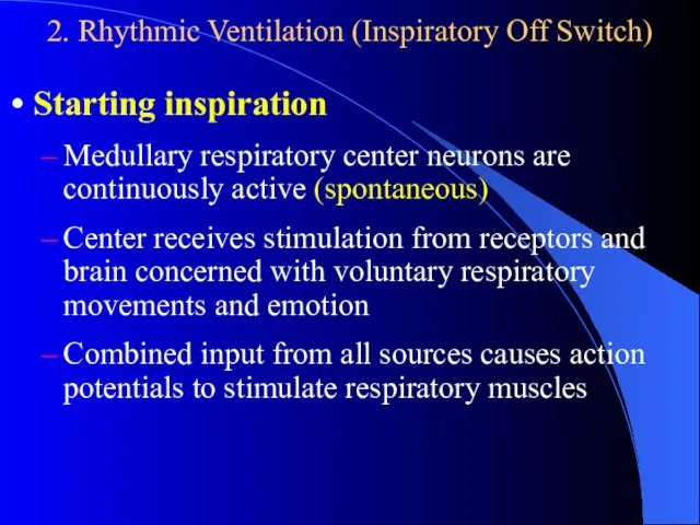
- 9. 2. Rhythmic Ventilation (Inspiratory Off Switch) Starting inspiration Medullary respiratory center neurons are continuously active (spontaneous)
- 10. Increasing inspiration More and more neurons are activated Stopping inspiration Neurons receive input from pontine group
- 11. 3. Higher Respiratory Centers Modulate the activity of the more primitive controlling centers in the medulla
- 12. II Pulmonary Reflex Chemoreceptor Reflex
- 13. Two Sets of Chemoreceptors Exist Central Chemoreceptors Responsive to increased arterial PCO2 Act by way of
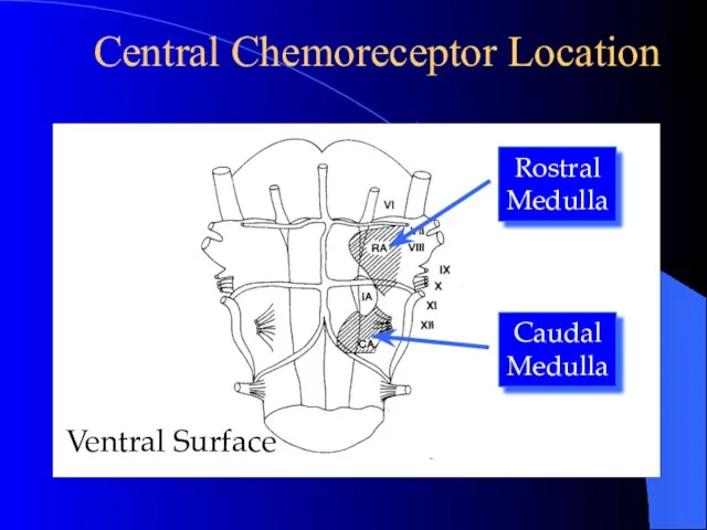
- 14. Central Chemoreceptor Location Rostral Medulla Caudal Medulla Ventral Surface
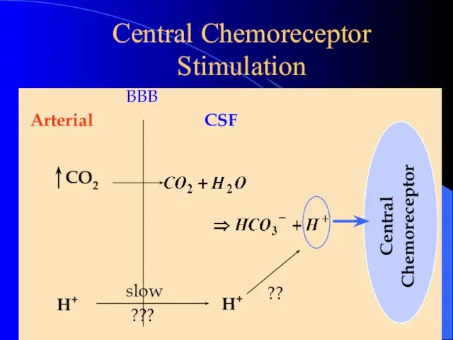
- 15. Central Chemoreceptor Stimulation
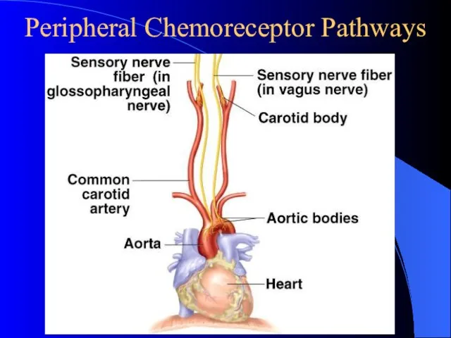
- 16. Peripheral Chemoreceptor Pathways
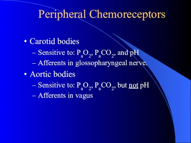
- 17. Peripheral Chemoreceptors Carotid bodies Sensitive to: PaO2, PaCO2, and pH Afferents in glossopharyngeal nerve. Aortic bodies
- 19. Carotid Body Function High flow per unit weight: (2 L/min/100 g) High carotid body VO2 consumption:
- 20. Carotid Body Response Critical PO2 Hypercapnea Acidosis Hypocapnea Alkalosis
- 21. Carbon Dioxide, Oxygen and pH Influence Ventilation (through peripheral receptor) Peripheral chemoreceptorssensitive to PO2, PCO2 and
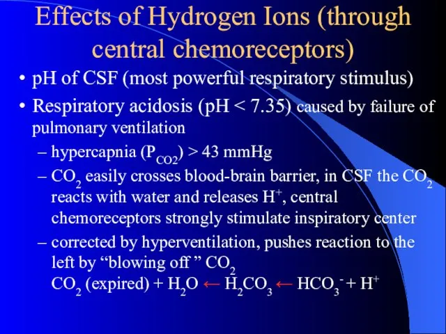
- 22. Effects of Hydrogen Ions (through central chemoreceptors) pH of CSF (most powerful respiratory stimulus) Respiratory acidosis
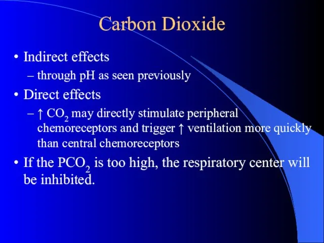
- 23. Carbon Dioxide Indirect effects through pH as seen previously Direct effects ↑ CO2 may directly stimulate
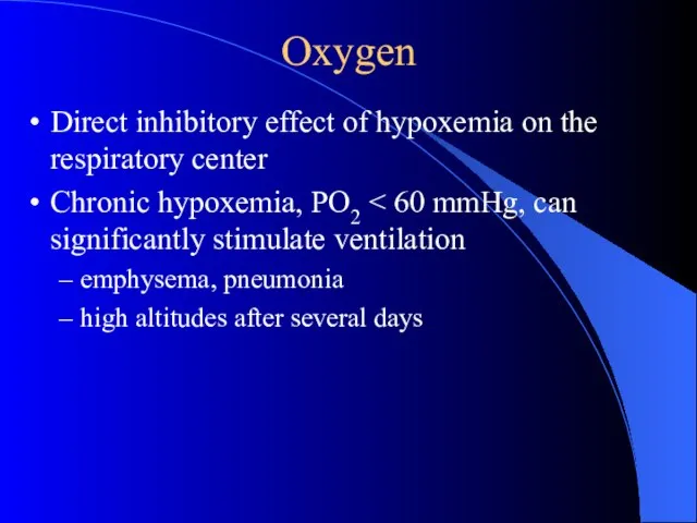
- 24. Oxygen Direct inhibitory effect of hypoxemia on the respiratory center Chronic hypoxemia, PO2 emphysema, pneumonia high
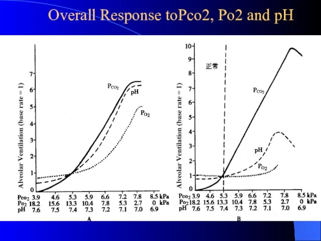
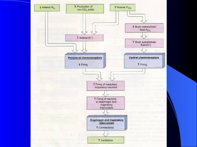
- 25. Overall Response toPco2, Po2 and pH
- 27. 2. Neuroreceptor reflex
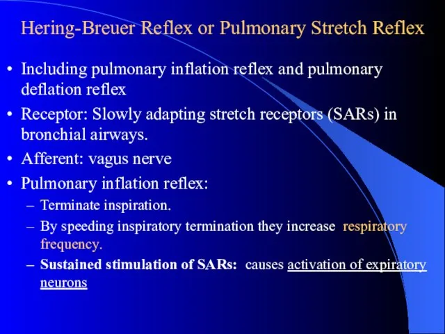
- 28. Hering-Breuer Reflex or Pulmonary Stretch Reflex Including pulmonary inflation reflex and pulmonary deflation reflex Receptor: Slowly
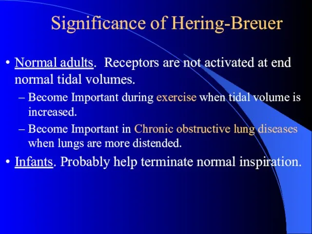
- 30. Significance of Hering-Breuer Normal adults. Receptors are not activated at end normal tidal volumes. Become Important
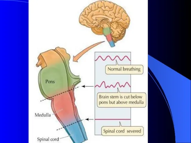
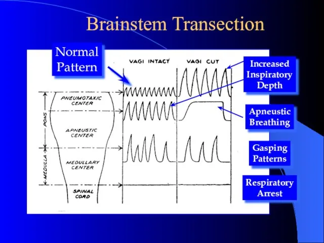
- 31. Brainstem Transection
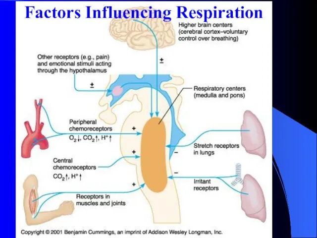
- 32. Factors Influencing Respiration
- 34. Скачать презентацию
Слайд 5Respiratory Centers
Respiratory Centers
Слайд 6Two respiratory nuclei in medulla oblongata
Expiratory center (ventral respiratory group, VRG)
involved in
Two respiratory nuclei in medulla oblongata
Expiratory center (ventral respiratory group, VRG)
involved in
Слайд 7Respiratory Centers in Pons
Apneustic center (lower pons)
Sends continual inhibitory impulses to
Respiratory Centers in Pons
Apneustic center (lower pons)
Sends continual inhibitory impulses to
Слайд 8Respiratory Structures in Brainstem
Respiratory Structures in Brainstem
Слайд 92. Rhythmic Ventilation (Inspiratory Off Switch)
Starting inspiration
Medullary respiratory center neurons are continuously
2. Rhythmic Ventilation (Inspiratory Off Switch)
Starting inspiration
Medullary respiratory center neurons are continuously
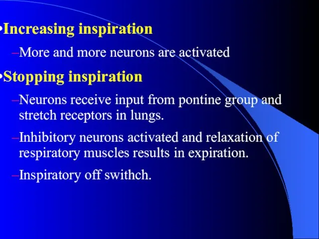
Слайд 10Increasing inspiration
More and more neurons are activated
Stopping inspiration
Neurons receive input from pontine
Increasing inspiration
More and more neurons are activated
Stopping inspiration
Neurons receive input from pontine
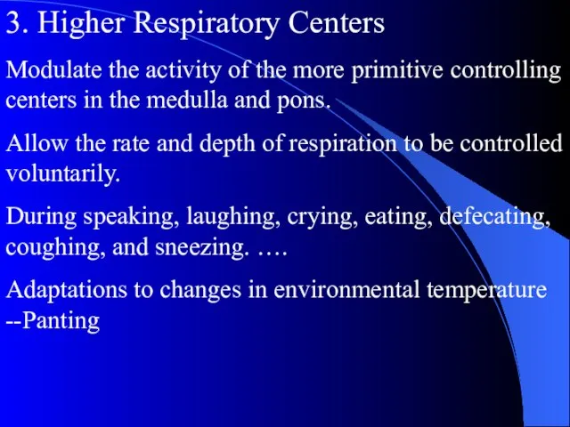
Слайд 113. Higher Respiratory Centers
Modulate the activity of the more primitive controlling centers
3. Higher Respiratory Centers
Modulate the activity of the more primitive controlling centers
Слайд 12II Pulmonary Reflex
Chemoreceptor Reflex
II Pulmonary Reflex
Chemoreceptor Reflex
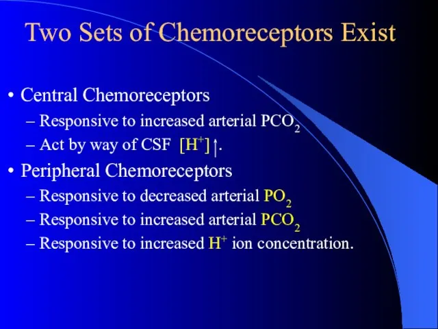
Слайд 13Two Sets of Chemoreceptors Exist
Central Chemoreceptors
Responsive to increased arterial PCO2
Act by
Two Sets of Chemoreceptors Exist
Central Chemoreceptors
Responsive to increased arterial PCO2
Act by
Слайд 14Central Chemoreceptor Location
Rostral
Medulla
Caudal
Medulla
Ventral Surface
Central Chemoreceptor Location
Rostral
Medulla
Caudal
Medulla
Ventral Surface
Слайд 15Central Chemoreceptor Stimulation
Central Chemoreceptor Stimulation
Слайд 16Peripheral Chemoreceptor Pathways
Peripheral Chemoreceptor Pathways
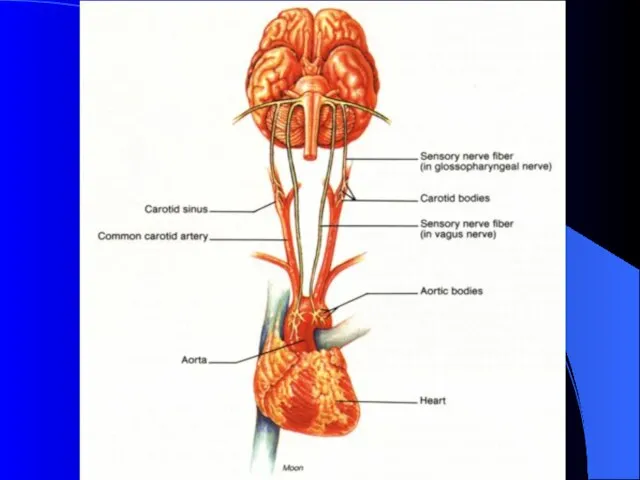
Слайд 17Peripheral Chemoreceptors
Carotid bodies
Sensitive to: PaO2, PaCO2, and pH
Afferents in glossopharyngeal nerve.
Aortic bodies
Sensitive
Peripheral Chemoreceptors
Carotid bodies
Sensitive to: PaO2, PaCO2, and pH
Afferents in glossopharyngeal nerve.
Aortic bodies
Sensitive
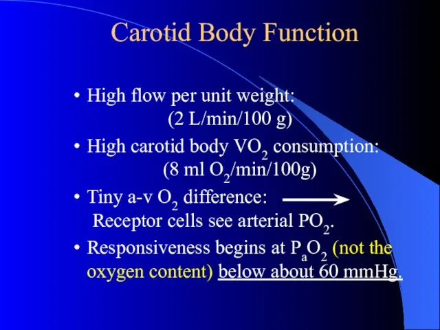
Слайд 19Carotid Body Function
High flow per unit weight:
(2 L/min/100 g)
High carotid body
Carotid Body Function
High flow per unit weight:
(2 L/min/100 g)
High carotid body
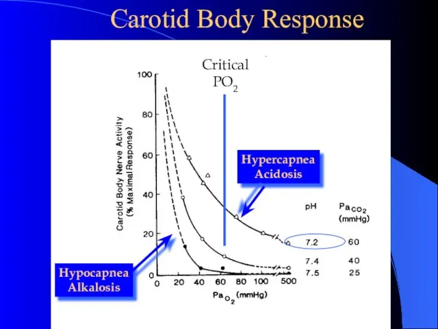
Слайд 20 Carotid Body Response
Critical
PO2
Hypercapnea
Acidosis
Hypocapnea
Alkalosis
Carotid Body Response
Critical
PO2
Hypercapnea
Acidosis
Hypocapnea
Alkalosis
Слайд 21
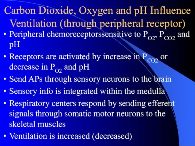
Carbon Dioxide, Oxygen and pH Influence Ventilation (through peripheral receptor)
Peripheral chemoreceptorssensitive to
Carbon Dioxide, Oxygen and pH Influence Ventilation (through peripheral receptor)
Peripheral chemoreceptorssensitive to
Слайд 22Effects of Hydrogen Ions (through central chemoreceptors)
pH of CSF (most powerful respiratory
Effects of Hydrogen Ions (through central chemoreceptors)
pH of CSF (most powerful respiratory
Слайд 23Carbon Dioxide
Indirect effects
through pH as seen previously
Direct effects
↑ CO2 may directly
Carbon Dioxide
Indirect effects
through pH as seen previously
Direct effects
↑ CO2 may directly
Слайд 24Oxygen
Direct inhibitory effect of hypoxemia on the respiratory center
Chronic hypoxemia, PO2 <
Oxygen
Direct inhibitory effect of hypoxemia on the respiratory center
Chronic hypoxemia, PO2 <
Слайд 25 Overall Response toPco2, Po2 and pH
Overall Response toPco2, Po2 and pH
Слайд 272. Neuroreceptor reflex
2. Neuroreceptor reflex
Слайд 28Hering-Breuer Reflex or Pulmonary Stretch Reflex
Including pulmonary inflation reflex and pulmonary deflation
Hering-Breuer Reflex or Pulmonary Stretch Reflex
Including pulmonary inflation reflex and pulmonary deflation
Слайд 30Significance of Hering-Breuer
Normal adults. Receptors are not activated at end normal tidal
Significance of Hering-Breuer
Normal adults. Receptors are not activated at end normal tidal
Слайд 31 Brainstem Transection
Brainstem Transection
Слайд 32Factors Influencing Respiration
Factors Influencing Respiration
 FSC - сертификация в Республике Коми – предложения по координации.
 ЭКОЛОГИЯ И БЕЗОПАСНОСТЬ ПИТАНИЯ
 ТАЙГА
ТАЙГА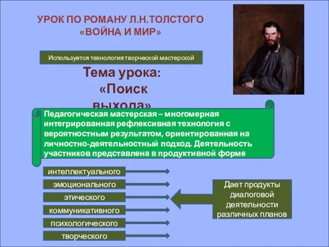 Тема урока: «Поиск выхода»
 Презентация на тему Искусство средневековой Руси
 Авиация МЧС РФ
 Интернет сервисы WEB.2 и формирование метапредметных и личностных результатов образования
 Возможности выпускников кафедры изобразительного искусства и дизайна в современной культурной сфере
 Видение перспектив ИТ-рынка 2012
 Магистерская диссертация Коротаевой Натальи Андреевны Научный руководитель: доцент Гусаковский Михаил Антонович
 Hello, sunshine!
 Сладкая жизнь. Постное ассорти
 Полуфабрикаты из птицы
 Политическая культура и поведение
 Информационно-технологическое обеспечение научной и образовательной деятельности в КГУ
 Времена английского глагола
 Башкирское национальное блюдо Вак бялеш
 Современные подходы и технологии социального воспитания
 Значения sin, cos, tg
 Урок 2
 Зачем пишу, чего же ради?
 Per aspera ad astra!Через тернии к звездам!
 Образ моря в литературе романтизма
 Вторая жизнь отслуживших вещей
 Моя семья
 Паралимпийцы Оренбургской области
 Этапы юридического интервьюирования
 Продукция General Electric включенная в морской регистр