- The integumentary system

Содержание
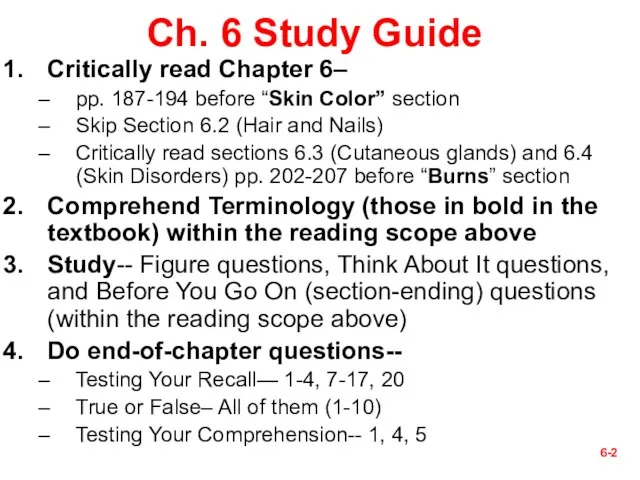
- 2. 6- Ch. 6 Study Guide Critically read Chapter 6– pp. 187-194 before “Skin Color” section Skip
- 3. 6- § Quotable Quotes (Skin) Some guys say beauty is only skin deep. But when you
- 4. 6- I. Introduction 6-
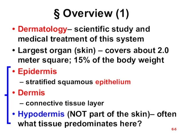
- 5. 6- § Overview (1) Dermatology– scientific study and medical treatment of this system Largest organ (skin)
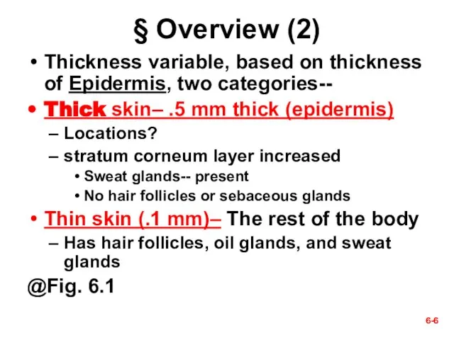
- 6. 6- 6- § Overview (2) Thickness variable, based on thickness of Epidermis, two categories-- Thick skin–
- 7. 6-
- 8. 6- 6- § Functions of the Skin Resistance to trauma/infection Why? (Fig. 5.28) acid mantle (pH
- 9. 6-
- 10. In hot environment In cold environment 6- vasodilation vasoconstriction Heat loss Less Heat loss Thermoregulation
- 11. 6- Social functions-- Figure 6.2 Skeletal muscles attach to dermal collagen fibers and produce expressions as
- 12. 6- II. Epidermis 6-
- 13. 6- 6- § Cells of the Epidermis (1) Five types of cells-- Keratinocytes – most of
- 14. The Epidermis— Fig. 6.2 6-
- 15. 6-
- 16. 6- 6- § Cells of the Epidermis (2) Location of the following types of cells— stratum
- 17. 6- Melanocyte Keratinocytes
- 18. 6- § Layers of the Epidermis— Next five slides (1-5) from deep to superficial and from
- 19. 6- 6- 1. Stratum Basale (deepest layer) Single layer cells on basement membrane (Fig. 6.3) Cell
- 20. Figure 6.2a 6-
- 21. 6- 6- 2. Stratum Spinosum– above stratum basale Several layers of keratinocytes (flattened as they cease
- 22. 6- 6- 3. Stratum Granulosum 3 to 5 layers flat keratinocytes: three developments occur to them--
- 23. 6- 6- 4. Stratum Lucidum— superficial to the stratum granulosum Thin translucent zone seen only in
- 24. 6-
- 25. 6- 6- 5. Stratum Corneum Up to 30 layers of dead, scaly, keratinized cells surface cells
- 26. 6- 6- § Life History of Keratinocytes Produced by stem cells in stratum basale New cells
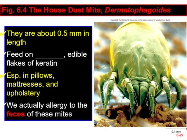
- 27. 6- Fig. 6.4 The House Dust Mite, Dermatophagoides They are about 0.5 mm in length Feed
- 28. 6- Questions (muddiest points)? Next section– III. Dermis & Hypodermis 6-
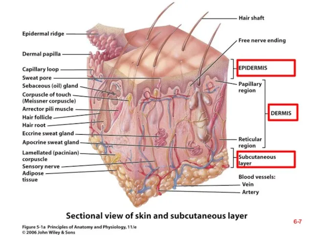
- 29. 6- 6- § Dermis- a C.T. layer Thickness = 0.2 to 4.0 mm Composition Collagen (mainly),
- 30. 6- Fig. 6.5 layers of the dermis Dermal papillae Epidermal ridges Areolar Tissue Dense irregular CT
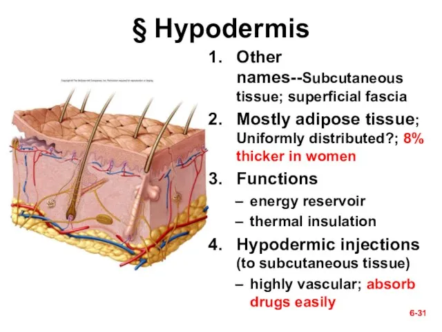
- 31. 6- 6- § Hypodermis Other names--Subcutaneous tissue; superficial fascia Mostly adipose tissue; Uniformly distributed?; 8% thicker
- 32. 6- Questions? Next section— IV. Cutaneous Glands 6-
- 33. 6- Table 6.2— summary of cutaneous glands 1. Sweat glands 2. Oil glands 3. Ceruminous glands
- 34. § Cutaneous Glands 6-
- 35. 6- 6- 1. Two kinds of Sweat Glands Filtrate of plasma and some waste products insensible
- 36. 6-
- 37. 6- 6- 2. Sebaceous (Oil) Glands Oily secretion called sebum that contains broken-down cells Due to
- 38. ID specific cutaneous glands (A & B). 6- A. B. Which specific kind?
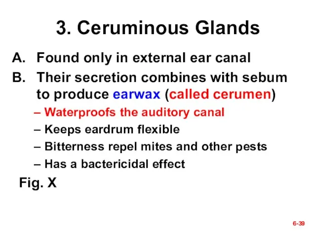
- 39. 6- 6- 3. Ceruminous Glands Found only in external ear canal Their secretion combines with sebum
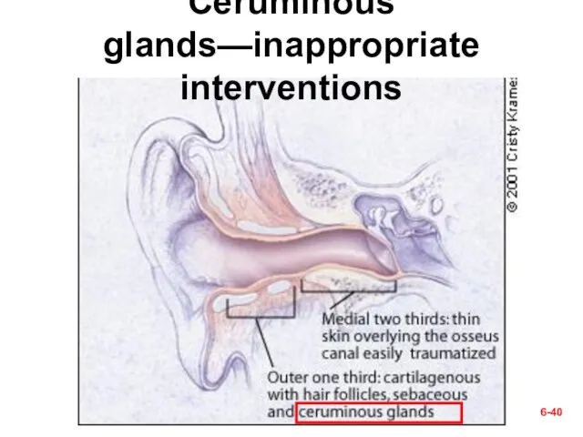
- 40. 6- Ceruminous glands—inappropriate interventions
- 41. 6- ? Cotton-tipped applicator (a no-no)
- 42. 6- ᵡ Ear Candling!?
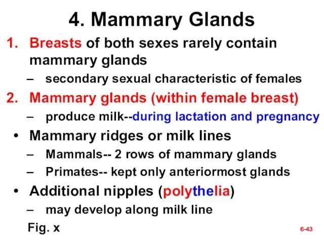
- 43. 6- 6- 4. Mammary Glands Breasts of both sexes rarely contain mammary glands secondary sexual characteristic
- 44. Mammary Glands 6- Areola Nipple
- 45. Check Point Questions (True/False) The three layers of the skin are the epidermis, dermis, and hypodermis.
- 46. 6- Questions (muddiest points)? Next section— V. Skin Disorders 6-
- 47. 6- 6- § Skin Cancer Cause– the ultraviolet rays of the sun There is no such
- 48. 6- 6- A. Basal cell carcinoma Most common type and the least dangerous one Origination- by
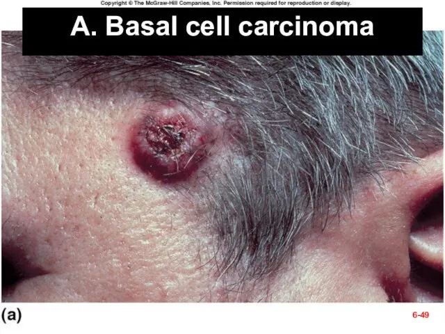
- 49. Fig. 6.12a A. Basal cell carcinoma 6-
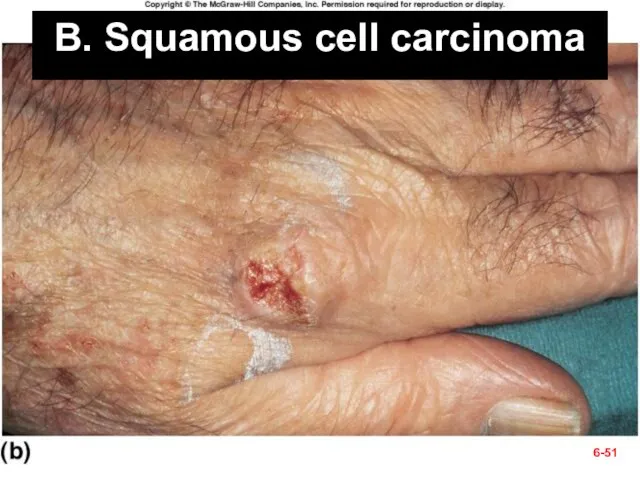
- 50. 6- 6- B. Squamous cell carcinoma Chance of recovery is good with early detection and surgical
- 51. B. Squamous cell carcinoma 6-
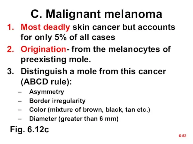
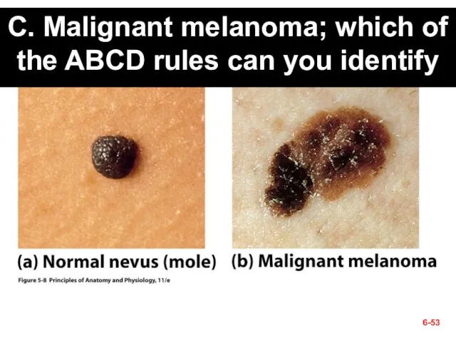
- 52. 6- 6- C. Malignant melanoma Most deadly skin cancer but accounts for only 5% of all
- 53. C. Malignant melanoma; which of the ABCD rules can you identify 6-
- 55. Скачать презентацию
Слайд 36-
§ Quotable Quotes (Skin)
Some guys say beauty is only skin deep. But
6-
§ Quotable Quotes (Skin)
Some guys say beauty is only skin deep. But
Слайд 46-
I. Introduction
6-
6-
I. Introduction
6-

Слайд 56-
§ Overview (1)
Dermatology– scientific study and medical treatment of this system
Largest organ
6-
§ Overview (1)
Dermatology– scientific study and medical treatment of this system
Largest organ
Слайд 66-
6-
§ Overview (2)
Thickness variable, based on thickness of Epidermis, two categories--
Thick skin–
6-
6-
§ Overview (2)
Thickness variable, based on thickness of Epidermis, two categories--
Thick skin–
Слайд 76-
6-
Слайд 86-
6-
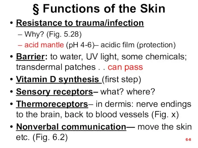
§ Functions of the Skin
Resistance to trauma/infection
Why? (Fig. 5.28)
acid mantle (pH 4-6)–
6-
6-
§ Functions of the Skin
Resistance to trauma/infection
Why? (Fig. 5.28)
acid mantle (pH 4-6)–
Слайд 96-
6-
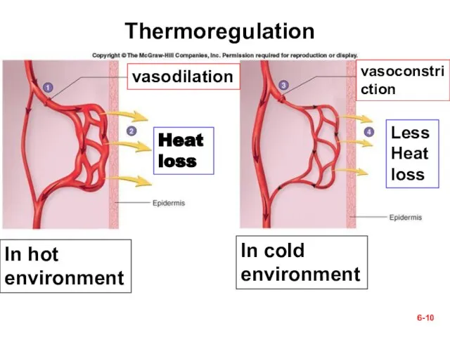
Слайд 10In hot environment
In cold environment
6-
vasodilation
vasoconstriction
Heat loss
Less
Heat loss
Thermoregulation
In hot environment
In cold environment
6-
vasodilation
vasoconstriction
Heat loss
Less
Heat loss
Thermoregulation
Слайд 116-
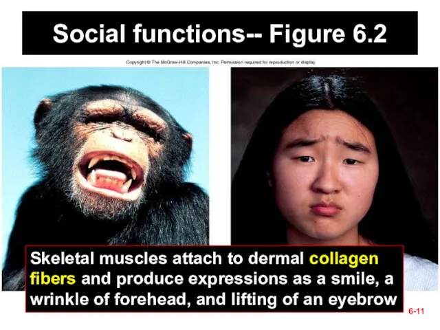
Social functions-- Figure 6.2
Skeletal muscles attach to dermal collagen fibers and produce
6-
Social functions-- Figure 6.2
Skeletal muscles attach to dermal collagen fibers and produce
Слайд 126-
II. Epidermis
6-
6-
II. Epidermis
6-
Слайд 136-
6-
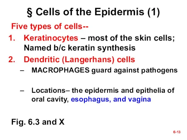
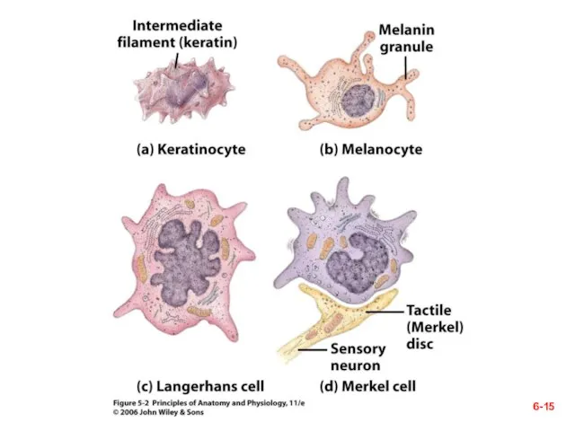
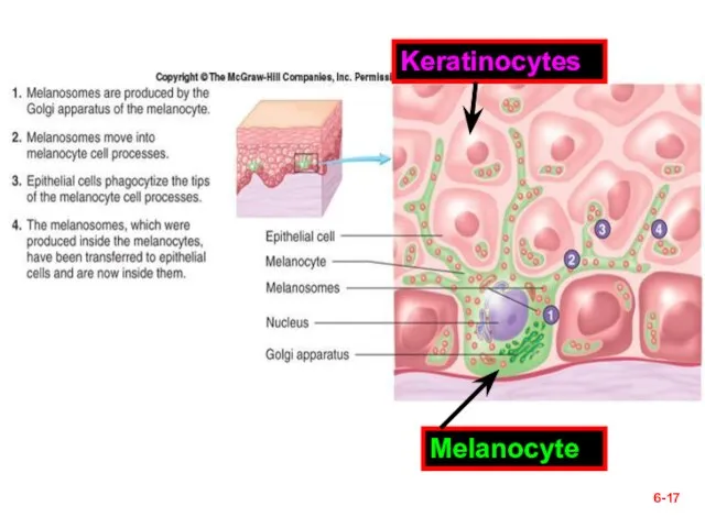
§ Cells of the Epidermis (1)
Five types of cells--
Keratinocytes – most of
6-
6-
§ Cells of the Epidermis (1)
Five types of cells--
Keratinocytes – most of
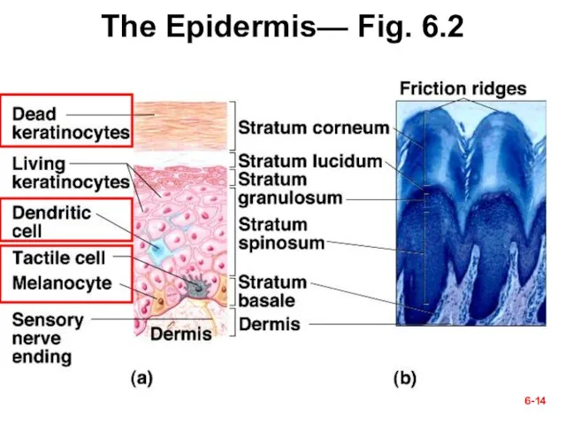
Слайд 14The Epidermis— Fig. 6.2
6-
The Epidermis— Fig. 6.2
6-
Слайд 156-
6-
Слайд 166-
6-
§ Cells of the Epidermis (2)
Location of the following types of cells—
6-
6-
§ Cells of the Epidermis (2)
Location of the following types of cells—
Слайд 176-
Melanocyte
Keratinocytes
6-
Melanocyte
Keratinocytes
Слайд 186-
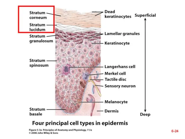
§ Layers of the Epidermis—
Next five slides (1-5)
from deep to superficial and
6-
§ Layers of the Epidermis— Next five slides (1-5) from deep to superficial and
Слайд 196-
6-
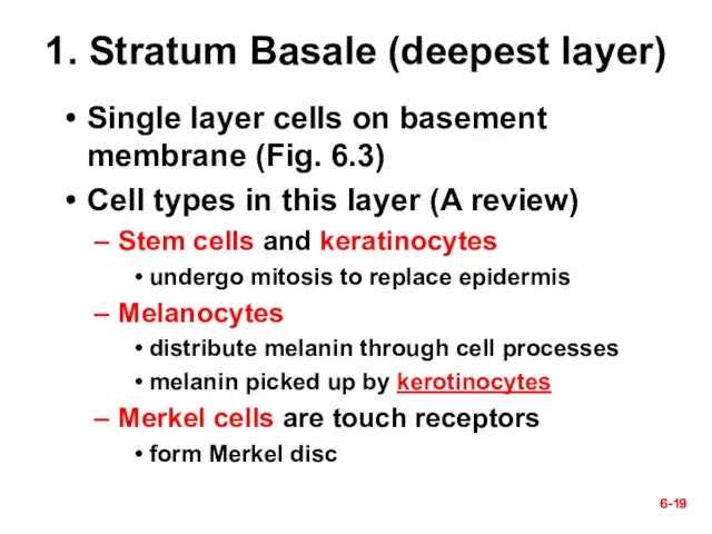
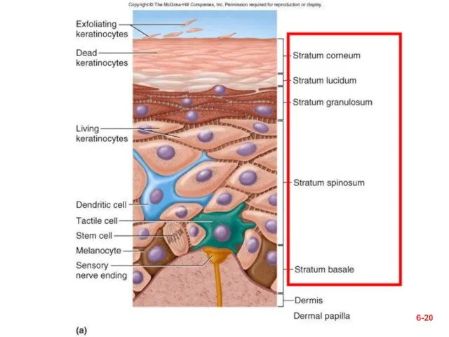
1. Stratum Basale (deepest layer)
Single layer cells on basement membrane (Fig. 6.3)
Cell
6-
6-
1. Stratum Basale (deepest layer)
Single layer cells on basement membrane (Fig. 6.3)
Cell
Слайд 20Figure 6.2a
6-
Figure 6.2a
6-
Слайд 216-
6-
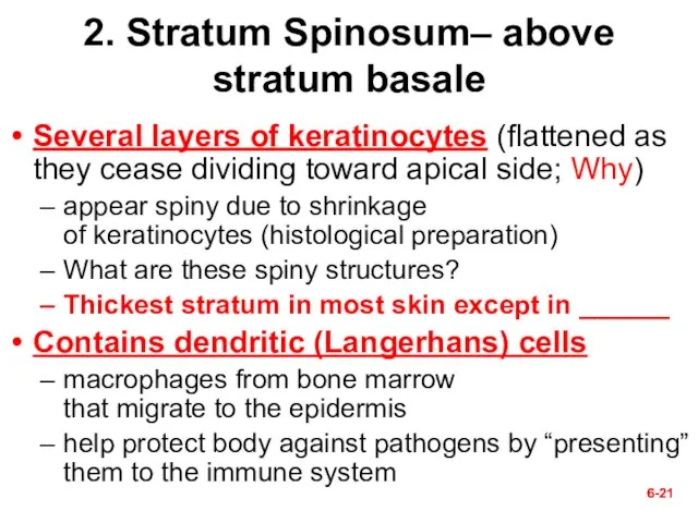
2. Stratum Spinosum– above stratum basale
Several layers of keratinocytes (flattened as they
6-
6-
2. Stratum Spinosum– above stratum basale
Several layers of keratinocytes (flattened as they
Слайд 226-
6-
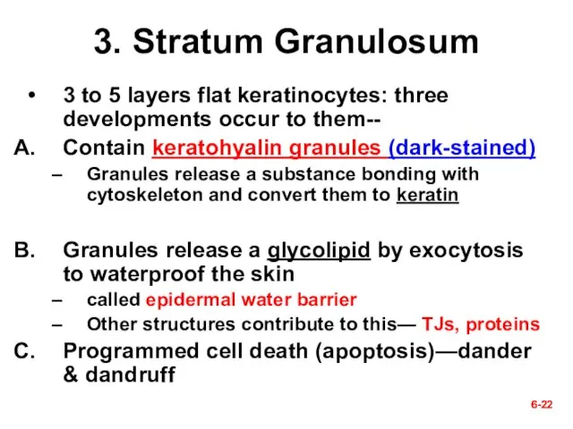
3. Stratum Granulosum
3 to 5 layers flat keratinocytes: three developments occur to
6-
6-
3. Stratum Granulosum
3 to 5 layers flat keratinocytes: three developments occur to
Слайд 236-
6-
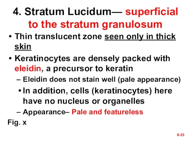
4. Stratum Lucidum— superficial to the stratum granulosum
Thin translucent zone seen only
6-
6-
4. Stratum Lucidum— superficial to the stratum granulosum
Thin translucent zone seen only
Слайд 246-
6-
Слайд 256-
6-
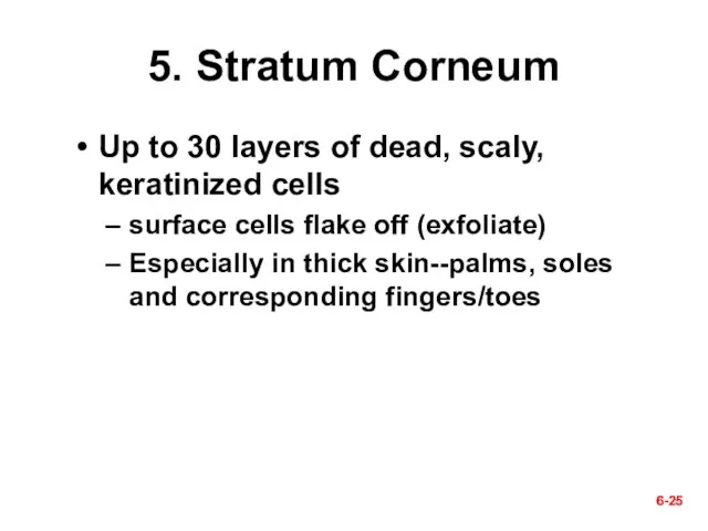
5. Stratum Corneum
Up to 30 layers of dead, scaly,
keratinized cells
surface cells flake
6-
6-
5. Stratum Corneum
Up to 30 layers of dead, scaly,
keratinized cells
surface cells flake
Слайд 266-
6-
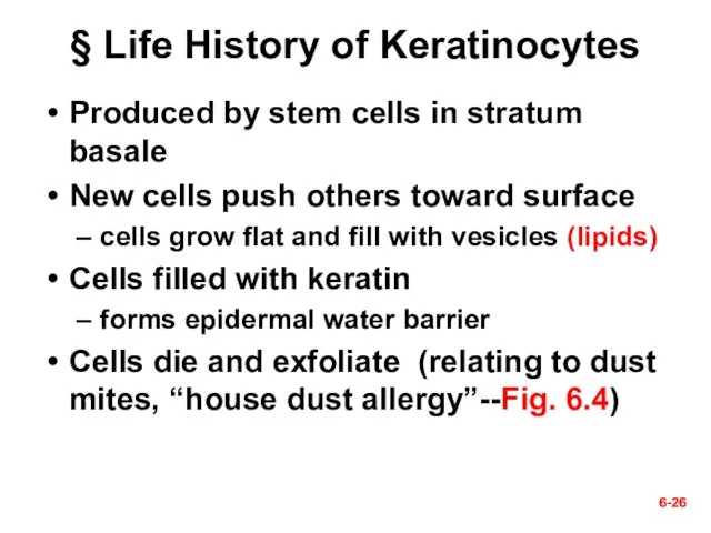
§ Life History of Keratinocytes
Produced by stem cells in stratum basale
New cells
6-
6-
§ Life History of Keratinocytes
Produced by stem cells in stratum basale
New cells
Слайд 276-
Fig. 6.4 The House Dust Mite, Dermatophagoides
They are about 0.5 mm in
6-
Fig. 6.4 The House Dust Mite, Dermatophagoides
They are about 0.5 mm in
Слайд 286-
Questions (muddiest points)?
Next section–
III. Dermis & Hypodermis
6-
6-
Questions (muddiest points)?
Next section–
III. Dermis & Hypodermis
6-
Слайд 296-
6-
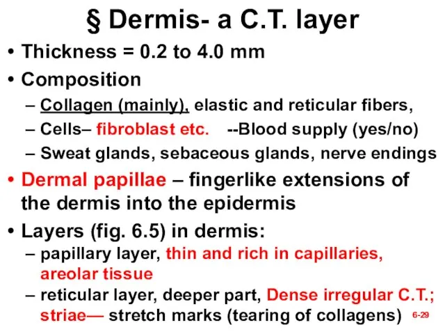
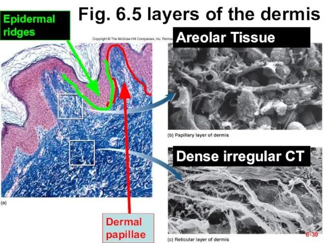
§ Dermis- a C.T. layer
Thickness = 0.2 to 4.0 mm
Composition
Collagen (mainly), elastic
6-
6-
§ Dermis- a C.T. layer
Thickness = 0.2 to 4.0 mm
Composition
Collagen (mainly), elastic
Слайд 306-
Fig. 6.5 layers of the dermis
Dermal papillae
Epidermal ridges
Areolar Tissue
Dense irregular CT
6-
6-
Fig. 6.5 layers of the dermis
Dermal papillae
Epidermal ridges
Areolar Tissue
Dense irregular CT
6-
Слайд 316-
6-
§ Hypodermis
Other names--Subcutaneous tissue; superficial fascia
Mostly adipose tissue; Uniformly distributed?; 8% thicker
6-
6-
§ Hypodermis
Other names--Subcutaneous tissue; superficial fascia
Mostly adipose tissue; Uniformly distributed?; 8% thicker
Слайд 326-
Questions?
Next section—
IV. Cutaneous Glands
6-
6-
Questions?
Next section—
IV. Cutaneous Glands
6-
Слайд 336-
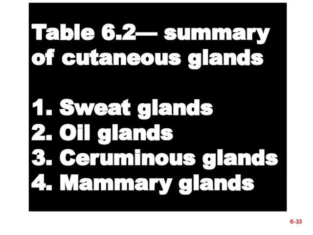
Table 6.2— summary of cutaneous glands
1. Sweat glands
2. Oil glands
3. Ceruminous glands
4.
6-
Table 6.2— summary of cutaneous glands 1. Sweat glands 2. Oil glands 3. Ceruminous glands 4.
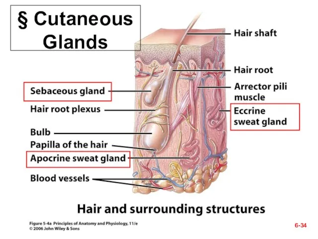
Слайд 34§ Cutaneous Glands
6-
§ Cutaneous Glands
6-
Слайд 356-
6-
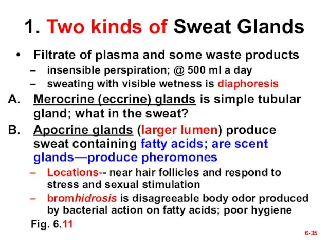
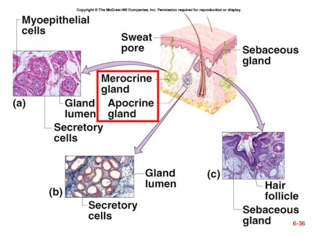
1. Two kinds of Sweat Glands
Filtrate of plasma and some waste products
insensible
6-
6-
1. Two kinds of Sweat Glands
Filtrate of plasma and some waste products
insensible
Слайд 366-
6-
Слайд 376-
6-
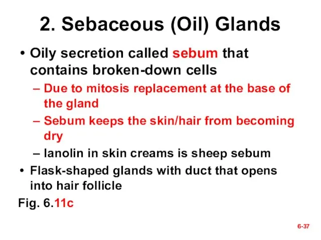
2. Sebaceous (Oil) Glands
Oily secretion called sebum that contains broken-down cells
Due to
6-
6-
2. Sebaceous (Oil) Glands
Oily secretion called sebum that contains broken-down cells
Due to
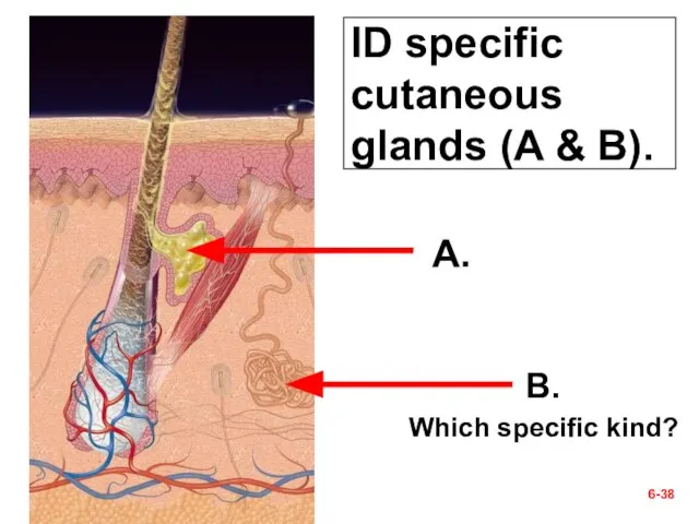
Слайд 38ID specific cutaneous glands (A & B).
6-
A.
B.
Which specific kind?
ID specific cutaneous glands (A & B).
6-
A.
B.
Which specific kind?
Слайд 396-
6-
3. Ceruminous Glands
Found only in external ear canal
Their secretion combines with sebum
6-
6-
3. Ceruminous Glands
Found only in external ear canal
Their secretion combines with sebum
Слайд 406-
Ceruminous glands—inappropriate interventions
6-
Ceruminous glands—inappropriate interventions
Слайд 416-
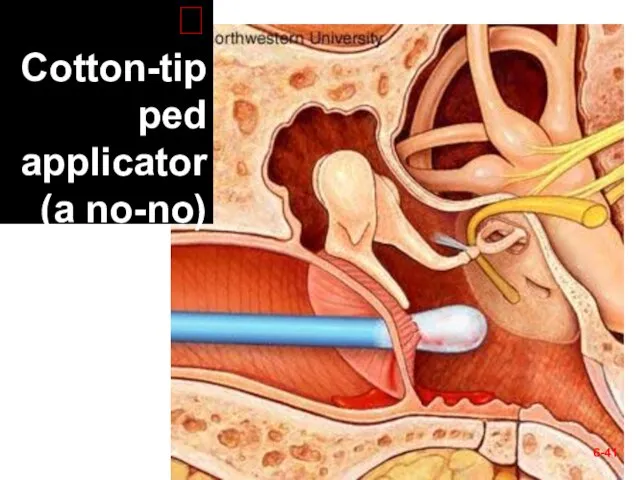
? Cotton-tipped applicator (a no-no)
6-
? Cotton-tipped applicator (a no-no)
Слайд 426-
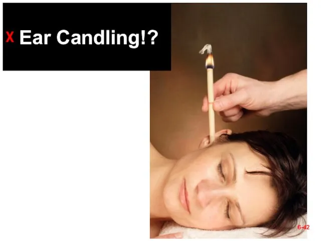
ᵡ Ear Candling!?
6-
ᵡ Ear Candling!?
Слайд 436-
6-
4. Mammary Glands
Breasts of both sexes rarely contain mammary glands
secondary sexual characteristic
6-
6-
4. Mammary Glands
Breasts of both sexes rarely contain mammary glands
secondary sexual characteristic
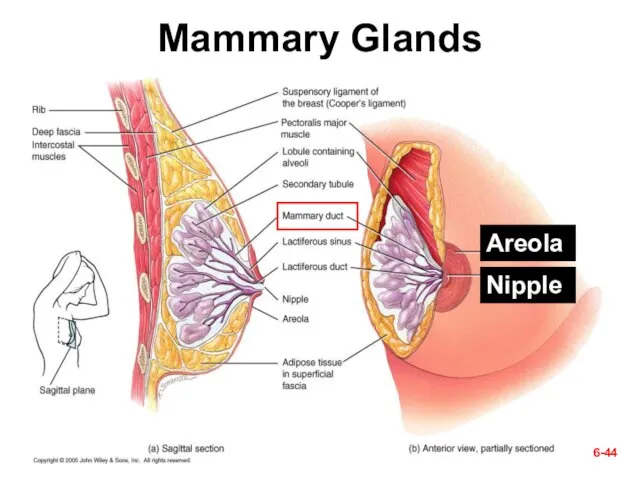
Слайд 44Mammary Glands
6-
Areola
Nipple
Mammary Glands
6-
Areola
Nipple
Слайд 45Check Point Questions
(True/False) The three layers of the skin are the epidermis,
Check Point Questions
(True/False) The three layers of the skin are the epidermis,
Слайд 466-
Questions (muddiest points)?
Next section—
V. Skin Disorders
6-
6-
Questions (muddiest points)?
Next section—
V. Skin Disorders
6-
Слайд 476-
6-
§ Skin Cancer
Cause– the ultraviolet rays of the sun
There is no
6-
6-
§ Skin Cancer
Cause– the ultraviolet rays of the sun
There is no
Слайд 486-
6-
A. Basal cell carcinoma
Most common type and the least dangerous one
Origination- by
6-
6-
A. Basal cell carcinoma
Most common type and the least dangerous one
Origination- by
Слайд 49Fig. 6.12a
A. Basal cell carcinoma
6-
Fig. 6.12a
A. Basal cell carcinoma
6-
Слайд 506-
6-
B. Squamous cell carcinoma
Chance of recovery is good with early detection and
6-
6-
B. Squamous cell carcinoma
Chance of recovery is good with early detection and
Слайд 51B. Squamous cell carcinoma
6-
B. Squamous cell carcinoma
6-
Слайд 526-
6-
C. Malignant melanoma
Most deadly skin cancer but accounts for only 5% of
6-
6-
C. Malignant melanoma
Most deadly skin cancer but accounts for only 5% of
Слайд 53C. Malignant melanoma; which of the ABCD rules can you identify
6-
C. Malignant melanoma; which of the ABCD rules can you identify
6-
 Консультации по написанию заявки РНФ
 Художественные поиски свободы в искусстве
 Презентация на тему Психолого-педагогическое использование сказкотерапии для воспитания положительных нравственных качеств лич
 Кадровое планирование в организации
 Педагогическая психология как наука Психология обучения
 Методические условия эффективности применения современных образовательных технологий
 8кл.23.09
 Рисуем героев любимых сказок
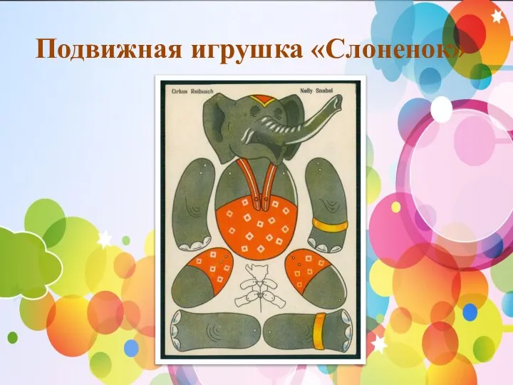 Подвижная игрушка Слоненок
 Кабы всё знал, так бы не учился
 Душа танца
 Формирование читательской компетентности младшего школьника
 Защита прав ребенка в школе. МОУ «СОШ № 8»
 Лучшие люди России
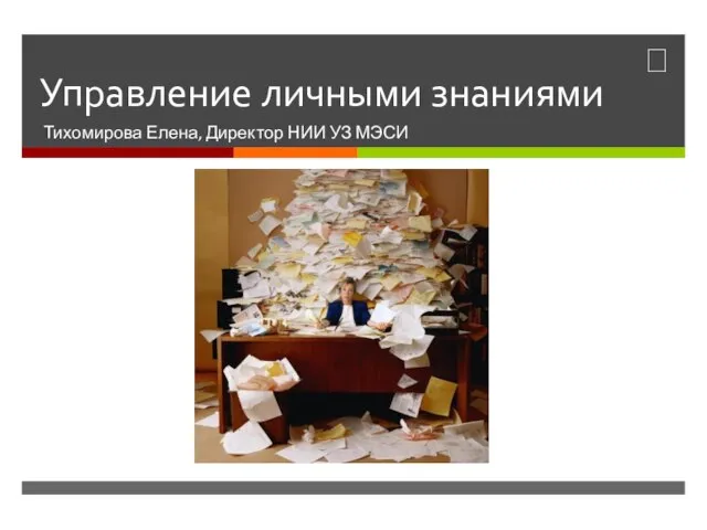 Управление личными знаниями
 Rezultate interviu
 Разработка высоконагруженных проектов(например – сайтов для сообществ)
 Техническое задание на фото
 Опасные природные явления
 Понятие об экономическом механизме функционирования фирмы и характеристика его основных элементов
 День Святого Валентина в США
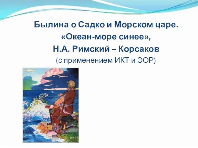 Былина о Садко и Морском царе. Океан-море синее, Н.А. Римский – Корсаков
 рабочая одежда
 Бардымскому району 90 лет
 Конструирование фартука
 Педагогический совет —коллегиальная форма управления
 Программа социального исследования
 Народная танцевальная культура