- What is the link

Содержание
- 2. The study of cells Dr. Sophia Semerdjieva [email protected] Office 2.203
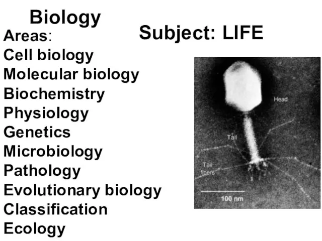
- 3. Biology Subject: LIFE Areas: Cell biology Molecular biology Biochemistry Physiology Genetics Microbiology Pathology Evolutionary biology Classification
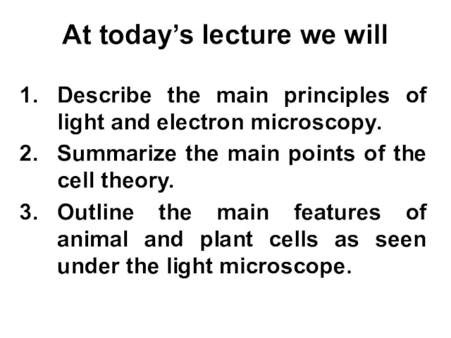
- 4. At today’s lecture we will Describe the main principles of light and electron microscopy. Summarize the
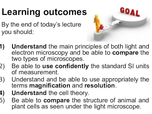
- 5. By the end of today’s lecture you should: Understand the main principles of both light and
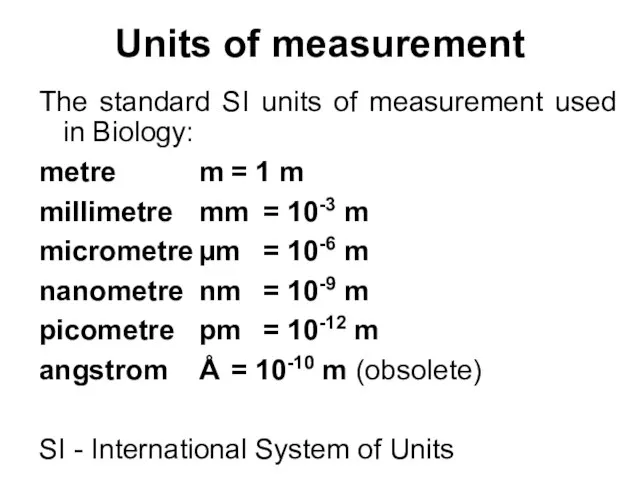
- 6. Units of measurement The standard SI units of measurement used in Biology: metre m = 1
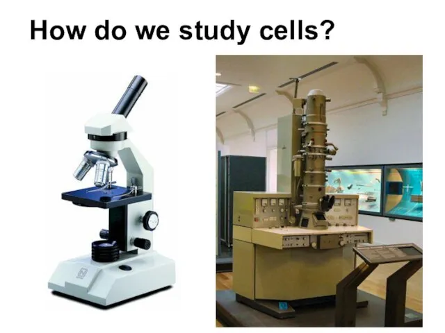
- 7. How do we study cells?
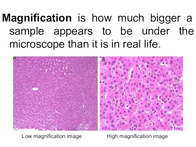
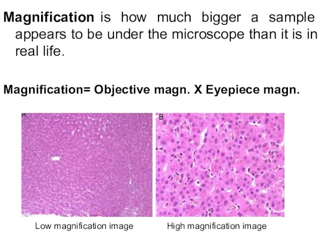
- 8. Magnification is how much bigger a sample appears to be under the microscope than it is
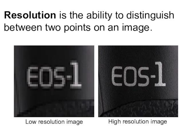
- 9. Resolution is the ability to distinguish between two points on an image.
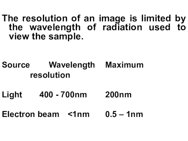
- 10. The resolution of an image is limited by the wavelength of radiation used to view the
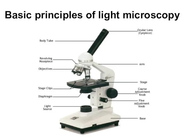
- 11. Basic principles of light microscopy
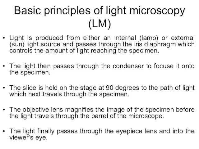
- 12. Basic principles of light microscopy (LM) Light is produced from either an internal (lamp) or external
- 13. Magnification is how much bigger a sample appears to be under the microscope than it is
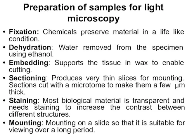
- 14. Preparation of samples for light microscopy Fixation: Chemicals preserve material in a life like condition. Dehydration:
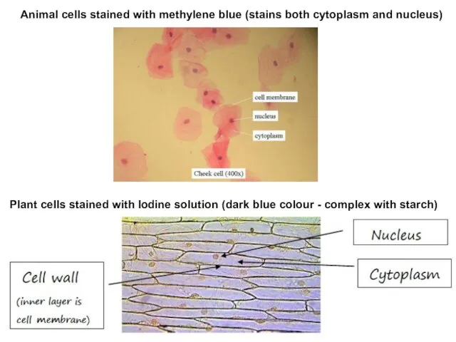
- 15. Animal cells stained with methylene blue (stains both cytoplasm and nucleus) Plant cells stained with Iodine
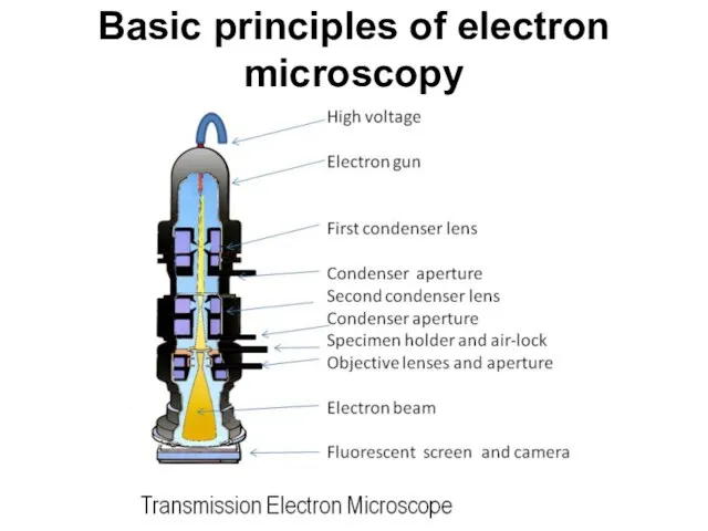
- 16. Basic principles of electron microscopy
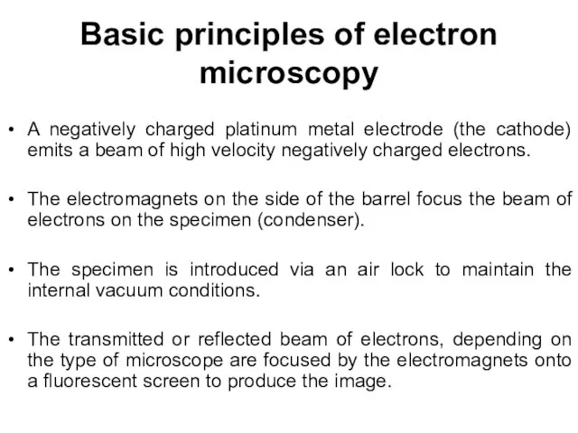
- 17. Basic principles of electron microscopy A negatively charged platinum metal electrode (the cathode) emits a beam
- 18. Transmission electron microscope (TEM) The beam of electrons passes through the specimen. The electrons that pass
- 19. Preparation of samples for electron microscopy Fixation. Dehydration: Particularly important for electron microscopy as water would
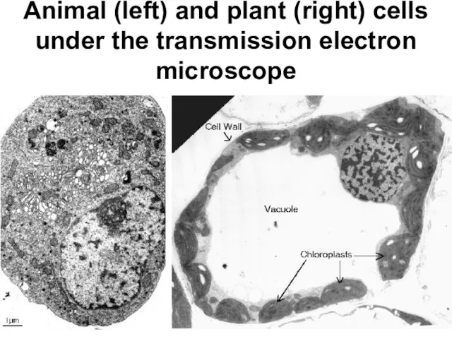
- 20. Animal (left) and plant (right) cells under the transmission electron microscope
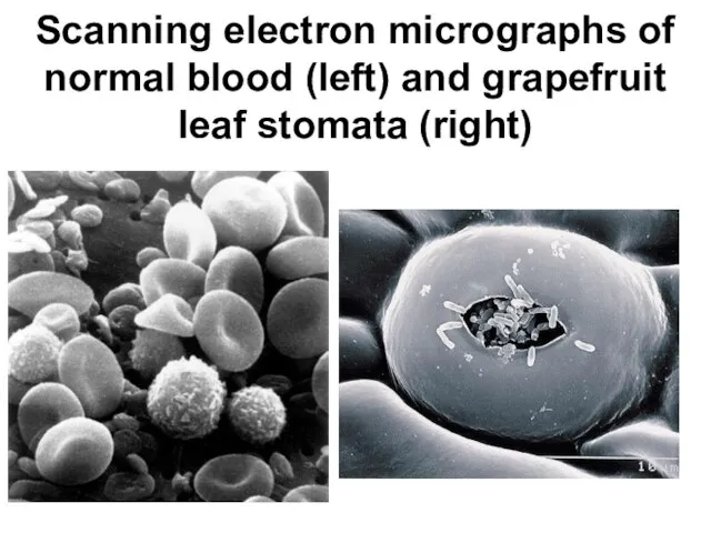
- 21. Scanning electron microscope (SEM) The beam of electrons is passed over the surface of the specimen
- 22. Scanning electron micrographs of normal blood (left) and grapefruit leaf stomata (right)
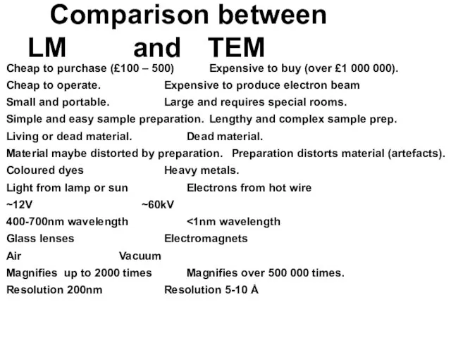
- 23. Cheap to purchase (£100 – 500) Expensive to buy (over £1 000 000). Cheap to operate.
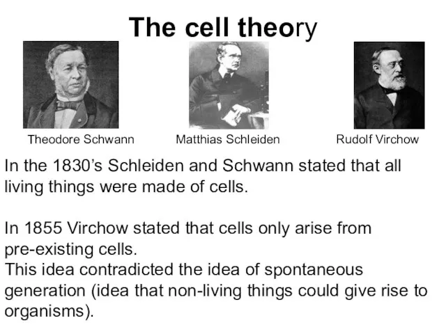
- 24. The cell theory In the 1830’s Schleiden and Schwann stated that all living things were made
- 25. The cell theory Cells form the building blocks of living organisms. Cells only form through division
- 26. Animal cell under the light microscope
- 27. Plant cell under the light microscope
- 28. Centrifugation Relative Centrifugal Force (g-force) (RCF) - suspended particles move towards the bottom and out of
- 29. S – Svedberg or sedimentation coefficient is a measure of the time taken by a particle
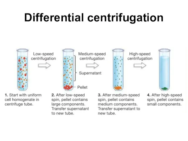
- 30. Differential centrifugation
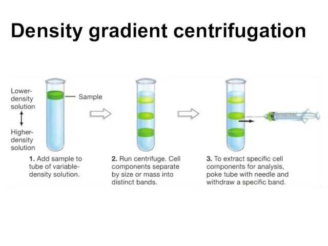
- 31. Density gradient centrifugation
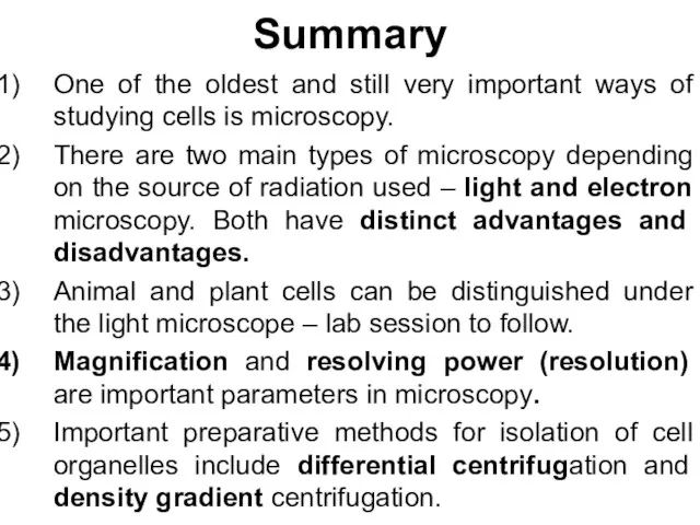
- 32. Summary One of the oldest and still very important ways of studying cells is microscopy. There
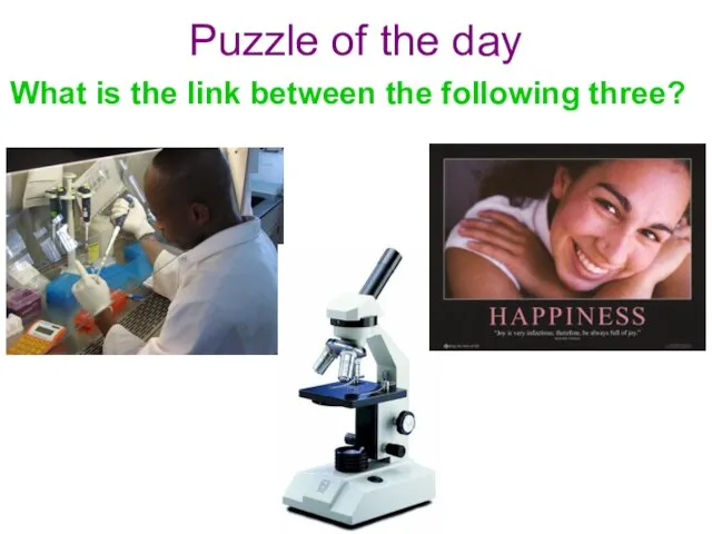
- 33. Puzzle of the day What is the link between the following three?
- 35. Скачать презентацию
Слайд 3Biology
Subject: LIFE
Areas:
Cell biology
Molecular biology
Biochemistry
Physiology
Genetics
Microbiology
Pathology
Evolutionary biology
Classification
Ecology
Biology
Subject: LIFE
Areas:
Cell biology
Molecular biology
Biochemistry
Physiology
Genetics
Microbiology
Pathology
Evolutionary biology
Classification
Ecology
Слайд 4At today’s lecture we will
Describe the main principles of light and electron
At today’s lecture we will
Describe the main principles of light and electron
Слайд 5By the end of today’s lecture
you should:
Understand the main principles of
By the end of today’s lecture
you should:
Understand the main principles of
Слайд 6Units of measurement
The standard SI units of measurement used in Biology:
metre m =
Units of measurement
The standard SI units of measurement used in Biology:
metre m =
Слайд 7How do we study cells?
How do we study cells?
Слайд 8Magnification is how much bigger a sample appears to be under the
Magnification is how much bigger a sample appears to be under the
Слайд 9Resolution is the ability to distinguish between two points on an image.
Resolution is the ability to distinguish between two points on an image.
Слайд 10The resolution of an image is limited by the wavelength of radiation
Слайд 11Basic principles of light microscopy
Basic principles of light microscopy
Слайд 12Basic principles of light microscopy (LM)
Light is produced from either an internal
Basic principles of light microscopy (LM)
Light is produced from either an internal
Слайд 13Magnification is how much bigger a sample appears to be under the
Magnification is how much bigger a sample appears to be under the
Слайд 14Preparation of samples for light microscopy
Fixation: Chemicals preserve material in a life
Preparation of samples for light microscopy
Fixation: Chemicals preserve material in a life
Слайд 15Animal cells stained with methylene blue (stains both cytoplasm and nucleus)
Plant cells
Animal cells stained with methylene blue (stains both cytoplasm and nucleus)
Plant cells
Слайд 16Basic principles of electron microscopy
Basic principles of electron microscopy
Слайд 17Basic principles of electron microscopy
A negatively charged platinum metal electrode (the cathode)
Basic principles of electron microscopy
A negatively charged platinum metal electrode (the cathode)
Слайд 18Transmission electron microscope (TEM)
The beam of electrons passes through the specimen. The
Transmission electron microscope (TEM)
The beam of electrons passes through the specimen. The
Слайд 19Preparation of samples for electron microscopy
Fixation.
Dehydration: Particularly important for electron microscopy as
Preparation of samples for electron microscopy
Fixation.
Dehydration: Particularly important for electron microscopy as
Слайд 20Animal (left) and plant (right) cells under the transmission electron microscope
Animal (left) and plant (right) cells under the transmission electron microscope
Слайд 21Scanning electron microscope (SEM)
The beam of electrons is passed over the surface
Scanning electron microscope (SEM)
The beam of electrons is passed over the surface
Слайд 22Scanning electron micrographs of normal blood (left) and grapefruit leaf stomata (right)
Scanning electron micrographs of normal blood (left) and grapefruit leaf stomata (right)
Слайд 23Cheap to purchase (£100 – 500) Expensive to buy (over £1 000
Слайд 24The cell theory
In the 1830’s Schleiden and Schwann stated that all living
The cell theory
In the 1830’s Schleiden and Schwann stated that all living
Слайд 25The cell theory
Cells form the building blocks of living organisms.
Cells only form
The cell theory
Cells form the building blocks of living organisms.
Cells only form
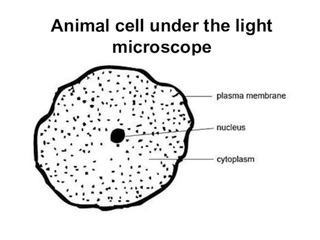
Слайд 26Animal cell under the light microscope
Animal cell under the light microscope
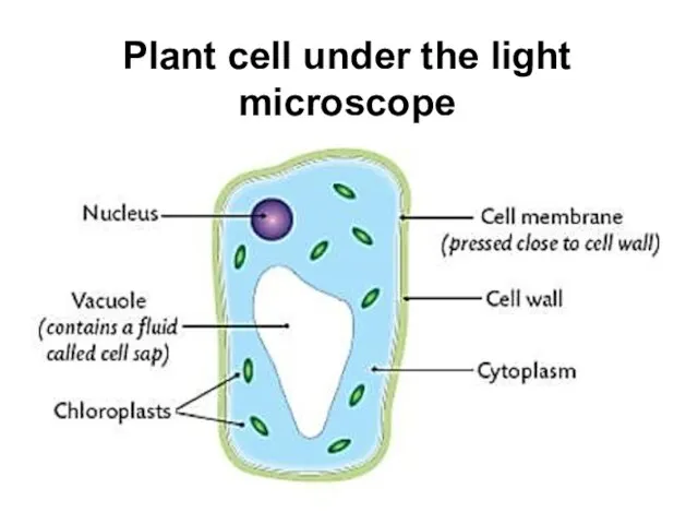
Слайд 27Plant cell under the light microscope
Plant cell under the light microscope
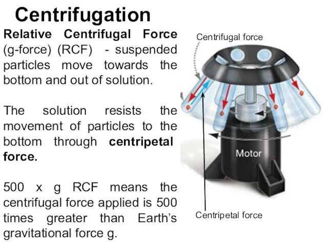
Слайд 28Centrifugation
Relative Centrifugal Force (g-force) (RCF) - suspended particles move towards the bottom
Centrifugation
Relative Centrifugal Force (g-force) (RCF) - suspended particles move towards the bottom
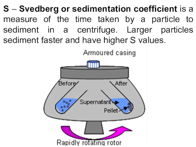
Слайд 29S – Svedberg or sedimentation coefficient is a measure of the time
S – Svedberg or sedimentation coefficient is a measure of the time
Слайд 30Differential centrifugation
Differential centrifugation
Слайд 31Density gradient centrifugation
Density gradient centrifugation
Слайд 32Summary
One of the oldest and still very important ways of studying cells
Summary
One of the oldest and still very important ways of studying cells
Слайд 33Puzzle of the day
What is the link between the following three?
Puzzle of the day
What is the link between the following three?
 Понятие трудового договора, его стороны и значение. Содержание трудового договора, порядок заключения и расторжения
 Автоматизация р
 All kinds of animals
 Экономика государства
 Компьютерные презентации
 Селекция2
 Презентация на тему Письменные буквы русского алфавита
  Суриков Сергей Григорьевич Ученик 9 б класса МБОУ СОШ № 9
  Художественное ремесло
 АСГОР «РГК»
 Знакомство с Богом
 Четыре живописца
 Терроризм как опаснейшее социально-политическое явление сегодняшнего мира
 Филиппины
  Программа поддержки многодетных семей в РФ
 Группа развития
 Презентация на тему Природные зоны Африки
  Тема доклада: «Обязательные виды страхования - драйвер роста или тупиковый путь развития?»
 Профориентационная работа в условиях школы-интерната
 Подводная угадайка
 Hafta 3-2Menderes Dönemi
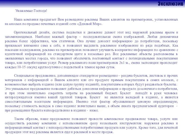 Эксклюзив
 ВРЕМЕНА ГОДА SEASONS
  Состав рабочих групп проектов окружного методического совета
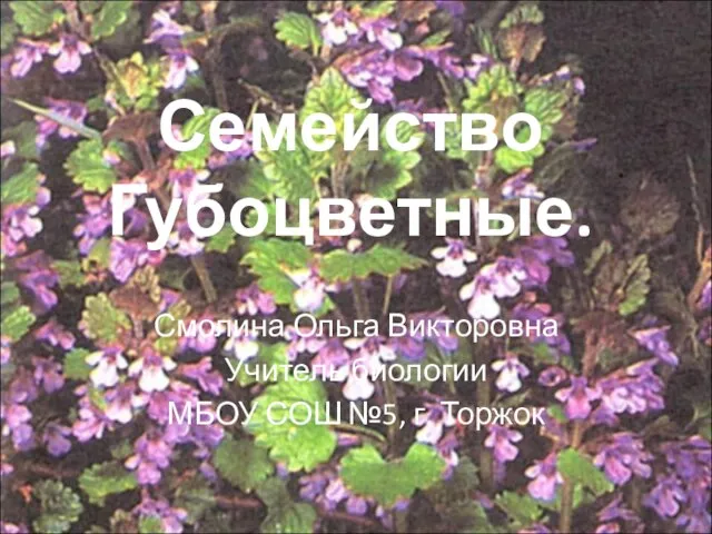 Презентация на тему Семейство Губоцветные
  Межрегиональный конкурс по проектной робототехнике РобоТех (Ярославль)
 О результатах государственной (итоговой) аттестации выпускников IX классов общеобразовательных учреждений, организуемой региона
 презентация