- Hemangioma

Содержание
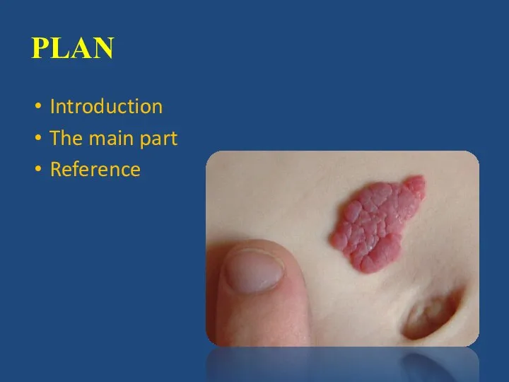
- 2. PLAN Introduction The main part Reference
- 5. Simple hemangioma is usually red or blue-purple color, is located on the surface, clearly delineated boundaries,
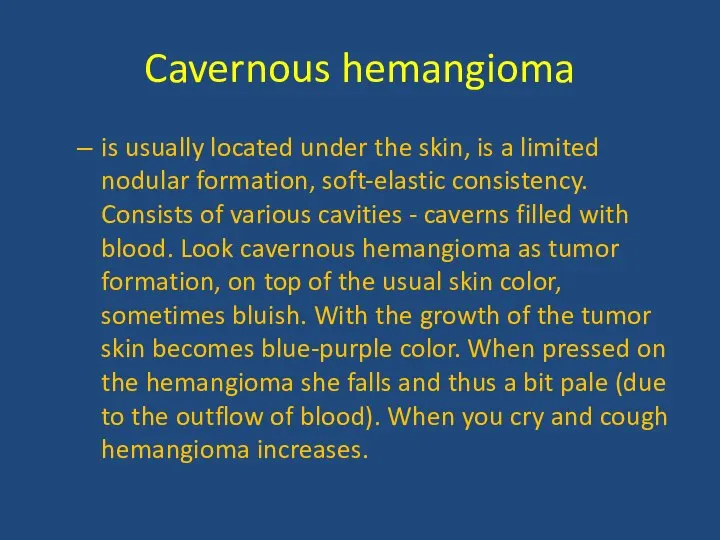
- 6. Cavernous hemangioma is usually located under the skin, is a limited nodular formation, soft-elastic consistency. Consists
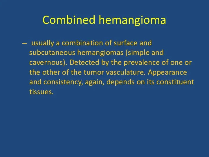
- 7. Combined hemangioma usually a combination of surface and subcutaneous hemangiomas (simple and cavernous). Detected by the
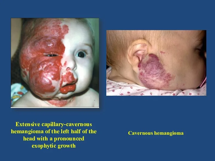
- 8. Extensive capillary-cavernous hemangioma of the left half of the head with a pronounced exophytic growth Cavernous
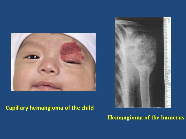
- 9. Capillary hemangioma of the child Hemangioma of the humerus
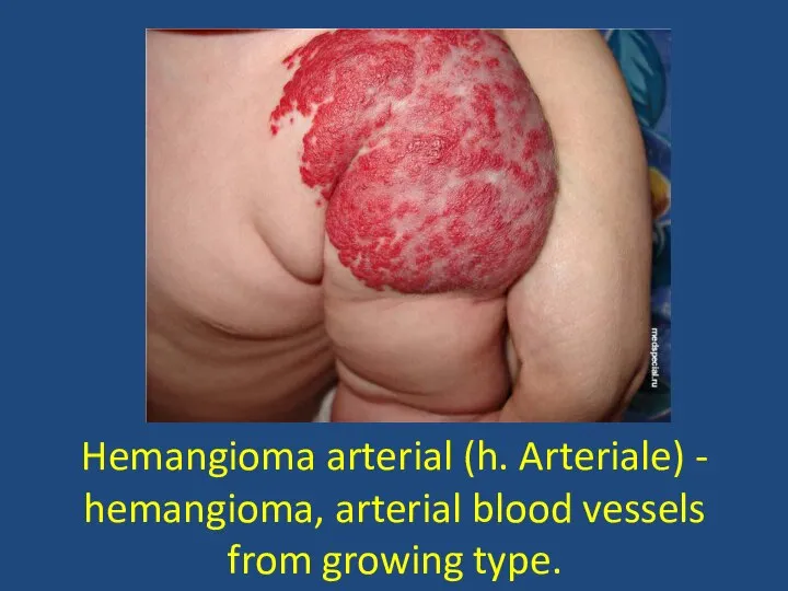
- 10. Hemangioma arterial (h. Arteriale) - hemangioma, arterial blood vessels from growing type.
- 11. HEMANGIOMA CAN BE LIVER KIDNEY VERTEBRAE LIPS
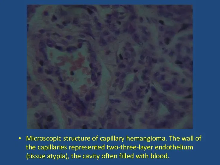
- 13. Microscopic structure of capillary hemangioma. The wall of the capillaries represented two-three-layer endothelium (tissue atypia), the
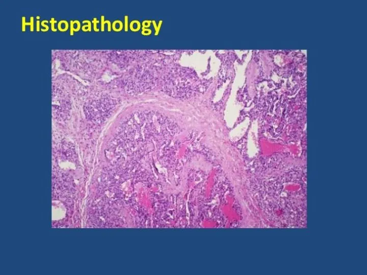
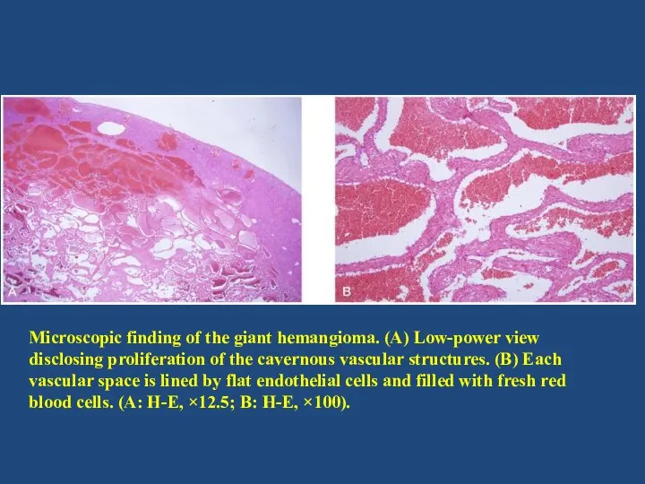
- 14. Microscopic finding of the giant hemangioma. (A) Low-power view disclosing proliferation of the cavernous vascular structures.
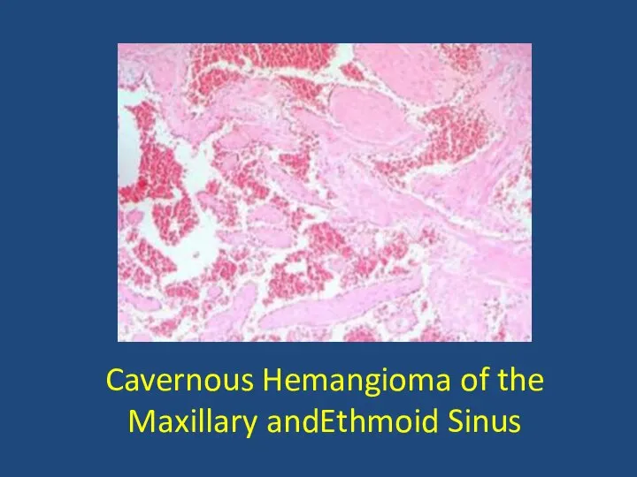
- 15. Cavernous Hemangioma of the Maxillary andEthmoid Sinus
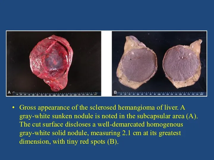
- 16. Gross appearance of the sclerosed hemangioma of liver. A gray-white sunken nodule is noted in the
- 20. Скачать презентацию
Слайд 5Simple hemangioma
is usually red or blue-purple color, is located on the
Simple hemangioma
is usually red or blue-purple color, is located on the
Слайд 6Cavernous hemangioma
is usually located under the skin, is a limited nodular
Cavernous hemangioma
is usually located under the skin, is a limited nodular
Слайд 7Combined hemangioma
usually a combination of surface and subcutaneous hemangiomas (simple
Combined hemangioma
usually a combination of surface and subcutaneous hemangiomas (simple
Слайд 8Extensive capillary-cavernous hemangioma of the left half of the head with a
Extensive capillary-cavernous hemangioma of the left half of the head with a
Слайд 9Capillary hemangioma of the child
Hemangioma of the humerus
Capillary hemangioma of the child
Hemangioma of the humerus
Слайд 10Hemangioma arterial (h. Arteriale) - hemangioma, arterial blood vessels from growing type.
Hemangioma arterial (h. Arteriale) - hemangioma, arterial blood vessels from growing type.
Слайд 11HEMANGIOMA CAN BE
LIVER
KIDNEY
VERTEBRAE
LIPS
HEMANGIOMA CAN BE
LIVER
KIDNEY
VERTEBRAE
LIPS
Слайд 13Microscopic structure of capillary hemangioma. The wall of the capillaries represented two-three-layer
Microscopic structure of capillary hemangioma. The wall of the capillaries represented two-three-layer
Слайд 14
Microscopic finding of the giant hemangioma. (A) Low-power view disclosing proliferation of
Microscopic finding of the giant hemangioma. (A) Low-power view disclosing proliferation of
Слайд 15Cavernous Hemangioma of the Maxillary andEthmoid Sinus
Cavernous Hemangioma of the Maxillary andEthmoid Sinus
Слайд 16Gross appearance of the sclerosed hemangioma of liver. A gray-white sunken nodule
Gross appearance of the sclerosed hemangioma of liver. A gray-white sunken nodule
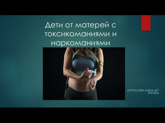 Дети от матерей с токсикоманиями и наркоманиями
 Пулороз (Тиф птиці)
 Миокард инфаркты
 Лечение и профилактика заболеваний тканей пародонта
 Симптомды жатыр миомасында жатыр артериясының эмболизациясы мен лапароскопиялық консервативті миомаэктомияның тиімділігі
 Выделительная система почек и кожи
 Инвалидность в России
 Поиск объективных физиологических индикаторов чувства тошноты
 Кардиологиялық науқастардың дәрігерлік- еңбектік сараптамасы
 Вакцина и вакцинация
 Сахарный диабет. Особенности у детей. Специфические осложнения
 Стоп ВИЧ и СПИД
 Vastsündinu-ja imikuiga
 lektsia_2_VBI_po_distsipline_sestrinskoe_delo
 Первая помощь при Кровотечениях
 Профессия моей мечты. Профессия окулист
 Характеристика различных видов повреждений (травм) организма человека и причины их вызывающие
 Ответ острой фазы. Лихорадка
 Доклад 2222по вторичной профилактике 2022 28.10
 Поликистозная болезнь почек
 Санитарно-эпидемиологические требования к условиям и организации обучения в общеобразовательных учреждениях
 Математические расчеты в деятельности медицинского работника по индивидуальной теме пневмония
 Аутизм. Характеристика
 Первая медицинская помощь при кровотечениях. Тема 18
 Бета-адреноблокаторы
 Логопедическое обследование по выявлению причин, обусловленности нарушений и проявления звукопроизношения
 Поверхностный кариес
 Защищенные аминопенициллины