- Placenta accreta

Содержание
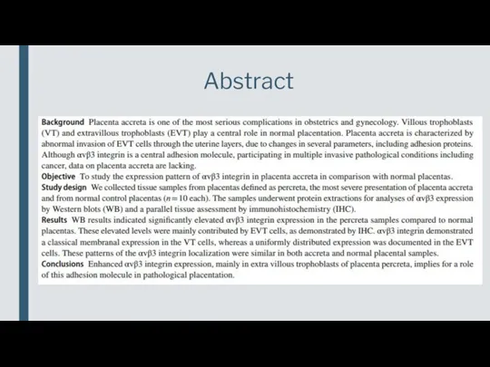
- 2. Abstract
- 3. Article and journal characteristics Introduction Materials and methods Results Discussion Conclusion Questions for discussion Outlines
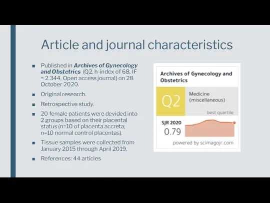
- 4. Published in Archives of Gynecology and Obstetrics (Q2, h-index of 68, IF = 2.344, Open access
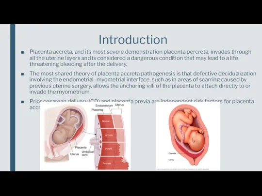
- 5. Placenta accreta, and its most severe demonstration placenta percreta, invades through all the uterine layers and
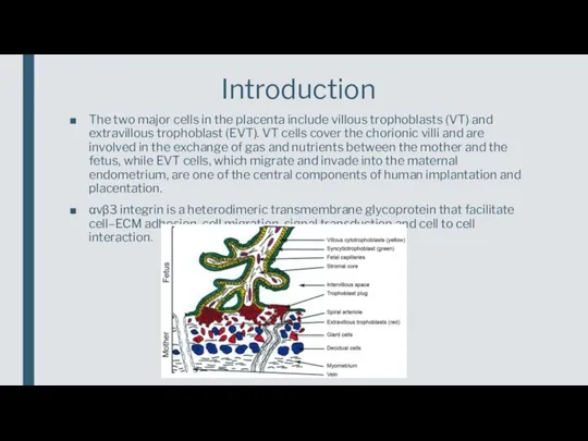
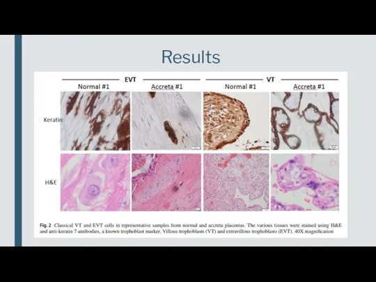
- 6. The two major cells in the placenta include villous trophoblasts (VT) and extravillous trophoblast (EVT). VT
- 7. Materials and methods
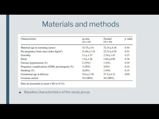
- 8. Baseline characteristics of the study group. Materials and methods
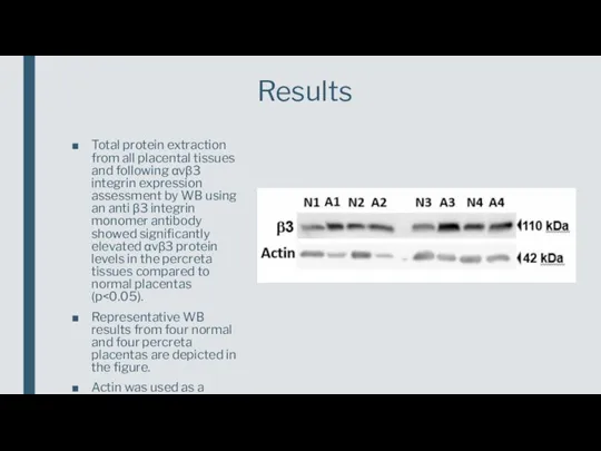
- 9. Total protein extraction from all placental tissues and following αvβ3 integrin expression assessment by WB using
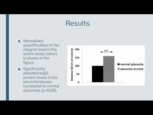
- 10. Normalized quantification of the integrin level in the entire study cohort is shown in the figure.
- 11. Results
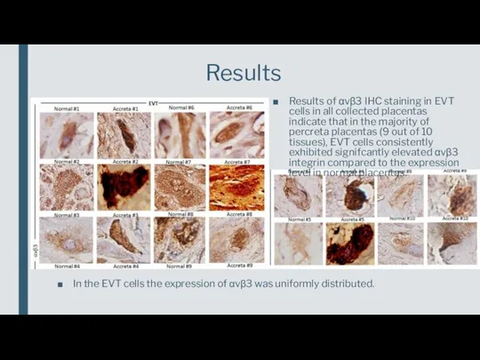
- 12. Results Results of αvβ3 IHC staining in EVT cells in all collected placentas indicate that in
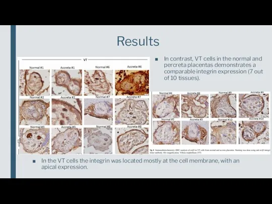
- 13. Results In contrast, VT cells in the normal and percreta placentas demonstrates a comparable integrin expression
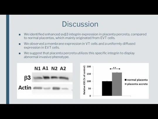
- 14. We identified enhanced αvβ3 integrin expression in placenta percreta, compared to normal placentas, which mainly originated
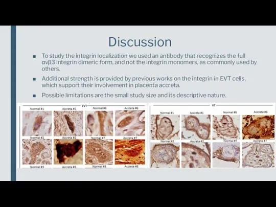
- 15. To study the integrin localization we used an antibody that recognizes the full αvβ3 integrin dimeric
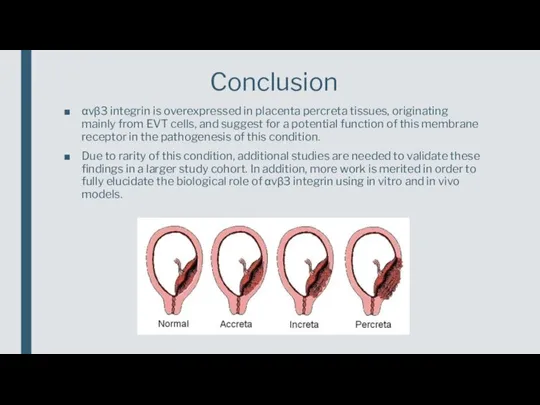
- 16. αvβ3 integrin is overexpressed in placenta percreta tissues, originating mainly from EVT cells, and suggest for
- 17. What are the potential benefits of using knowledge of αvβ3 integrin overexpression in treatment and diagnosis
- 19. Скачать презентацию
Слайд 3Article and journal characteristics
Introduction
Materials and methods
Results
Discussion
Conclusion
Questions for discussion
Outlines
Article and journal characteristics
Introduction
Materials and methods
Results
Discussion
Conclusion
Questions for discussion
Outlines
Слайд 4Published in Archives of Gynecology and Obstetrics (Q2, h-index of 68, IF
Published in Archives of Gynecology and Obstetrics (Q2, h-index of 68, IF
Слайд 5Placenta accreta, and its most severe demonstration placenta percreta, invades through all
Placenta accreta, and its most severe demonstration placenta percreta, invades through all
Слайд 6The two major cells in the placenta include villous trophoblasts (VT) and
The two major cells in the placenta include villous trophoblasts (VT) and
Слайд 7Materials and methods
Materials and methods
Слайд 8Baseline characteristics of the study group.
Materials and methods
Baseline characteristics of the study group.
Materials and methods
Слайд 9Total protein extraction from all placental tissues and following αvβ3 integrin expression
Total protein extraction from all placental tissues and following αvβ3 integrin expression
Слайд 10Normalized quantification of the integrin level in the entire study cohort is
Normalized quantification of the integrin level in the entire study cohort is
Слайд 11Results
Results
Слайд 12Results
Results of αvβ3 IHC staining in EVT cells in all collected placentas
Results
Results of αvβ3 IHC staining in EVT cells in all collected placentas
Слайд 13Results
In contrast, VT cells in the normal and percreta placentas demonstrates a
Results
In contrast, VT cells in the normal and percreta placentas demonstrates a
Слайд 14We identified enhanced αvβ3 integrin expression in placenta percreta, compared to normal
We identified enhanced αvβ3 integrin expression in placenta percreta, compared to normal
Слайд 15To study the integrin localization we used an antibody that recognizes the
To study the integrin localization we used an antibody that recognizes the
Слайд 16αvβ3 integrin is overexpressed in placenta percreta tissues, originating mainly from EVT
αvβ3 integrin is overexpressed in placenta percreta tissues, originating mainly from EVT
Слайд 17What are the potential benefits of using knowledge of αvβ3 integrin overexpression
What are the potential benefits of using knowledge of αvβ3 integrin overexpression
 Челюстно-лицевая ортопедия. Цель, задачи. Классификация переломов челюстей. Причины и механизм смещения отломков. (Тема 4)
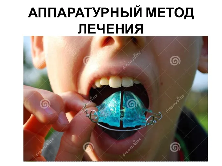 Аппаратурный метод лечения в стоматологии
 b9176ef5-aa6a-464c-ba1d-b03a8995427f
 Заболеваемость подросткового населения в 21 веке
 Мозговой штурм
 ОКИ возбудители сальмонеллёза, брюшного тифа
 Миеломенингоцеле. Синонимы
 Анатомия и физиология челюстно-лицевой области
 Сравнительная перкуссия легких (1)
 Актуальная информация о коронавирусе
 Техника интубации трахеи
 Терминальные состояния. Первая помощь при утоплении
 Патофизиология поджелудочной железы
 Психологическая семиотика
 Функциональная анатомия аппарата дыхания
 Пуринергиялық синапста қозудың берілуіне әсер ететін заттар
 Персонифицированная диагностика в онкологии
 Хирургический шов
 Бронхолегочная дисплазия
 Психопатологическая семиотика
 Синдром ранней реполяризации желудочков. Гипертрофия
 ухудшение состояния здоровья приводит к снижению успеваемости по учебным предметам
 Регуляция энергетического обмена в клетке гормонами
 Тыныштандырғыш заттар
 Національна нормативна база. Конституція України. Стандарти у сфері охорони здоров’я
 Туберкулез половых органов. Диагностика
 Организация работы эндоскопического подразделения медицинской организации
 Клинико-этические особенности научных исследований