- External Anatomy of the Eye

Содержание
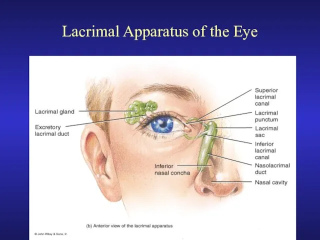
- 2. Lacrimal Apparatus of the Eye
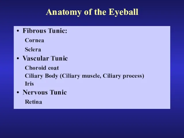
- 3. Anatomy of the Eyeball Fibrous Tunic: Cornea Sclera Vascular Tunic Choroid coat Ciliary Body (Ciliary muscle,
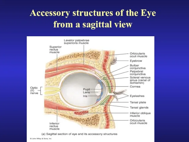
- 4. Accessory structures of the Eye from a sagittal view
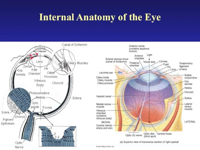
- 5. Internal Anatomy of the Eye
- 6. Detail view of the anterior anatomy of the eye
- 7. Production of Aqueous Humor and Intraocular pressure Ciliary Process: Produces Aqueous Humor 2. Posterior Chamber: Aqueous
- 8. Opthalmoscopic view of the retina showing the location of the Macula to the Optic Disc
- 9. Histology of the retina of the eye
- 10. Photomicroscopic view of the Histology of the Eye S = Sclera C = Choroid coat PE
- 11. Photomicroscopic view of the Histology of the Eye showing the location of the central fovea
- 12. Intrinsic Eye Muscles and their response to light
- 13. The Visual Pathway
- 14. Light Refractory Pathway: Bulbar Conjunctiva Cornea Aqueous Humor Lens Vitreous Humor Ganglion Cell Layer Inner Synaptic
- 15. Abnormalities of The Eye: Myopic - nearsighted Hypermetropic - Farsighted Presbyopia - age-related failure of lens
- 16. Accommodation of the Lens for near vision Ciliary muscles contract Ciliary body pulls forward and inward
- 17. Accommodation of the Lens for far vision Ciliary muscles relaxes Ciliary body returns to its resting
- 18. Anatomy of Rods and Cones
- 19. Physiology of Rods and Photopigments
- 21. Скачать презентацию
Слайд 3Anatomy of the Eyeball
Fibrous Tunic:
Cornea
Sclera
Vascular Tunic
Choroid coat
Ciliary Body (Ciliary muscle,
Anatomy of the Eyeball
Fibrous Tunic:
Cornea
Sclera
Vascular Tunic
Choroid coat
Ciliary Body (Ciliary muscle,
Слайд 4Accessory structures of the Eye
from a sagittal view
Accessory structures of the Eye
from a sagittal view
Слайд 5Internal Anatomy of the Eye
Internal Anatomy of the Eye
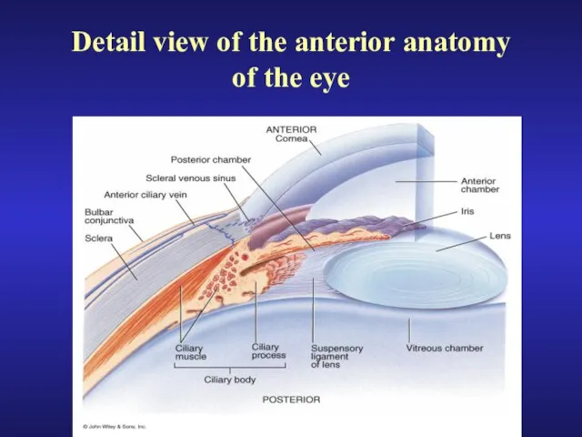
Слайд 6Detail view of the anterior anatomy
of the eye
Detail view of the anterior anatomy
of the eye
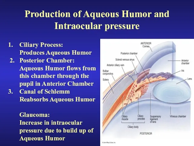
Слайд 7Production of Aqueous Humor and Intraocular pressure
Ciliary Process:
Produces Aqueous Humor
2. Posterior
Production of Aqueous Humor and Intraocular pressure
Ciliary Process:
Produces Aqueous Humor
2. Posterior
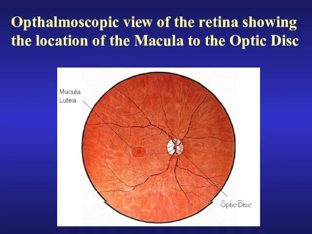
Слайд 8Opthalmoscopic view of the retina showing the location of the Macula to
Opthalmoscopic view of the retina showing the location of the Macula to
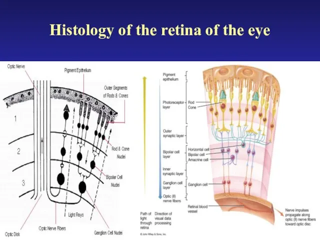
Слайд 9Histology of the retina of the eye
Histology of the retina of the eye
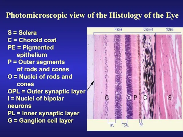
Слайд 10 Photomicroscopic view of the Histology of the Eye
S = Sclera
C =
Photomicroscopic view of the Histology of the Eye
S = Sclera
C =
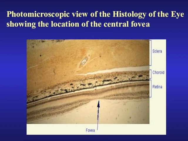
Слайд 11Photomicroscopic view of the Histology of the Eye
showing the location of the
Photomicroscopic view of the Histology of the Eye
showing the location of the
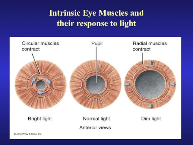
Слайд 12Intrinsic Eye Muscles and
their response to light
Intrinsic Eye Muscles and
their response to light
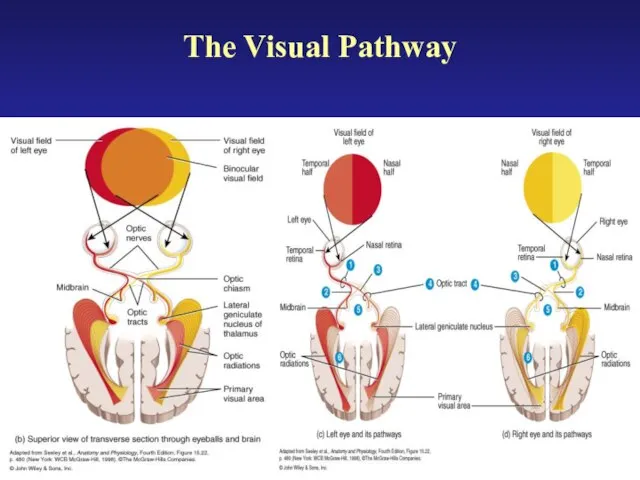
Слайд 13The Visual Pathway
The Visual Pathway
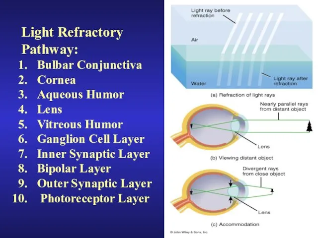
Слайд 14Light Refractory
Pathway:
Bulbar Conjunctiva
Cornea
Aqueous Humor
Lens
Vitreous Humor
Ganglion Cell Layer
Inner Synaptic Layer
Bipolar Layer
Outer Synaptic Layer
Light Refractory
Pathway:
Bulbar Conjunctiva
Cornea
Aqueous Humor
Lens
Vitreous Humor
Ganglion Cell Layer
Inner Synaptic Layer
Bipolar Layer
Outer Synaptic Layer
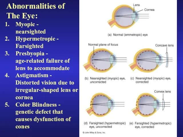
Слайд 15Abnormalities of
The Eye:
Myopic -
nearsighted
Hypermetropic -
Farsighted
Presbyopia -
age-related failure of
lens
Abnormalities of
The Eye:
Myopic -
nearsighted
Hypermetropic -
Farsighted
Presbyopia -
age-related failure of
lens
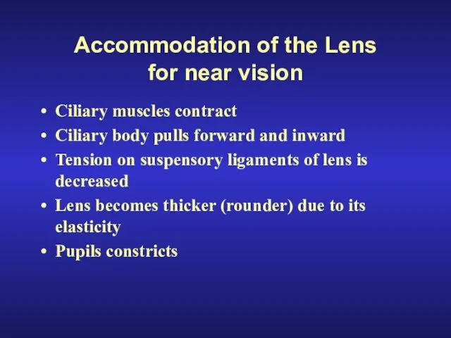
Слайд 16Accommodation of the Lens
for near vision
Ciliary muscles contract
Ciliary body pulls forward
Accommodation of the Lens
for near vision
Ciliary muscles contract
Ciliary body pulls forward
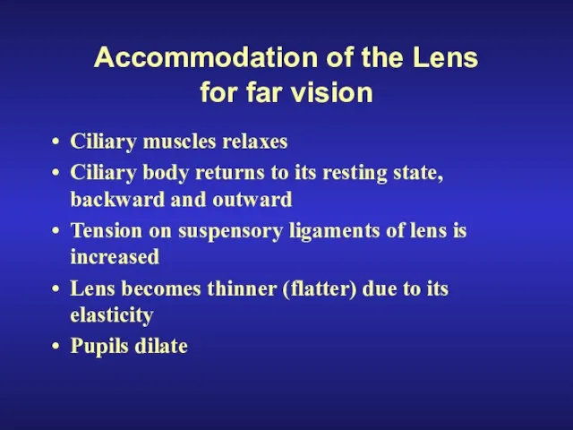
Слайд 17Accommodation of the Lens
for far vision
Ciliary muscles relaxes
Ciliary body returns to
Accommodation of the Lens
for far vision
Ciliary muscles relaxes
Ciliary body returns to
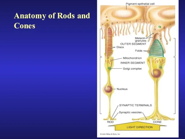
Слайд 18Anatomy of Rods and
Cones
Anatomy of Rods and
Cones
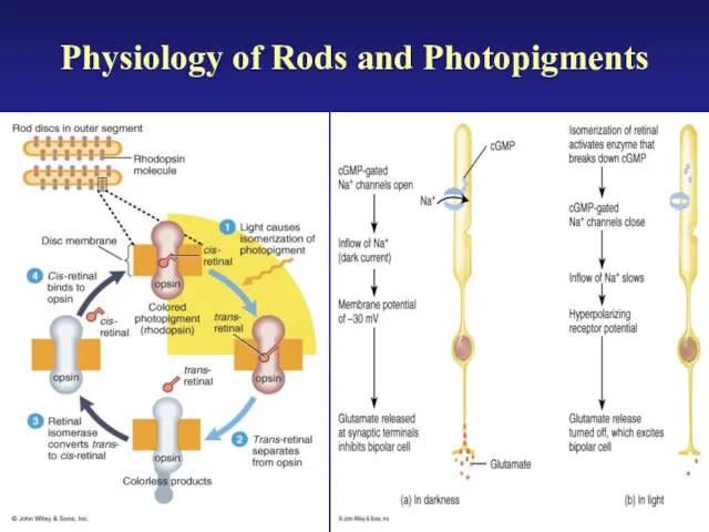
Слайд 19Physiology of Rods and Photopigments
Physiology of Rods and Photopigments
 Perfect Vision Gallery
 Тема: «Парадигма информационного риска»
 Баскетбол
 Деятельность ТК 23 в международном проекте «Баренц-2020». Результаты и планы
 Мастер-класс. Реальное дипломное проектирование с привлечением талантливой молодежи
 Презентация на тему Образ Богини-Матери
 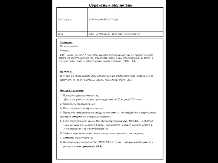 Bulletin MPS
 Национальные виды спорта и игр народов мира
 Свободное время моей семьи. Арина Мурадян
 Презентация на тему Поль Гоген жизнь в творчестве (1848-1903 гг.)
  Деревянные конструкции
 Что такое «Рейтинг доверия» E-xecutive? Это результат голосования участников Сообщества с помощью сервиса WishList. Именно этим компаниям
  Современное лечение острого коронарного синдрома
 Визитка лайфстайл издания Вuro 24/7
 КОНКУРС «ПРОФЕССИОНАЛ2009»
 Презентація-4 (2)
 Цифровое право: основные научные подходы
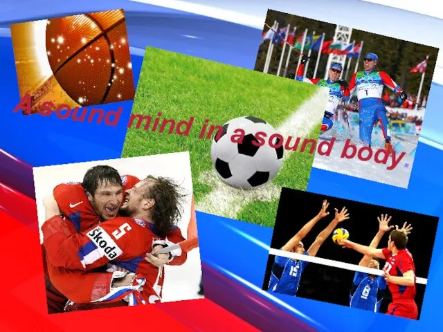 A sound mind in a sound body
 Лекция Специализированные машины, оборудование и механизмы
 Тема: Природные экосистемы, взаимоотношения между компонентами экосистемы. Антропологические аксиомы
 - растворитель
 Контур.EDI
 Азбука цветов
 Родовые знаки Лиевых
 Медицинское образование в России и Германии
 Архитектура и дизайн
 О Компании:
 1 ГОСУДАРСТВЕННАЯ КОРПОРАЦИЯ «БАНК РАЗВИТИЯ И ВНЕШНЕЭКОНОМИЧЕСКОЙ ДЕЯТЕЛЬНОСТИ (ВНЕШЭКОНОМБАНК)» ВЗАИМОДЕЙСТВИЕ ПРЕДСТАВИТЕЛЬС