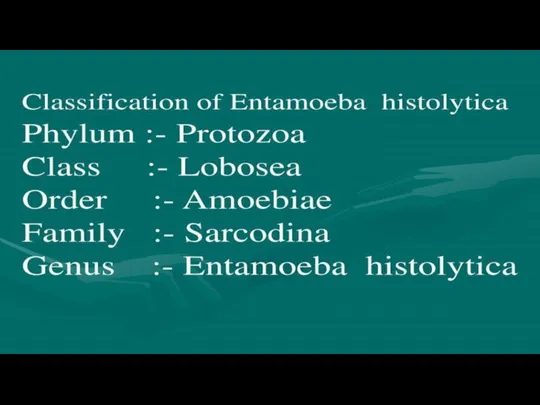
- Biological bases of parasism class Sarcodina

Содержание
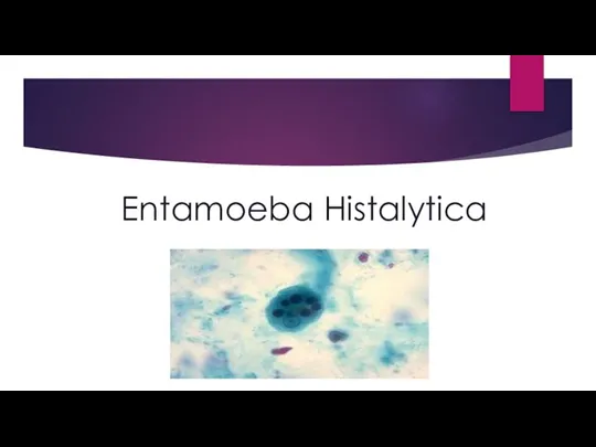
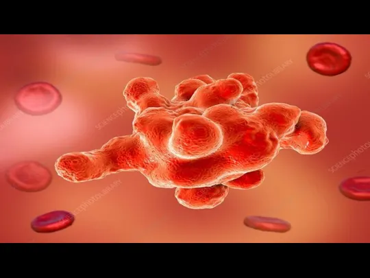
- 2. Entamoeba Histalytica
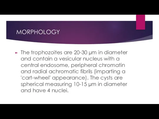
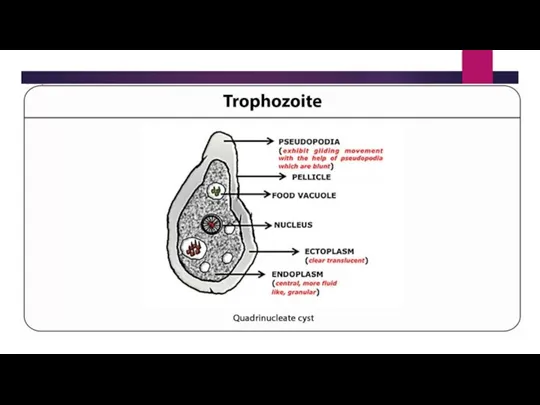
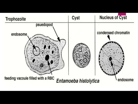
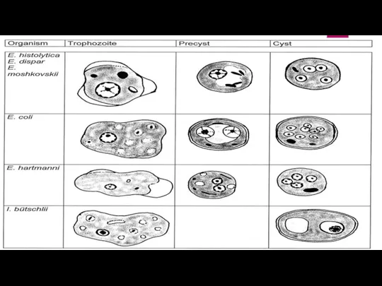
- 5. MORPHOLOGY The trophozoites are 20-30 µm in diameter and contain a vesicular nucleus with a central
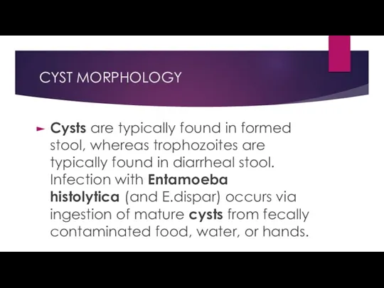
- 7. CYST MORPHOLOGY Cysts are typically found in formed stool, whereas trophozoites are typically found in diarrheal
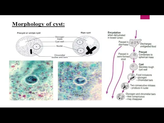
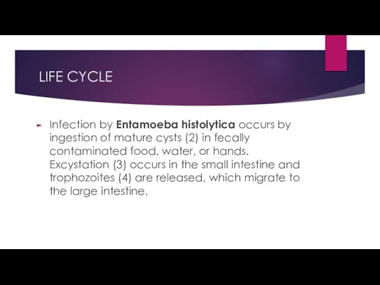
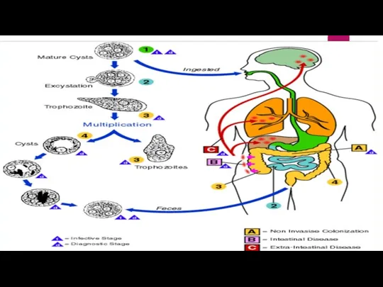
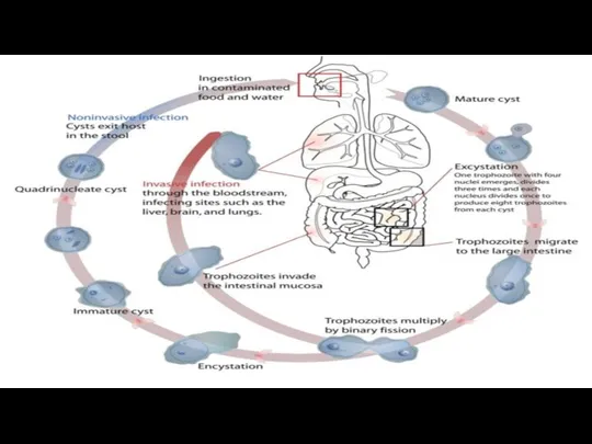
- 11. LIFE CYCLE Infection by Entamoeba histolytica occurs by ingestion of mature cysts (2) in fecally contaminated
- 14. PATHOGENECITY Entamoeba histolytica, a protozoan parasite, is the etiologic agent of amoebiasis in humans. It exists
- 15. DISEASE Entamoeba histolytica is an anaerobic parasitic amoebozoan, part of the genus Entamoeba. Predominantly infecting humans
- 16. DIAGNOSIS A single stool examination has a low sensitivity of detecting the parasite . The best
- 17. TREATMENT To treat invasive amebiasis, metronidazole (Flagyl, MetroGel, Noritate) is recommended even for amoebic liver abscesses
- 18. PREVENTION and CONTROL Improved sanitation will help to reduce the liklihood of transmission. Travelers to endemic
- 21. Скачать презентацию
Слайд 5MORPHOLOGY
The trophozoites are 20-30 µm in diameter and contain a vesicular nucleus
MORPHOLOGY
The trophozoites are 20-30 µm in diameter and contain a vesicular nucleus
Слайд 7CYST MORPHOLOGY
Cysts are typically found in formed stool, whereas trophozoites are typically found
CYST MORPHOLOGY
Cysts are typically found in formed stool, whereas trophozoites are typically found
Слайд 11LIFE CYCLE
Infection by Entamoeba histolytica occurs by ingestion of mature cysts (2) in fecally
LIFE CYCLE
Infection by Entamoeba histolytica occurs by ingestion of mature cysts (2) in fecally
Слайд 14PATHOGENECITY
Entamoeba histolytica, a protozoan parasite, is the etiologic agent of amoebiasis in
PATHOGENECITY
Entamoeba histolytica, a protozoan parasite, is the etiologic agent of amoebiasis in
Слайд 15DISEASE
Entamoeba histolytica is an anaerobic parasitic amoebozoan, part of the genus Entamoeba.
DISEASE
Entamoeba histolytica is an anaerobic parasitic amoebozoan, part of the genus Entamoeba.
Слайд 16DIAGNOSIS
A single stool examination has a low sensitivity of detecting the parasite
DIAGNOSIS
A single stool examination has a low sensitivity of detecting the parasite
Слайд 17TREATMENT
To treat invasive amebiasis, metronidazole (Flagyl, MetroGel, Noritate) is recommended even for amoebic liver
TREATMENT
To treat invasive amebiasis, metronidazole (Flagyl, MetroGel, Noritate) is recommended even for amoebic liver
Слайд 18PREVENTION and CONTROL
Improved sanitation will help to reduce the liklihood of transmission. Travelers
PREVENTION and CONTROL
Improved sanitation will help to reduce the liklihood of transmission. Travelers

 Острый аппендицит
 Краниотомия. Показания
 Классификация климактерических расстройств по времени проявления. Вазомоторные расстройства. Патогенез приливов
 Әлеуметтік міндетті медициналық сақтандыруды енгізу кезінде өндірістерде жұмыс атқаратын репродуктивті жастағы әйелдер
 Көмейдің иннервациясы
 Арсеникум альбум (Яд). Общее действие
 Лечебная физическая культура
 Дети с нарушениями интеллекта
 Бордетеллалар. Көкжөтел және паракөкжөтел
 Учение об иммунитете
 Адаптация костного таза в родах. Остеопатическое сопровождение родов во втором периоде
 Миелин. Нарушение миелиновой оболочки
 Сестринская помощь при нарушении функции опорно-двигательного аппарата
 Имуномодуляторы. В каких случаях назначают имуномодуляторы
 Функциональная активность человека и взаимосвязь физической и умственной деятельности
 Интеллект и интеллектуальная недостаточность. Глубокая умственная отсталость (идиотия)
 Оказание первой помощи при несчастных случаях на производстве
 Moderni ošetřovatelská praxe
 Правила внутреннего трудового распорядка в аптеке
 Стресс-ЭКГ Велоэргометрия и тредмил-тест
 Деятельность медицинской сестры при выполнении различных видов физиотерапевтических процедур
 Bronchial asthma
 Понятие о шоке и меры его предупреждения. Простейшие способы реанимации на поле боя
 Четырехугольники
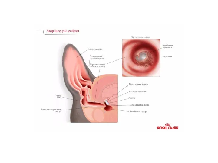 Причины поражений ушной раковины и наружного слухового прохода
 Общая анестезия в акушерстве
 Врожденная патология лица. Клаcсификация, этиология, патогенез, клиника, диагностика. Сроки и принципы комплексного лечения