- Histolytica

Содержание
- 4. Anaerobic parasitic amoebozoan, part of the genus Entamoeba.[1] Predominantly infecting humans and other primates causing amoebiasis,
- 7. It was thought that 10% of the world population was infected, but these figures predate the
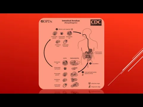
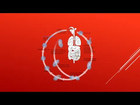
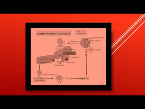
- 9. TRANSMISSION The active (trophozoite) stage exists only in the host and in fresh loose feces; cysts
- 11. The cysts are readily killed by heat and by freezing temperatures, and survive for only a
- 14. Symptoms can include fulminating dysentery, bloody diarrhea, weight loss, fatigue, abdominal pain, and amoeboma. The amoeba
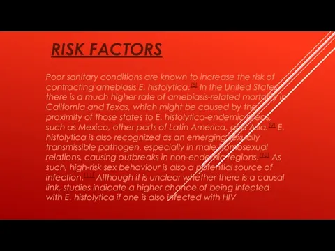
- 16. RISK FACTORS Poor sanitary conditions are known to increase the risk of contracting amebiasis E. histolytica.[8]
- 18. PATHOGEN INTERACTION E. histolytica may modulate the virulence of certain human viruses and is itself a
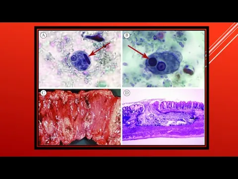
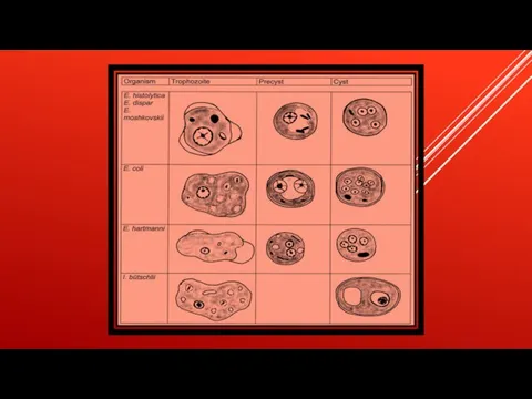
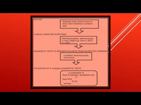
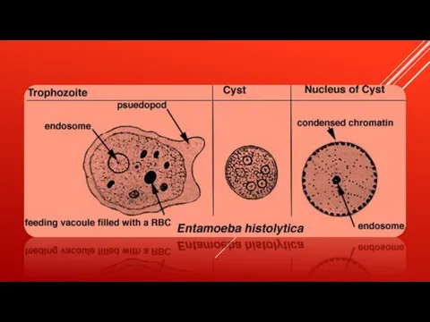
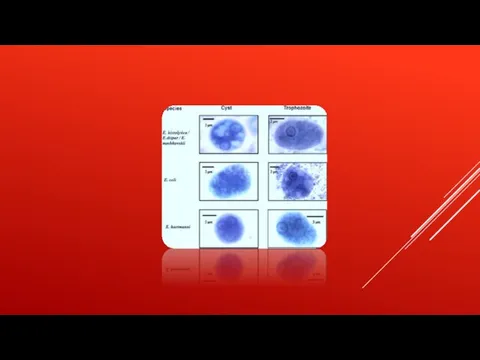
- 19. DIAGNOSIS Diagnosis is confirmed by microscopic examination for trophozoites or cysts in fresh or suitably preserved
- 21. Скачать презентацию

Слайд 4 Anaerobic parasitic amoebozoan, part of the genus Entamoeba.[1] Predominantly infecting humans and other primates causing amoebiasis, E. histolytica is estimated
Anaerobic parasitic amoebozoan, part of the genus Entamoeba.[1] Predominantly infecting humans and other primates causing amoebiasis, E. histolytica is estimated
![Anaerobic parasitic amoebozoan, part of the genus Entamoeba.[1] Predominantly infecting humans and](/_ipx/f_webp&q_80&fit_contain&s_1440x1080/imagesDir/jpg/914432/slide-3.jpg)
Слайд 7
It was thought that 10% of the world population was infected, but
It was thought that 10% of the world population was infected, but
Слайд 9TRANSMISSION
The active (trophozoite) stage exists only in the host and in fresh
TRANSMISSION
The active (trophozoite) stage exists only in the host and in fresh
Слайд 11The cysts are readily killed by heat and by freezing temperatures, and
The cysts are readily killed by heat and by freezing temperatures, and
Слайд 14Symptoms can include fulminating dysentery, bloody diarrhea, weight loss, fatigue, abdominal pain,
Symptoms can include fulminating dysentery, bloody diarrhea, weight loss, fatigue, abdominal pain,
Слайд 16RISK FACTORS
Poor sanitary conditions are known to increase the risk of contracting
RISK FACTORS
Poor sanitary conditions are known to increase the risk of contracting
Слайд 18PATHOGEN INTERACTION
E. histolytica may modulate the virulence of certain human viruses and is
PATHOGEN INTERACTION
E. histolytica may modulate the virulence of certain human viruses and is
Слайд 19DIAGNOSIS
Diagnosis is confirmed by microscopic examination for trophozoites or cysts in fresh
DIAGNOSIS
Diagnosis is confirmed by microscopic examination for trophozoites or cysts in fresh
 Оценка образа жизни
 Режим дня
 Особенности развития познавательной сферы у детей при ДЦП (детский церебральный паралич)
 Профилактика сердечных заболеваний
 Врожденная патология лица. Клаcсификация, этиология, патогенез, клиника, диагностика. Сроки и принципы комплексного лечения
 Издания по стоматологии (книги, журналы)
 Дерматозойный бред, паразитофобия, акарофобия, синдром Экбома или синдром Берса Конрада
 Игра. Средства, влияющие на функцию мочевыделительной системы
 Сrest-синдром
 Диабетическая кардиомиопатия
 Биологическое значение растворов
 Використання засобів фізичної культури і реабілітації в акушерсько-гінекологічній практиці
 USPTF. Новые рекомендации по применению статинов
 Белки-ферменты
 Детская инфекционная болезнь корь
 Шаблон презентации - 2022 врачи
 Наследственно-дегенеративные заболевания нервной системы
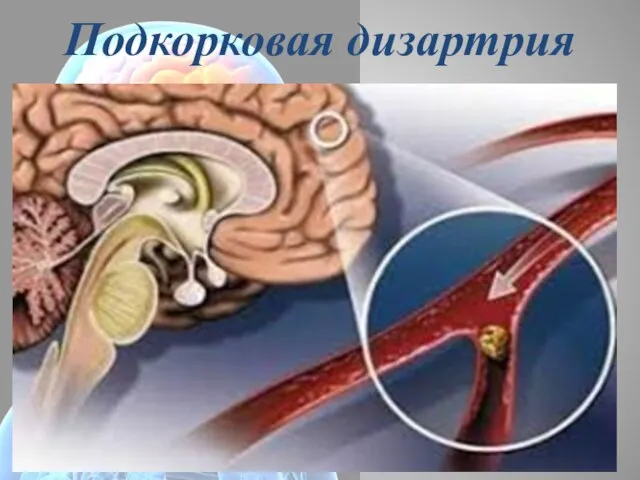 Подкорковая дизартрия
 Эпилепсия. Определение, история, распространенность, этиология, патогенез, классификация
 Использование приемов нейрофункциональной терапии в работе с детьми раннего возраста для развития артикуляционного аппарата
 Бескровная хирургия – миф или реальность XXI века
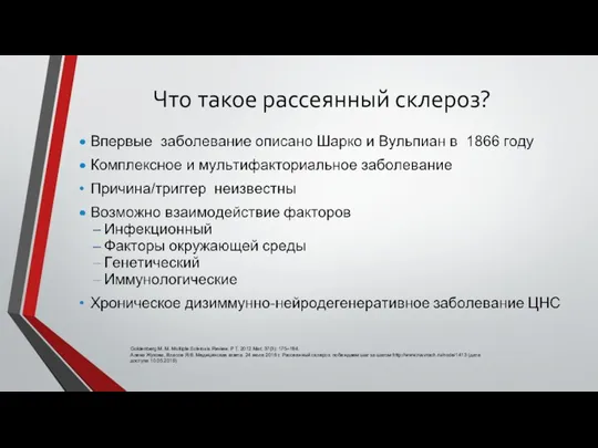 Что такое рассеянный склероз?
 Первая помощь при ранениях
 Филадельфийская хромосома
 Influenza
 Дифтерия
 Характеристики диет
 Выделительная система