Слайд 2Introduction to Demodicosis
Canine demodicosis is one of the well known skin
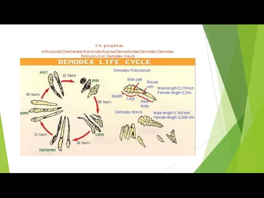
diseases encountered in veterinary practice. It is a dermatologic disease that occurs when mites colonize the hair follicles, sebaceous glands. Dermatological changes include erythema, alopecia, comedones, follicular hyperkeratosis, pustules, crusts and seborrhea. Often, a secondary pyoderma further complicates the disease (Scott et al. 2001). Demodex canis was the main causative agent of canine demodicosis and it is characterized by the presence of large numbers of Demodex mites. The three recognized canine Demodex mites are: Demodex canis, Demodexinjai, and the unnamed short-bodied mite. Demodex caniswas the first to be identified and named the two additional Demodex mites may be mutations of Demodex canis, or separate species.
Слайд 9Signs and symptoms
Minor cases of demodectic mange usually do not cause much
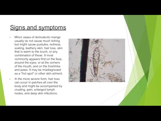
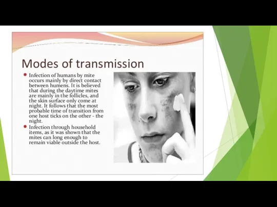
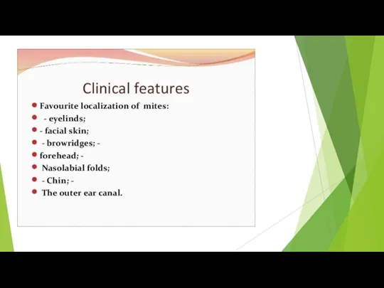
itching but might cause pustules, redness, scaling, leathery skin, hair loss, skin that is warm to the touch, or any combination of these. It most commonly appears first on the face, around the eyes, or at the corners of the mouth, and on the forelimbs and paws. It may be misdiagnosed as a "hot spot" or other skin ailment.
In the more severe form, hair loss can occur in patches all over the body and might be accompanied by crusting, pain, enlarged lymph nodes, and deep skin infections.
Слайд 10Materials and methods
Two mongrel dogs aged between 7 and 9 months belongs to

a same house was brought to the Veterinary Hospital, Proddatur with a history of skin lesions associated with pruritus from One month. Upon clinical examination, dogs exhibited papules, pustules, erythema, alopecia, hyperpigmentation, erosions, lichenification and cellulitis. Distribution of lesions observed on face, around the eyes and ears, chin region, fore limbs, neck and lateral abdomen (Fig. 1). Skin scrapings, tape impression smears and hair plucks was collected from the affected dogs for laboratory examination. Scrapings were collected with scalpel blade dipped in liquid paraffin and collection of scrapings was continued until there was slight ooze of blood from dermal capillaries
Слайд 11Material was suspended in a few drops of liquid paraffin on a

microscopic slide, a coverslip was applied and the preparation was examined under low and high power (10X, 40X) of microscope. The acetate tape impression smears was used to investigate superficial mites. The sticky surface of the tape was pressed on the suspected lesions, and tape was then mounted directly on a glass slide. The glass slides were examined under compound microscopes with 10X and 40X of magnification. Few tape impression smears were stained with new methylene blue for 1 min and examined under 100X (Rosenkrantz 2008).
Слайд 12Results and discussion
Skin scrapings collected from the head region, revealed different stages
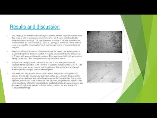
of Demodexmites (Fig. 2) along with few ovigerous female mites (Fig. 4a). D. canis were found in hair pluck examination technique. The tape impression technique of the dogs revealed more number of short-tail Demodexmites (D. cornei). Cytology of impression smears revealed cocci, cocci engulfed by neutrophils which indicate involvement of secondary bacterial infection.
Based on the history, lesions and laboratory findings, the present case was diagnosed as generalized superficial demodicosis of D. cornei and generalized follicular demodicosis of D. canis with secondary bacterial pyoderma. Dogs were treated with oral ivermectin at 500 μg/kg/day for 45 days by regular monitoring for the side effects.
Ampicillin at 25 mg/kg twice a day orally, BID for 14 days was given to control secondary bacterial infection. After one week of antibiotic therapy, amitraz (2 ml in 1 litre of water) was given weekly twice as topical application followed by bath with benzyl peroxide (petben) shampoo up to the recovery period.
One week after therapy moist lesions and scales was disappeared and dogs had mild pruritus. 2 weeks after treatment, the number of surface Demodexmites detected by the tape preparation technique was gradually decreased and the dogs were free from pruritus, erythema, erosions, and ulcers. One month after treatment, the general skin condition was improved; absence of pruritus was noticed and number of surface Demodex mites was also decreased. Complete disappearance of mites and re-growth of hair was noticed after 45 days of after therapy.
Слайд 13Mites with short tail were identified as D. cornei based on other morphological characteristics.
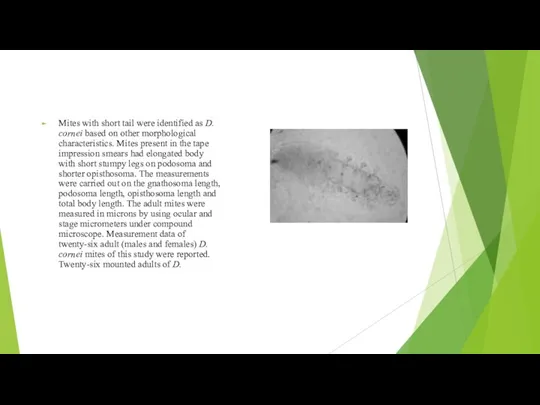
Mites present in the tape impression smears had elongated body with short stumpy legs on podosoma and shorter opisthosoma. The measurements were carried out on the gnathosoma length, podosoma length, opisthosoma length and total body length. The adult mites were measured in microns by using ocular and stage micrometers under compound microscope. Measurement data of twenty-six adult (males and females) D. cornei mites of this study were reported. Twenty-six mounted adults of D.
Слайд 15Conclusion
The short tailed Demodex mites collected from the two dermatitis dogs in this study

were D. cornei. They had short opisthosoma and blunted posterior end when compared with D. canis. The mean total body length of short form of Demodex spp. was 132.21 microns while the mean total body length of D. canis was 214.32 microns.


 Функциональные обязанности и его компетенции семейного врача
 Sterilizatsia
 Винирлердің астаны тістің апроксимальды беткейін егеу
 Характеристика обстоятельств заражения и первых проявлений лейшманиоза
 Чем занимается врач-репродуктолог?
 Ēdienkarte
 Электр станцияларының тарату құрылғылары
 Рентгенологические синдромы заболеваний легких
 СРС: Хламидиоз
 Бешенство. Источники вируса
 Выписка ЭЛН
 Cанитарно-эпидемиологические требования к организациям общественного питания
 Инфузионная терапия
 Тест на тему: Микрофлора тела человека, цели и способы антимикробных мероприятий
 Инфрақызыл радиацияның гигиеналық маңызы. Ауа температурасының гигиеналық маңызы
 Миома матки
 Обмен холестерола
 Неспецифический язвенный колит. Разбор клинического случая
 Плавание как средство коррекции
 Ампициллин
 Профилактика коронавируса для школы
 ДВС - синдром. Классификация
 Противомикробные препараты
 Современные проблемы биофармации
 Фигура груша. Характеристика, метаболизм и отношение к спорту. Рацион
 Миология. Академия фитнеса Корус
 Иммунопатология при гранулематозах
 ВПС, протекающие с обогащением малого круга кровообращения. Лекция №3