- Radiological research methods and radiological semiotics of acute cerebrovascular accident

Содержание
- 2. ACUTE CEREBROVASCULAR ACCIDENT(CVA, STROKE) – acute and severe brain disease. Blood may be interrupted or stop
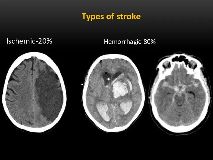
- 3. Types of stroke Ischemic-20% Hemorrhagic-80%
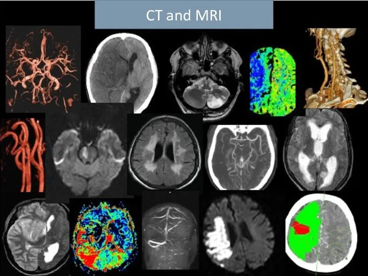
- 4. CT and MRI
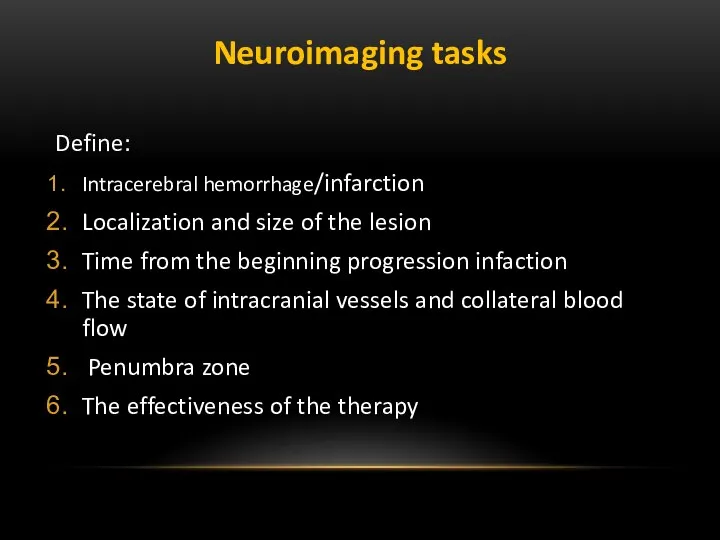
- 5. Neuroimaging tasks Define: Intracerebral hemorrhage/infarction Localization and size of the lesion Time from the beginning progression
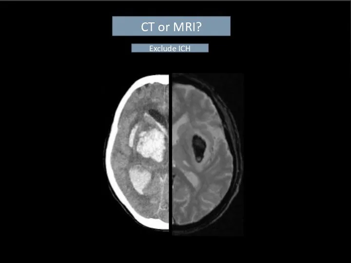
- 6. CT or MRI? Exclude ICH
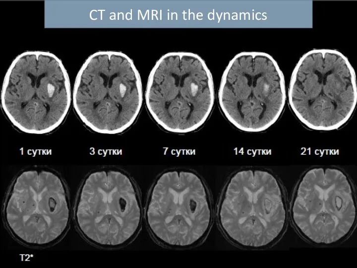
- 7. CT and MRI in the dynamics
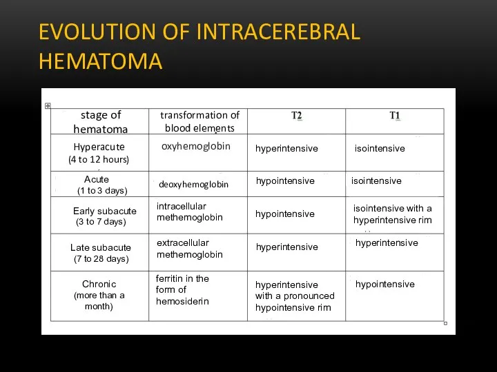
- 8. EVOLUTION OF INTRACEREBRAL HEMATOMA stage of hematoma transformation of blood elements oxyhemoglobin Hyperacute (4 to 12
- 9. Hemorrhagic strokes CT - is a “gold standard” of diagnosis. Hemorrhage is a focus of increased
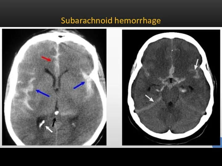
- 10. SUBARACHNOID HEMORRHAGE The sensitivity of CT to the presence of blood in the subarachnoid spaces is
- 11. Subarachnoid hemorrhage
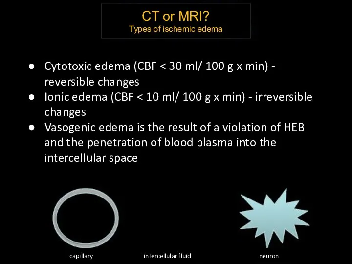
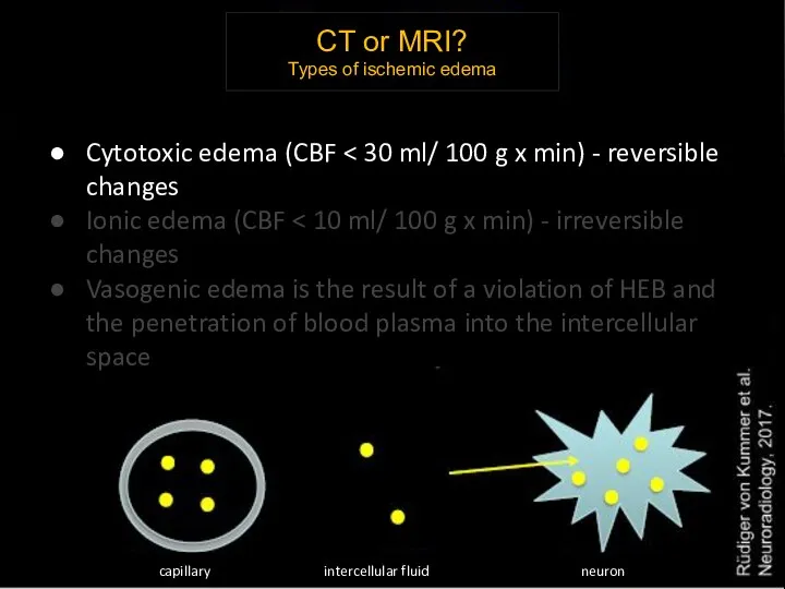
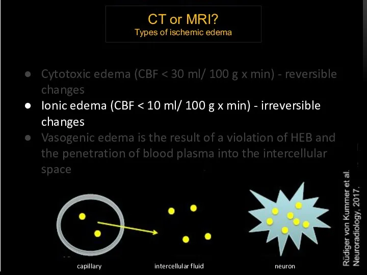
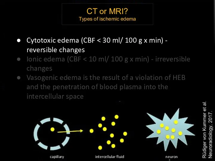
- 12. CT or MRI? Types of ischemic edema Cytotoxic edema (CBF Ionic edema (CBF Vasogenic edema is
- 13. CT or MRI? Types of ischemic edema Cytotoxic edema (CBF Ionic edema (CBF Vasogenic edema is
- 14. CT or MRI? Types of ischemic edema Cytotoxic edema (CBF Ionic edema (CBF Vasogenic edema is
- 15. CT or MRI? Types of ischemic edema Cytotoxic edema (CBF Ionic edema (CBF Vasogenic edema is
- 16. Early СТ signs CVA Arterial hyperdensity (a sign of intravascular thrombosis: hyperdensive middle cerebral artery, a
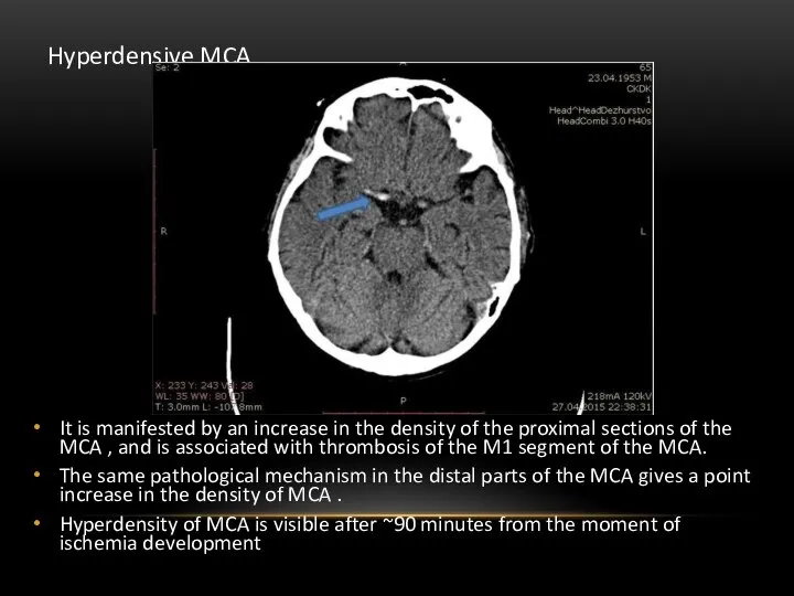
- 17. Hyperdensive MCA It is manifested by an increase in the density of the proximal sections of
- 18. ASPECTS SCORE The Alberta stroke programme early CT score (ASPECTS) 1 is a 10-point quantitative topographic
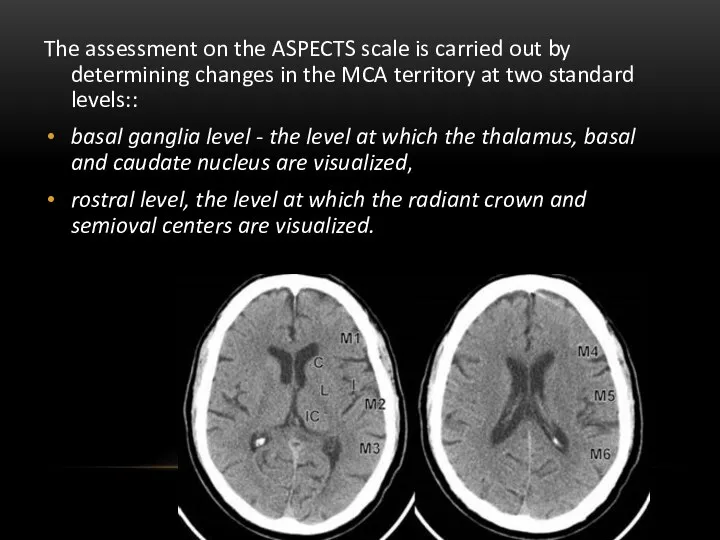
- 19. The assessment on the ASPECTS scale is carried out by determining changes in the MCA territory
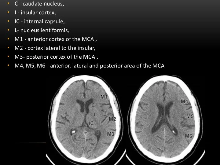
- 20. C - caudate nucleus, I - insular cortex, IC - internal capsule, L- nucleus lentiformis, M1
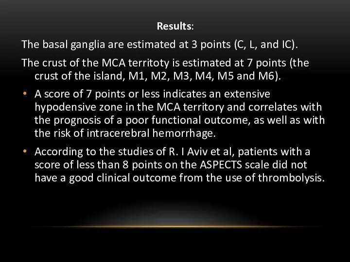
- 21. Results: The basal ganglia are estimated at 3 points (C, L, and IC). The crust of
- 22. PATHOPHYSIOLOGY AND MR-PICTURE OF CEREBRAL ISCHEMIC STROKE I acute stage(0-3 days). Pathoanatomically-focal cytotoxic edema, macroscopically-thickening of
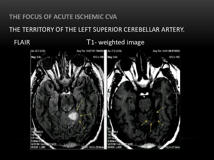
- 23. THE FOCUS OF ACUTE ISCHEMIC CVA THE TERRITORY OF THE LEFT SUPERIOR CEREBELLAR ARTERY. FLAIR Т1-
- 24. PATHOPHYSIOLOGY AND MR-PICTURE OF CEREBRAL ISCHEMIC STROKE Subacute stage (3 days-10-14 days). The combination of cytotoxic
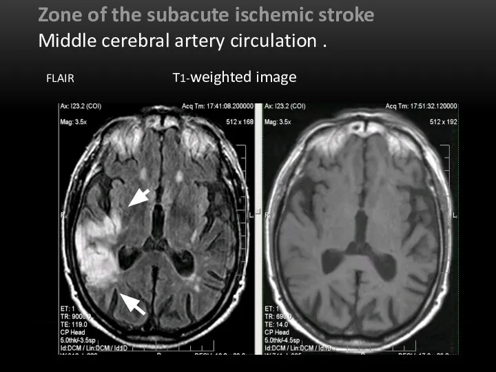
- 25. Zone of the subacute ischemic stroke Middle cerebral artery circulation . FLAIR Т1-weighted image
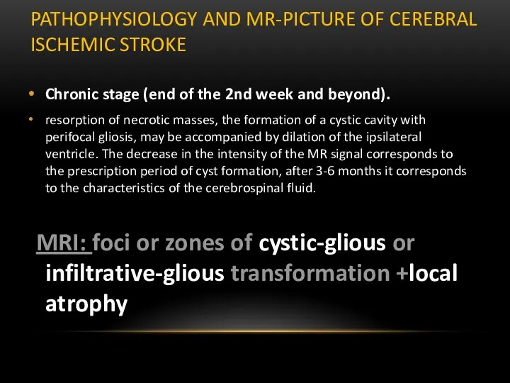
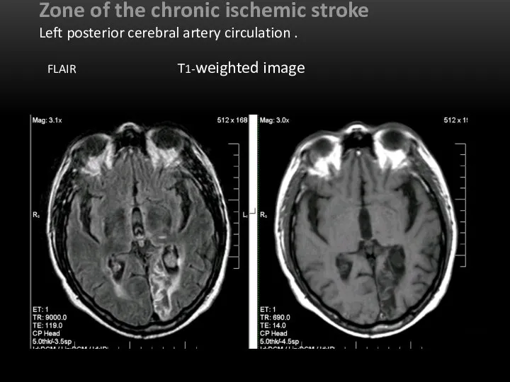
- 26. PATHOPHYSIOLOGY AND MR-PICTURE OF CEREBRAL ISCHEMIC STROKE Chronic stage (end of the 2nd week and beyond).
- 27. Zone of the chronic ischemic stroke Left posterior cerebral artery circulation . FLAIR Т1-weighted image
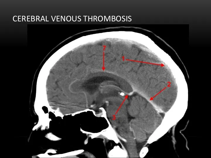
- 28. Direct signs(⅓ cases ): Triangle sign – visualization of a blood clot on the contrast-free part
- 29. CEREBRAL VENOUS THROMBOSIS
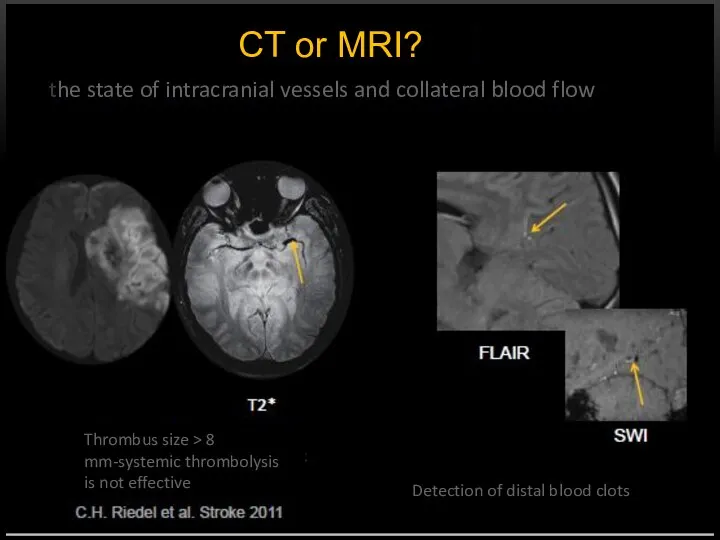
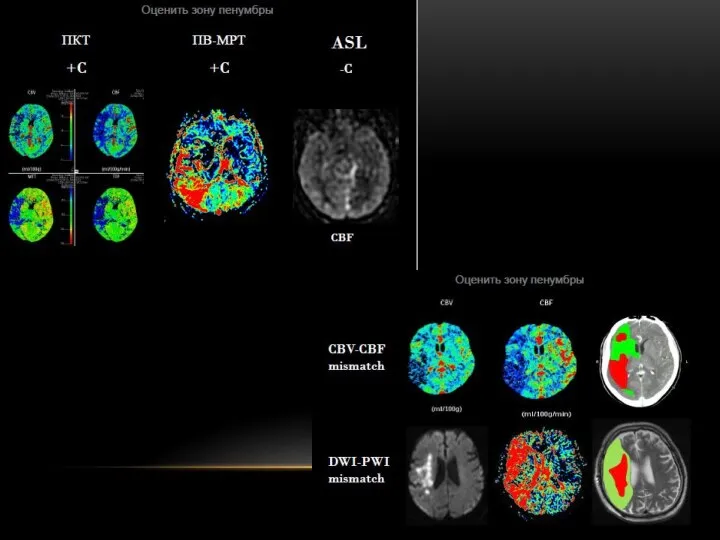
- 30. CT or MRI? the state of intracranial vessels and collateral blood flow Thrombus size > 8
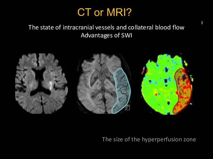
- 32. CT or MRI? The state of intracranial vessels and collateral blood flow Advantages of SWI The
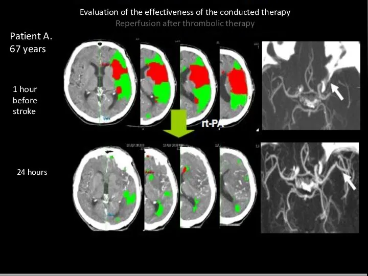
- 35. Evaluation of the effectiveness of the conducted therapy Reperfusion after thrombolic therapy Patient A. 67 years
- 37. Скачать презентацию
Слайд 3Types of stroke
Ischemic-20%
Hemorrhagic-80%
Types of stroke
Ischemic-20%
Hemorrhagic-80%
Слайд 4CT and MRI
CT and MRI
Слайд 5Neuroimaging tasks
Define:
Intracerebral hemorrhage/infarction
Localization and size of the lesion
Time from the beginning progression
Neuroimaging tasks
Define:
Intracerebral hemorrhage/infarction
Localization and size of the lesion
Time from the beginning progression
Слайд 6CT or MRI?
Exclude ICH
CT or MRI?
Exclude ICH
Слайд 7CT and MRI in the dynamics
CT and MRI in the dynamics
Слайд 8EVOLUTION OF INTRACEREBRAL HEMATOMA
stage of hematoma
transformation of blood elements
oxyhemoglobin
Hyperacute
(4 to 12 hours)
deoxyhemoglobin
EVOLUTION OF INTRACEREBRAL HEMATOMA
stage of hematoma
transformation of blood elements
oxyhemoglobin
Hyperacute
(4 to 12 hours)
deoxyhemoglobin
Слайд 9Hemorrhagic strokes
CT - is a “gold standard” of diagnosis.
Hemorrhage is a focus
Hemorrhagic strokes
CT - is a “gold standard” of diagnosis.
Hemorrhage is a focus
Слайд 10SUBARACHNOID HEMORRHAGE
The sensitivity of CT to the presence of blood in the
SUBARACHNOID HEMORRHAGE
The sensitivity of CT to the presence of blood in the
Слайд 11Subarachnoid hemorrhage
Subarachnoid hemorrhage
Слайд 12CT or MRI?
Types of ischemic edema
Cytotoxic edema (CBF < 30 ml/ 100
CT or MRI?
Types of ischemic edema
Cytotoxic edema (CBF < 30 ml/ 100
Слайд 13CT or MRI?
Types of ischemic edema
Cytotoxic edema (CBF < 30 ml/ 100
CT or MRI?
Types of ischemic edema
Cytotoxic edema (CBF < 30 ml/ 100
Слайд 14CT or MRI?
Types of ischemic edema
Cytotoxic edema (CBF < 30 ml/ 100
CT or MRI?
Types of ischemic edema
Cytotoxic edema (CBF < 30 ml/ 100
Слайд 15CT or MRI?
Types of ischemic edema
Cytotoxic edema (CBF < 30 ml/ 100
CT or MRI?
Types of ischemic edema
Cytotoxic edema (CBF < 30 ml/ 100
Слайд 16Early СТ signs CVA
Arterial hyperdensity (a sign of intravascular thrombosis: hyperdensive middle
Early СТ signs CVA
Arterial hyperdensity (a sign of intravascular thrombosis: hyperdensive middle
Слайд 17Hyperdensive MCA
It is manifested by an increase in the density of the
Hyperdensive MCA
It is manifested by an increase in the density of the
Слайд 18ASPECTS SCORE
The Alberta stroke programme early CT score (ASPECTS) 1 is a
ASPECTS SCORE
The Alberta stroke programme early CT score (ASPECTS) 1 is a
Слайд 19The assessment on the ASPECTS scale is carried out by determining changes
The assessment on the ASPECTS scale is carried out by determining changes
Слайд 20C - caudate nucleus,
I - insular cortex,
IC - internal capsule,
L- nucleus lentiformis,
M1
C - caudate nucleus,
I - insular cortex,
IC - internal capsule,
L- nucleus lentiformis,
M1
Слайд 21Results:
The basal ganglia are estimated at 3 points (C, L, and IC).
Results:
The basal ganglia are estimated at 3 points (C, L, and IC).
Слайд 22
PATHOPHYSIOLOGY AND MR-PICTURE OF CEREBRAL ISCHEMIC STROKE
I
acute stage(0-3 days).
Pathoanatomically-focal
PATHOPHYSIOLOGY AND MR-PICTURE OF CEREBRAL ISCHEMIC STROKE
I
acute stage(0-3 days).
Pathoanatomically-focal
Слайд 23THE FOCUS OF ACUTE ISCHEMIC CVA
THE TERRITORY OF THE LEFT SUPERIOR
THE FOCUS OF ACUTE ISCHEMIC CVA THE TERRITORY OF THE LEFT SUPERIOR
Слайд 24
PATHOPHYSIOLOGY AND MR-PICTURE OF CEREBRAL ISCHEMIC STROKE
Subacute stage (3 days-10-14 days).
The
PATHOPHYSIOLOGY AND MR-PICTURE OF CEREBRAL ISCHEMIC STROKE
Subacute stage (3 days-10-14 days).
The
Слайд 25Zone of the subacute ischemic stroke
Middle cerebral artery circulation . FLAIR
Zone of the subacute ischemic stroke
Middle cerebral artery circulation . FLAIR
Слайд 26PATHOPHYSIOLOGY AND MR-PICTURE OF CEREBRAL ISCHEMIC STROKE
Chronic stage (end of the 2nd
PATHOPHYSIOLOGY AND MR-PICTURE OF CEREBRAL ISCHEMIC STROKE
Chronic stage (end of the 2nd
Слайд 27
Zone of the chronic ischemic stroke
Left posterior cerebral artery circulation . FLAIR
Zone of the chronic ischemic stroke Left posterior cerebral artery circulation . FLAIR
Слайд 28Direct signs(⅓ cases ):
Triangle sign – visualization of
a blood clot on the
Direct signs(⅓ cases ):
Triangle sign – visualization of
a blood clot on the
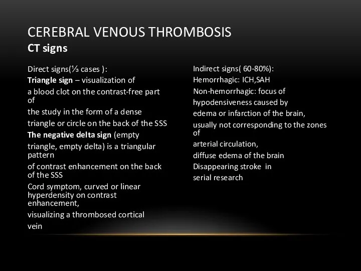
Слайд 29CEREBRAL VENOUS THROMBOSIS
CEREBRAL VENOUS THROMBOSIS
Слайд 30CT or MRI?
the state of intracranial vessels and collateral blood flow
Thrombus size
CT or MRI?
the state of intracranial vessels and collateral blood flow
Thrombus size
Слайд 32CT or MRI?
The state of intracranial vessels and collateral blood flow
Advantages of
CT or MRI?
The state of intracranial vessels and collateral blood flow
Advantages of
Слайд 35Evaluation of the effectiveness of the conducted therapy
Reperfusion after thrombolic therapy
Patient
Evaluation of the effectiveness of the conducted therapy
Reperfusion after thrombolic therapy
Patient
 Роль медсестры гастроэнтерологического отделения стационара в лечении заболеваний желудочно- кишечного тракта
 Пневмония 2021 Манищенкова
 Лечение гиперурикемии
 Профилактика инфекционных болезней плотоядных животных
 Медицинское освидетельствование при первоначальной постановке на воинский учет
 Трихинеллез животных
 Репродуктивные органы
 Черепно-мозговая травма. Протокол действий
 Опорно-двигательный аппарат
 Концепция нейропсихологического синдрома и симптома
 Шизофрения. Продуктивная симптоматика
 Нейровоспаление. Микроглия
 Анэспум_ВОПРОСЫ-ОТВЕТЫ
 Операции при свищах и опухолях околоушной слюнной железы
 История развития психопатологии в зарубежный странах
 Род Corynebacterium
 Чума м,ясоїдних
 Брюшной тиф. Шигеллёз. Сестринское дело
 профилактика 1 лекция
 Общие положения о щитовидной железе. Лекция 5
 Особенноси ухода за больными с заболеваниями дыхательной системы
 Профилактика туберкулеза. Лекция №11
 Айырша бездің топографиялық анатомиясы. Балалардағы ерекшеліктері
 Экстренная реанимационная помощь
 Особенности клинических исследований в кардиологии
 Помповая инсулинотерапия. Технические аспекты
 дезінфекція2
 Что такое СПИД