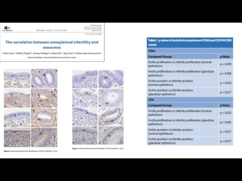
- Role of exosomes in pregnancy loss (miscarriage) CD 9, 81, 63

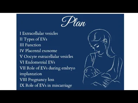
Содержание
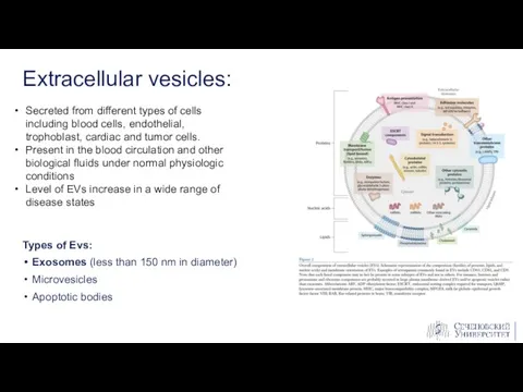
- 3. Types of Evs: Exosomes (less than 150 nm in diameter) Microvesicles Apoptotic bodies Extracellular vesicles: Secreted
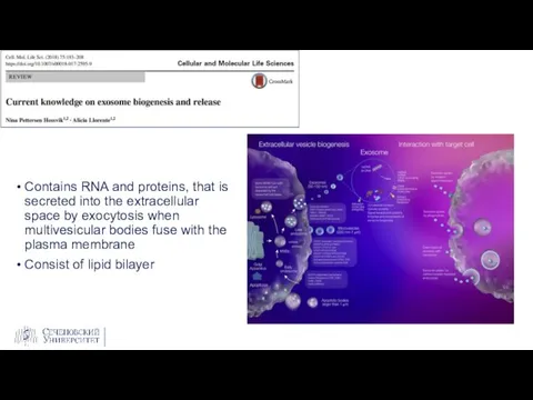
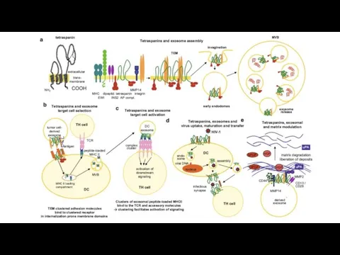
- 4. Contains RNA and proteins, that is secreted into the extracellular space by exocytosis when multivesicular bodies
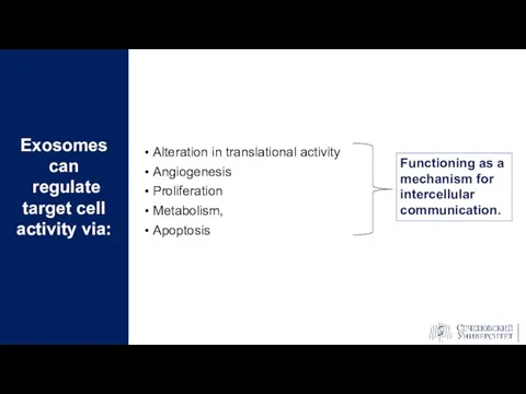
- 5. Alteration in translational activity Angiogenesis Proliferation Metabolism, Apoptosis Exosomes can regulate target cell activity via: Functioning
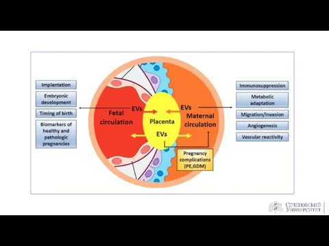
- 8. Placenta secretes exosomes into maternal circulation and play important roles in several different aspects of pregnancy
- 9. Can be detected in maternal plasma from as early as 6 weeks of gestation Number increases
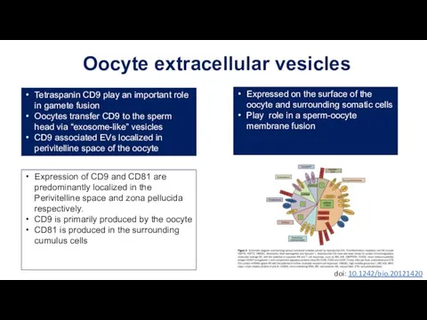
- 10. Oocyte extracellular vesicles Tetraspanin CD9 play an important role in gamete fusion Oocytes transfer CD9 to
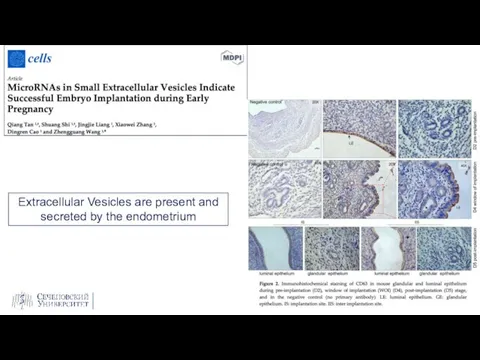
- 11. Extracellular Vesicles are present and secreted by the endometrium
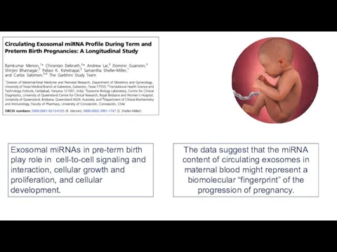
- 12. The data suggest that the miRNA content of circulating exosomes in maternal blood might represent a
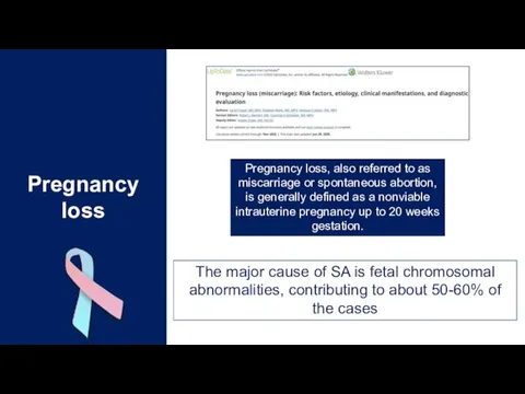
- 14. Pregnancy loss, also referred to as miscarriage or spontaneous abortion, is generally defined as a nonviable
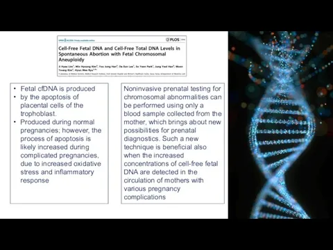
- 16. Fetal cfDNA is produced by the apoptosis of placental cells of the trophoblast. Produced during normal
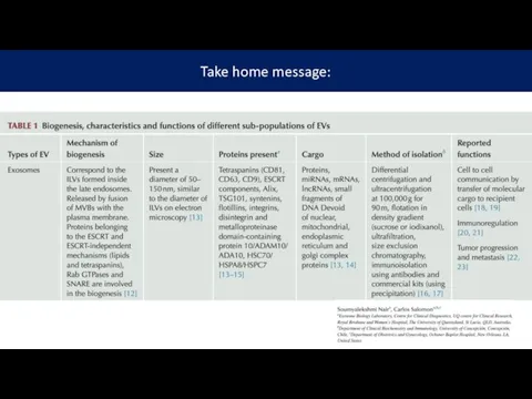
- 17. Take home message:
- 19. Скачать презентацию
Слайд 3Types of Evs:
Exosomes (less than 150 nm in diameter)
Microvesicles
Apoptotic bodies
Extracellular vesicles:
Secreted
Types of Evs:
Exosomes (less than 150 nm in diameter)
Microvesicles
Apoptotic bodies
Extracellular vesicles:
Secreted
Слайд 4Contains RNA and proteins, that is secreted into the extracellular space by
Contains RNA and proteins, that is secreted into the extracellular space by
Слайд 5Alteration in translational activity
Angiogenesis
Proliferation
Metabolism,
Apoptosis
Exosomes can
regulate target cell activity via:
Functioning
Alteration in translational activity
Angiogenesis
Proliferation
Metabolism,
Apoptosis
Exosomes can
regulate target cell activity via:
Functioning
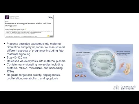
Слайд 8Placenta secretes exosomes into maternal circulation and play important roles in several
Placenta secretes exosomes into maternal circulation and play important roles in several
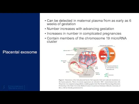
Слайд 9Can be detected in maternal plasma from as early as 6 weeks
Can be detected in maternal plasma from as early as 6 weeks
Слайд 10Oocyte extracellular vesicles
Tetraspanin CD9 play an important role in gamete fusion
Oocytes transfer
Oocyte extracellular vesicles
Tetraspanin CD9 play an important role in gamete fusion
Oocytes transfer
Слайд 11Extracellular Vesicles are present and
secreted by the endometrium
Extracellular Vesicles are present and
secreted by the endometrium
Слайд 12The data suggest that the miRNA content of circulating exosomes in maternal
The data suggest that the miRNA content of circulating exosomes in maternal
Слайд 14Pregnancy loss, also referred to as miscarriage or spontaneous abortion, is generally
Pregnancy loss, also referred to as miscarriage or spontaneous abortion, is generally
Слайд 16Fetal cfDNA is produced
by the apoptosis of placental cells of the
Fetal cfDNA is produced
by the apoptosis of placental cells of the
Слайд 17Take home message:
Take home message:
 Грибок ногтей (онихомикоз)
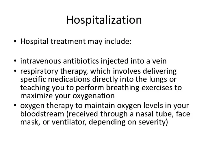 Hospitalization. Госпитализация
 Философия и медицина
 Реанимация
 Антибиотики
 Нейродефектология. Клинические основы нейродефектологии и нейрореабилитации
 Чистка лица
 Пчёлы. Опасность укуса
 Әсемділікті қалыптастыратын факторлар
 Особые свойства минеральной воды и оздоравливающее воздействие воды на организм человека
 Линименты. Гомогенные линименты
 Двигательные расстройства
 История нейрохирургии. Черепно-мозговая травма: классификация, клиника. Лекция №1
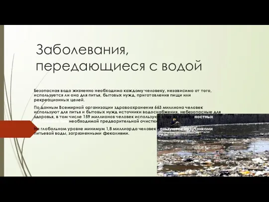 Заболевания, передающиеся с водой
 Мырыш жақпа майы
 Продукты и блюда не разрешённые к использованию в возрасте 1-3 года, в дошкольном учреждении
 Анальгетики, определение понятия, классификация
 Принципы профилактики и лечения эндокринопатий
 Атоиммунный полигландулярный синдром 2-ого типа
 Острая дыхытельная недостаточность
 Особенности репродукции человека в связи с его биосоциальной сущностью
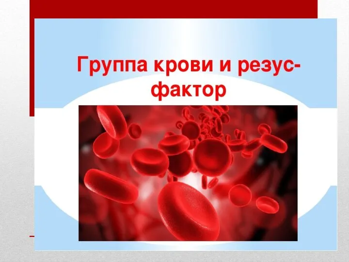 Группа крови
 Функциональные обязанности санитарки-буфетчицы
 antiseptiki
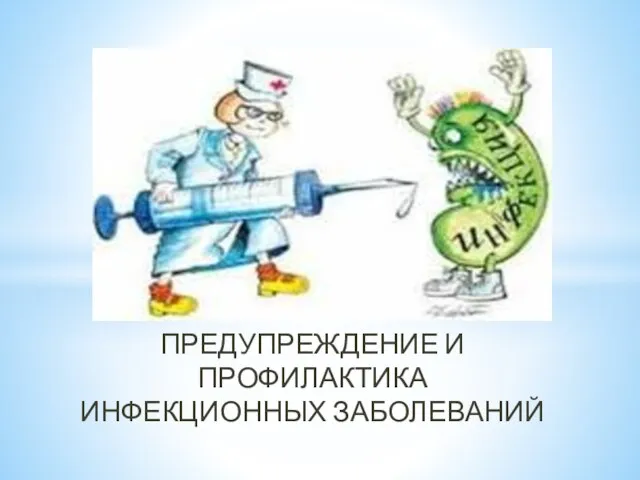 Предупреждение и профилактика инфекционных заболеваний. Гигиенические правила
 заболевания волос
 Die Familie Schrot
 Ассоциация медицинских (клинических) психологов. Отчет 2020