- Neurobiology and neurophysiology of schizophrenia

Содержание
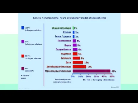
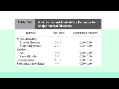
- 2. •Schizophrenia is a chronic mental illness affecting approximately 1 percent of the population •several neurotransmitter systems
- 3. •Other neurotransmitters have also been implicated, including glutamate, serotonin, and γaminobutyric acid (GABA). • Также были
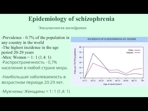
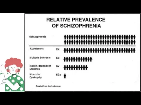
- 4. Epidemiology of schizophrenia -Prevalence - 0.7% of the population in any country in the world -The
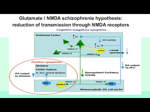
- 10. Glutamate / NMDA schizophrenia hypothesis: reduction of transmission through NMDA receptors
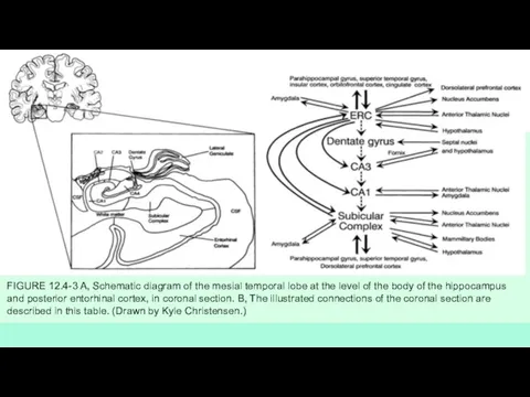
- 11. FIGURE 12.4-3 A, Schematic diagram of the mesial temporal lobe at the level of the body
- 12. STRUCTURAL AND FUNCTIONAL NEUROIMAGING СТРУКТУРНО-ФУНКЦИОНАЛЬНОЕ НЕЙРО ИЗОБРАЖЕНИЕ
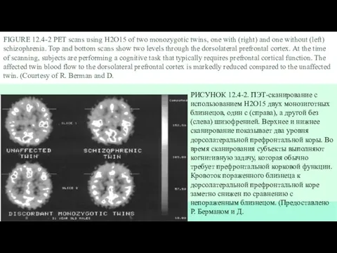
- 13. FIGURE 12.4-2 PET scans using H2O15 of two monozygotic twins, one with (right) and one without
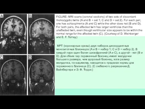
- 14. FIGURE: MRI scans (coronal sections) of two sets of discordant monozygotic twins (A and B =
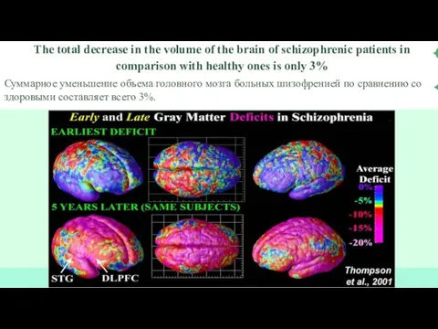
- 15. The total decrease in the volume of the brain of schizophrenic patients in comparison with healthy
- 19. Скачать презентацию
Слайд 3•Other neurotransmitters have also been implicated, including glutamate, serotonin, and γaminobutyric acid
•Other neurotransmitters have also been implicated, including glutamate, serotonin, and γaminobutyric acid
Слайд 4Epidemiology of schizophrenia
-Prevalence - 0.7% of the population in any country in
Epidemiology of schizophrenia
-Prevalence - 0.7% of the population in any country in
Слайд 10Glutamate / NMDA schizophrenia hypothesis:
reduction of transmission through NMDA receptors
Glutamate / NMDA schizophrenia hypothesis:
reduction of transmission through NMDA receptors
Слайд 11FIGURE 12.4-3 A, Schematic diagram of the mesial temporal lobe at the
FIGURE 12.4-3 A, Schematic diagram of the mesial temporal lobe at the
Слайд 12STRUCTURAL AND FUNCTIONAL NEUROIMAGING
СТРУКТУРНО-ФУНКЦИОНАЛЬНОЕ НЕЙРО ИЗОБРАЖЕНИЕ
STRUCTURAL AND FUNCTIONAL NEUROIMAGING
СТРУКТУРНО-ФУНКЦИОНАЛЬНОЕ НЕЙРО ИЗОБРАЖЕНИЕ
Слайд 13FIGURE 12.4-2 PET scans using H2O15 of two monozygotic twins, one with
FIGURE 12.4-2 PET scans using H2O15 of two monozygotic twins, one with
Слайд 14FIGURE: MRI scans (coronal sections) of two sets of discordant monozygotic twins
FIGURE: MRI scans (coronal sections) of two sets of discordant monozygotic twins
Слайд 15The total decrease in the volume of the brain of schizophrenic patients
The total decrease in the volume of the brain of schizophrenic patients

 Прогрессивная мышечная релаксация по Э. Джекобсону
 Уход за пациентами с нарушениями мочеиспускания
 Формулирование клинической проблемы с использованием принципа PICO
 Первая помощь при ранениях, переломах костей, ожогах, отморожениях (тема 2)
 lean әдіснамасы ұқыпты технологиялар
 Гемолитическая болезнь новорожденных
 Атеросклероз
 Инфаркт миокарда и его осложнения
 Тубоовариалды түзілісі бар науқастарды жүргізу тактикасы
 Воспалительные заболевания женских половых органов
 Хроническая обструктивная болезнь легких
 Національна настанова і документи з епідеміологічного нагляду за ГВП в Україні
 Лікарські засоби, що діють переважно на ПНС. Частина 1. Лекція 13
 Токсикомания
 Дыхание – важнейшая функция организма
 Ожоги. Классификация ожогов
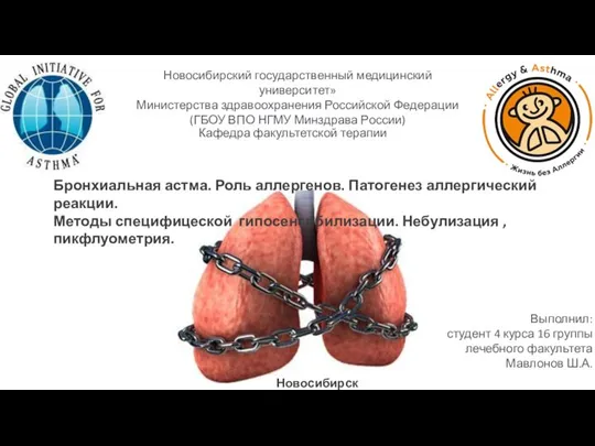 Бронхиальная астма. Роль аллергенов. Патогенез аллергической реакции. Методы специфической гипосенсибилизации
 Оценка общего сердечно-сосудистого риска
 Муковисцидоз
 Антисептик
 Способы борьбы со старением в 21 веке
 ЭКГ при жизнеугрожающих нарушениях ритма
 Стационарлардағы медициналық көмектің сапасын қамтамасыз етудегі дәрілік формулярлар жүйесі
 Определение риска сердечно сосудистый заболеваний по шкале SCORE
 Кампилобактериозы, иерсиниозы, листериозы
 Пролактин, андрогены (тестостерон)
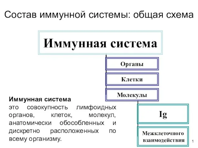 Состав иммунной системы: общая схема
 Злоякісні пухлини: легені, молочної залози, урогенітальної зони. Лекція №2