- Phylogenetic. Disorders of Human

Содержание
- 2. Topic; Phylogenetic Disorders of Human Excretory System Guided By: Prof. Anna Zhukova
- 3. CONTENTS INTRODUCTION Normal structure and Function of excretory System. Phylogenetic Disorders Example and images
- 4. INTRODUCTION The excretory system is a passive biological system that removes excess, unnecessary materials from the
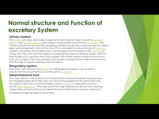
- 5. Normal structure and Function of excretory System Urinary system The kidneys are large, bean-shaped organs which
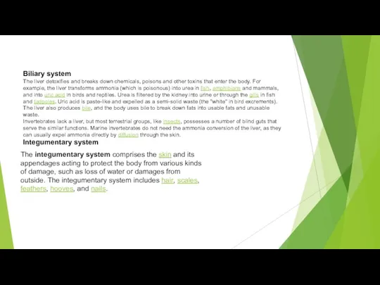
- 6. Biliary system The liver detoxifies and breaks down chemicals, poisons and other toxins that enter the
- 7. Phylogenetic disorders of Excretory System Horseshoe Kidney. Polycystic Kidney. Renal Agenesis. Renal Hypoplasia. Epispadias. Renal Ectopia
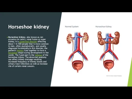
- 8. Horseshoe kidney Horseshoe kidney, also known as ren arcuatus (in Latin), renal fusion or super kidney,
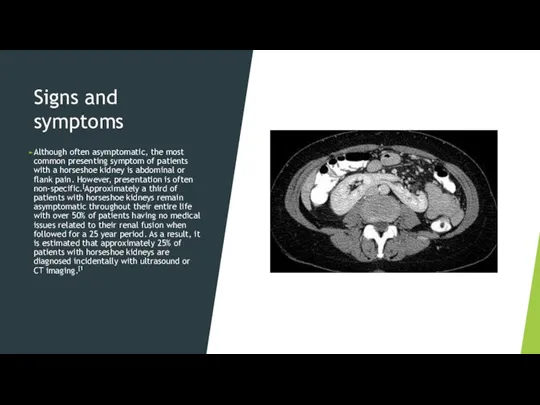
- 9. Signs and symptoms Although often asymptomatic, the most common presenting symptom of patients with a horseshoe
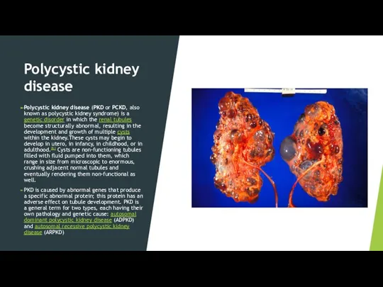
- 10. Polycystic kidney disease Polycystic kidney disease (PKD or PCKD, also known as polycystic kidney syndrome) is
- 11. Polycystic kidney disease symptoms can include: 1. High blood pressure 2. Back or side pain 3.
- 12. Polycystic kidney disease can be ascertained via a CT scan of abdomen, as well as, an
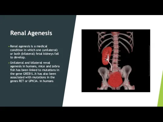
- 13. Renal Agenesis Renal agenesis is a medical condition in which one (unilateral) or both (bilateral) fetal
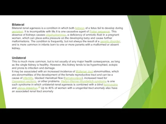
- 14. Bilateral Bilateral renal agenesis is a condition in which both kidneys of a fetus fail to
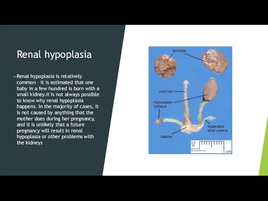
- 15. Renal hypoplasia Renal hypoplasia is relatively common – it is estimated that one baby in a
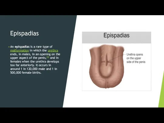
- 16. Epispadias An epispadias is a rare type of malformation in which the urethra ends, in males,
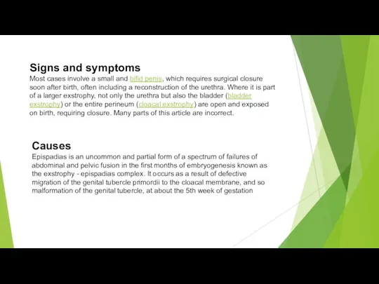
- 17. Signs and symptoms Most cases involve a small and bifid penis, which requires surgical closure soon
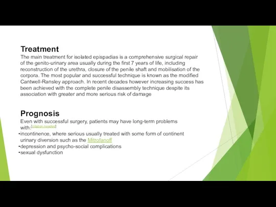
- 18. Treatment The main treatment for isolated epispadias is a comprehensive surgical repair of the genito-urinary area
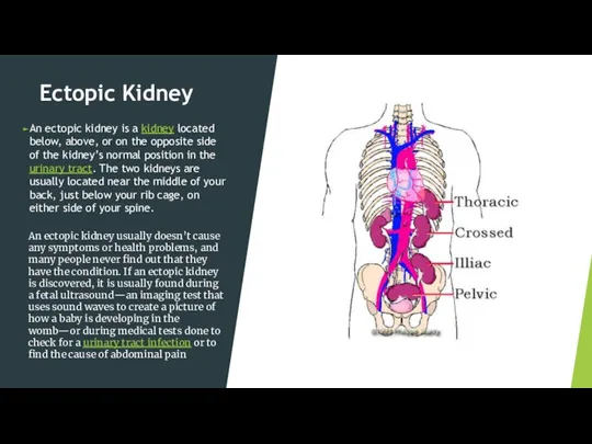
- 19. Ectopic Kidney An ectopic kidney is a kidney located below, above, or on the opposite side
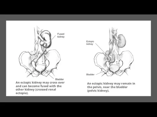
- 20. An ectopic kidney may remain in the pelvis, near the bladder (pelvic kidney). An ectopic kidney
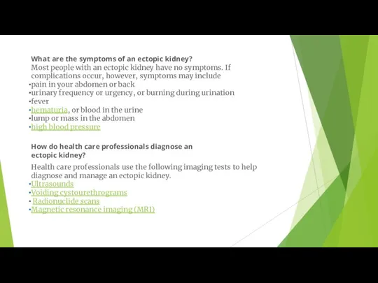
- 21. What are the symptoms of an ectopic kidney? Most people with an ectopic kidney have no
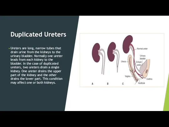
- 22. Duplicated Ureters Ureters are long, narrow tubes that drain urine from the kidneys to the urinary
- 23. Duplicated ureters can take one of two forms: Incomplete: Two separate ureters are attached to the
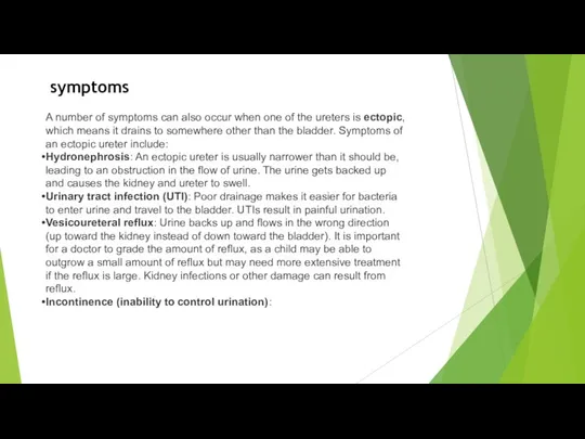
- 24. A number of symptoms can also occur when one of the ureters is ectopic, which means
- 26. Скачать презентацию

Слайд 3CONTENTS
INTRODUCTION
Normal structure and Function of excretory System.
Phylogenetic Disorders
Example and images
CONTENTS
INTRODUCTION
Normal structure and Function of excretory System.
Phylogenetic Disorders
Example and images
Слайд 4INTRODUCTION
The excretory system is a passive biological system that removes excess, unnecessary
INTRODUCTION
The excretory system is a passive biological system that removes excess, unnecessary

Слайд 5Normal structure and Function of excretory System
Urinary system
The kidneys are large, bean-shaped
Normal structure and Function of excretory System
Urinary system
The kidneys are large, bean-shaped
Слайд 6Biliary system
The liver detoxifies and breaks down chemicals, poisons and other toxins
Biliary system
The liver detoxifies and breaks down chemicals, poisons and other toxins
Слайд 7Phylogenetic disorders of
Excretory System
Horseshoe Kidney.
Polycystic Kidney.
Renal Agenesis.
Renal Hypoplasia.
Epispadias.
Renal Ectopia [ECTOPIC KIDNEY].
Doubling
Phylogenetic disorders of
Excretory System
Horseshoe Kidney.
Polycystic Kidney.
Renal Agenesis.
Renal Hypoplasia.
Epispadias.
Renal Ectopia [ECTOPIC KIDNEY].
Doubling
Слайд 8Horseshoe kidney
Horseshoe kidney, also known as ren arcuatus (in Latin), renal fusion
Horseshoe kidney
Horseshoe kidney, also known as ren arcuatus (in Latin), renal fusion
Слайд 9Signs and symptoms
Although often asymptomatic, the most common presenting symptom of patients
Signs and symptoms
Although often asymptomatic, the most common presenting symptom of patients
Слайд 10Polycystic kidney disease
Polycystic kidney disease (PKD or PCKD, also known as
Polycystic kidney disease
Polycystic kidney disease (PKD or PCKD, also known as
Слайд 11Polycystic kidney disease symptoms can include:
1. High blood pressure 2. Back
Polycystic kidney disease symptoms can include:
1. High blood pressure 2. Back

Слайд 12Polycystic kidney disease can be ascertained via a CT scan of abdomen,
Polycystic kidney disease can be ascertained via a CT scan of abdomen,
Слайд 13Renal Agenesis
Renal agenesis is a medical condition in which one (unilateral) or
Renal Agenesis
Renal agenesis is a medical condition in which one (unilateral) or
Слайд 14Bilateral
Bilateral renal agenesis is a condition in which both kidneys of a
Bilateral
Bilateral renal agenesis is a condition in which both kidneys of a
Слайд 15Renal hypoplasia
Renal hypoplasia is relatively common – it is estimated that
Renal hypoplasia
Renal hypoplasia is relatively common – it is estimated that
Слайд 16Epispadias
An epispadias is a rare type of malformation in which the urethra
Epispadias
An epispadias is a rare type of malformation in which the urethra
Слайд 17Signs and symptoms
Most cases involve a small and bifid penis, which requires
Signs and symptoms
Most cases involve a small and bifid penis, which requires
Слайд 18Treatment
The main treatment for isolated epispadias is a comprehensive surgical repair of
Treatment
The main treatment for isolated epispadias is a comprehensive surgical repair of
Слайд 19Ectopic Kidney
An ectopic kidney is a kidney located below, above, or on
Ectopic Kidney
An ectopic kidney is a kidney located below, above, or on
Слайд 20An ectopic kidney may remain in the pelvis, near the bladder (pelvic
An ectopic kidney may remain in the pelvis, near the bladder (pelvic
Слайд 21What are the symptoms of an ectopic kidney?
Most people with an ectopic
What are the symptoms of an ectopic kidney?
Most people with an ectopic
Слайд 22Duplicated Ureters
Ureters are long, narrow tubes that drain urine from the
Duplicated Ureters
Ureters are long, narrow tubes that drain urine from the
Слайд 23Duplicated ureters can take one of two forms:
Incomplete: Two separate ureters are
Duplicated ureters can take one of two forms:
Incomplete: Two separate ureters are

Слайд 24A number of symptoms can also occur when one of the ureters
A number of symptoms can also occur when one of the ureters
 Il corpo umano. Come ti senti
 Наркотики
 Противотуберкулезные, противосифилитические, противопротозойные, противомикозные, противовирусные средства
 Влияние факторов окружающей среды на овогенез, менструальный цикл, фертильность
 Прогрессирующая мультифокальная лейкоэнцефалопатия
 Ксабаны. Типы ксабанов
 Показания к резекции отделов кишки. Правила определения жизнеспособности кишки
 Пневмония. Классификация
 Болезни, передающиеся половым путем
 Нарушение потребности выделять. Запор
 Синдром дефицита эстрогенов
 Остеоартроз
 Давление в дуге аорты
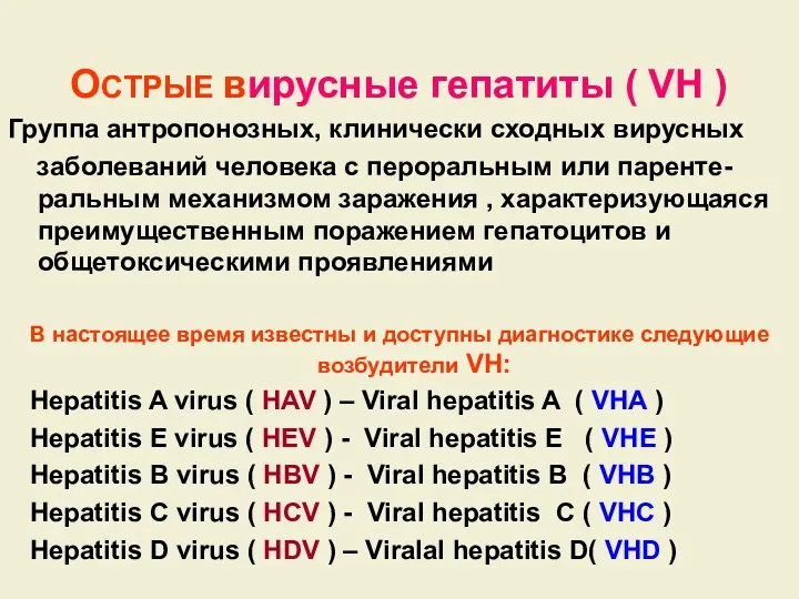 Вирусные гепатиты ( VH )
 Патоморфология первичного роста
 Гипоксемическая ДН
 Резекция верхушки корня зуба
 Тест. Клиникалық жағдай
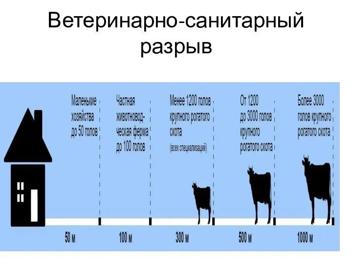 Ветеринарно-санитарный разрыв
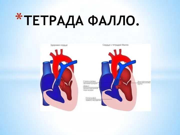 Тетрада Фалло
 диф.дз при очаговых заболеваниях легких
 Инфекционные заболевания у детей
 Дренирование гнойно-воспалительных процессов плеча
 Молекулярно-генетические основы поведения
 Межзубные ершики
 Омыртқа ауруларының сәулелі диагностикасы
 Особенности жирового и углеводного обмена у детей
 Бронхоэктазии