- ThinPrep® Pap Test Diagnostic Challenges and Differential

Содержание
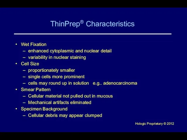
- 3. ThinPrep® Characteristics Wet Fixation enhanced cytoplasmic and nuclear detail variability in nuclear staining Cell Size proportionately
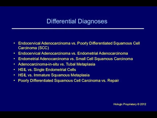
- 4. Differential Diagnoses Endocervical Adenocarcinoma vs. Poorly Differentiated Squamous Cell Carcinoma (SCC) Endocervical Adenocarcinoma vs. Endometrial Adenocarcinoma
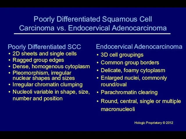
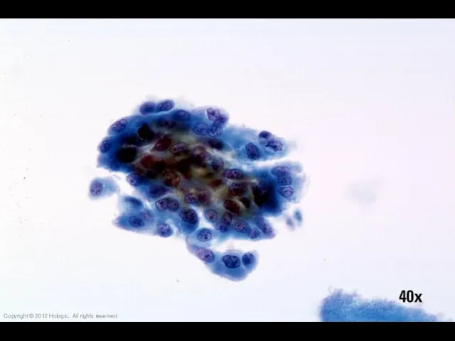
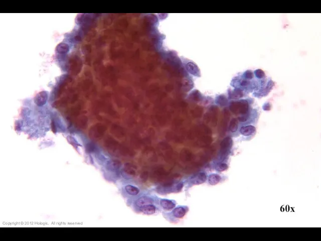
- 5. Poorly Differentiated Squamous Cell Carcinoma vs. Endocervical Adenocarcinoma Poorly Differentiated SCC 2D sheets and single cells
- 6. Copyright © 2012 Hologic, All rights reserved
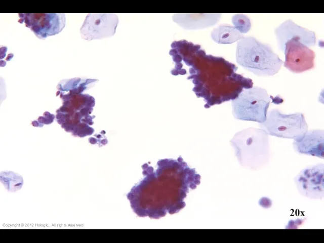
- 7. 20x Copyright © 2012 Hologic, All rights reserved
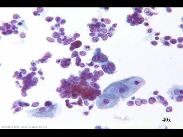
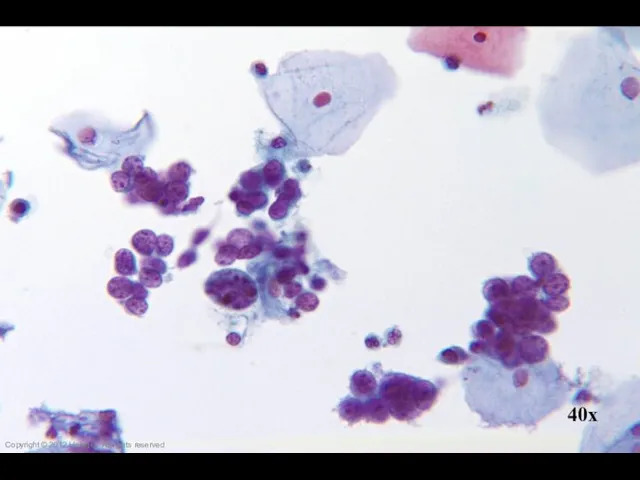
- 8. 40x Copyright © 2012 Hologic, All rights reserved
- 9. Copyright © 2012 Hologic, All rights reserved
- 10. 60x Copyright © 2012 Hologic, All rights reserved
- 11. 60x Copyright © 2012 Hologic, All rights reserved
- 12. Copyright © 2012 Hologic, All rights reserved
- 13. 60x Copyright © 2012 Hologic, All rights reserved
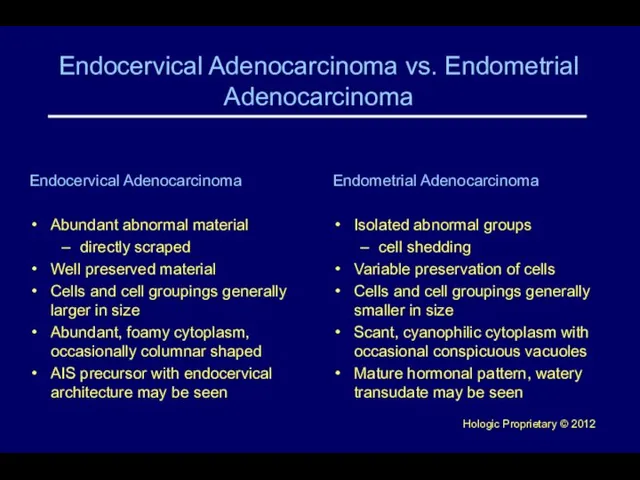
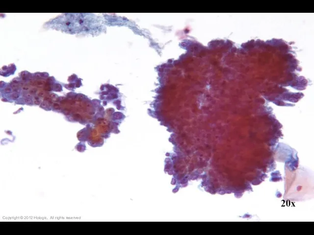
- 14. Endocervical Adenocarcinoma vs. Endometrial Adenocarcinoma Endocervical Adenocarcinoma Abundant abnormal material directly scraped Well preserved material Cells
- 15. 20x Copyright © 2012 Hologic, All rights reserved
- 16. 60x Copyright © 2012 Hologic, All rights reserved
- 17. 40x Copyright © 2012 Hologic, All rights reserved
- 18. 60x Copyright © 2012 Hologic, All rights reserved
- 19. 40x Copyright © 2012 Hologic, All rights reserved
- 20. 60x Copyright © 2012 Hologic, All rights reserved
- 21. Copyright © 2012 Hologic, All rights reserved
- 22. Copyright © 2012 Hologic, All rights reserved
- 23. 60x Copyright © 2012 Hologic, All rights reserved
- 24. 40x Copyright © 2012 Hologic, All rights reserved
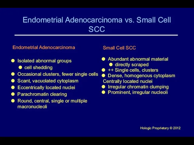
- 25. Endometrial Adenocarcinoma vs. Small Cell SCC Endometrial Adenocarcinoma Isolated abnormal groups cell shedding Occasional clusters, fewer
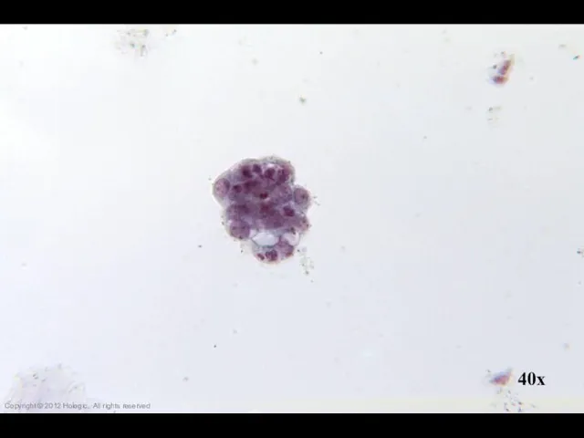
- 26. 40x Copyright © 2012 Hologic, All rights reserved
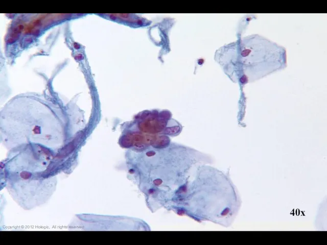
- 27. 40x Copyright © 2012 Hologic, All rights reserved
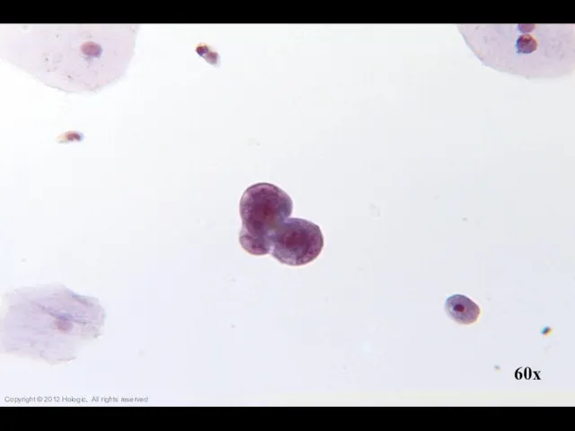
- 28. 60x Copyright © 2012 Hologic, All rights reserved
- 29. 40x Copyright © 2012 Hologic, All rights reserved
- 30. 40x Copyright © 2012 Hologic, All rights reserved
- 31. 20x Copyright © 2012 Hologic, All rights reserved
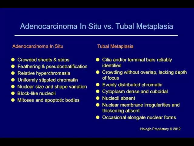
- 32. Adenocarcinoma In Situ vs. Tubal Metaplasia Adenocarcinoma In Situ Crowded sheets & strips Feathering & pseudostratification
- 33. 40x Copyright © 2012 Hologic, All rights reserved
- 34. 40x Copyright © 2012 Hologic, All rights reserved
- 35. 40x Copyright © 2012 Hologic, All rights reserved
- 36. Copyright © 2012 Hologic, All rights reserved
- 37. 60x Copyright © 2012 Hologic, All rights reserved
- 38. 40x Copyright © 2012 Hologic, All rights reserved
- 39. 40x Copyright © 2012 Hologic, All rights reserved
- 40. 40x Copyright © 2012 Hologic, All rights reserved
- 41. 40x Copyright © 2012 Hologic, All rights reserved
- 42. 40x Copyright © 2012 Hologic, All rights reserved
- 43. HSIL vs. Endometrial Cells HSIL Sheets, syncitia; thick plaques rather than 3D ball-like clusters Hyperchromasia Irregular
- 44. Copyright © 2012 Hologic, All rights reserved
- 45. 40x Copyright © 2012 Hologic, All rights reserved
- 46. 40x Copyright © 2012 Hologic, All rights reserved
- 47. 40x Copyright © 2012 Hologic, All rights reserved
- 48. 40x Copyright © 2012 Hologic, All rights reserved
- 49. Copyright © 2012 Hologic, All rights reserved
- 50. 40x Copyright © 2012 Hologic, All rights reserved
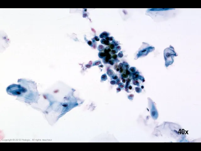
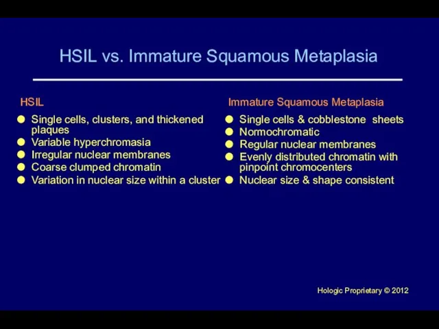
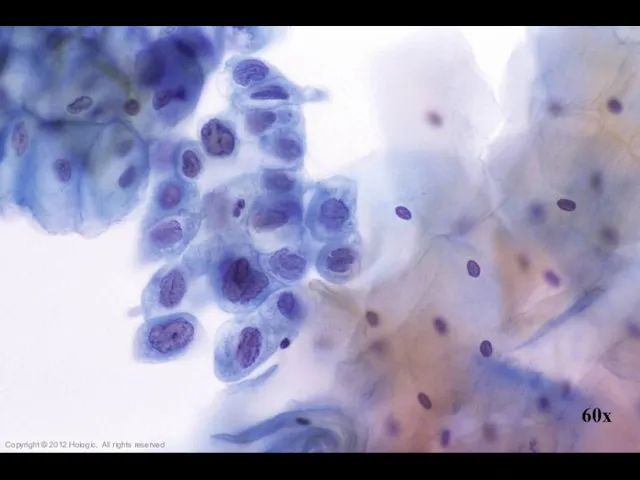
- 51. HSIL vs. Immature Squamous Metaplasia HSIL Single cells, clusters, and thickened plaques Variable hyperchromasia Irregular nuclear
- 52. 60x Copyright © 2012 Hologic, All rights reserved
- 53. 40x Copyright © 2012 Hologic, All rights reserved
- 54. 40x 40x Copyright © 2012 Hologic, All rights reserved
- 55. 40x 40x Copyright © 2012 Hologic, All rights reserved
- 56. 40x Copyright © 2012 Hologic, All rights reserved
- 57. 40x Copyright © 2012 Hologic, All rights reserved
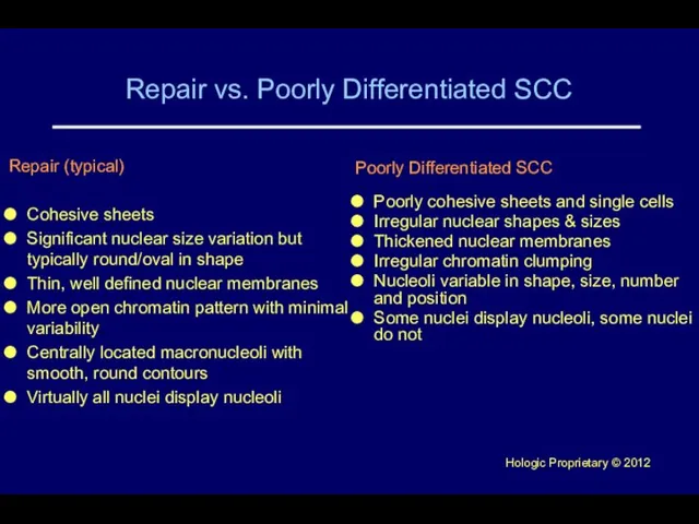
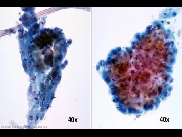
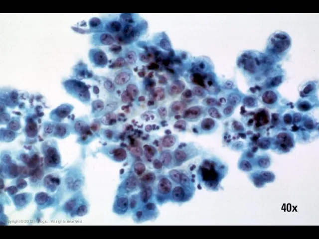
- 58. Repair vs. Poorly Differentiated SCC Poorly Differentiated SCC Poorly cohesive sheets and single cells Irregular nuclear
- 59. Copyright © 2012 Hologic, All rights reserved
- 60. Copyright © 2012 Hologic, All rights reserved
- 61. 40x Copyright © 2012 Hologic, All rights reserved
- 62. 40x Copyright © 2012 Hologic, All rights reserved
- 63. 40x Copyright © 2012 Hologic, All rights reserved
- 64. 60x Copyright © 2012 Hologic, All rights reserved
- 65. Trademark Statement CytoLyt, Hologic, PreservCyt, ThinPrep, and UroCyte are registered trademarks of Hologic, Inc. and/or its
- 67. Скачать презентацию

Слайд 3ThinPrep® Characteristics
Wet Fixation
enhanced cytoplasmic and nuclear detail
variability in nuclear staining
Cell Size
proportionately smaller
single
ThinPrep® Characteristics
Wet Fixation
enhanced cytoplasmic and nuclear detail
variability in nuclear staining
Cell Size
proportionately smaller
single
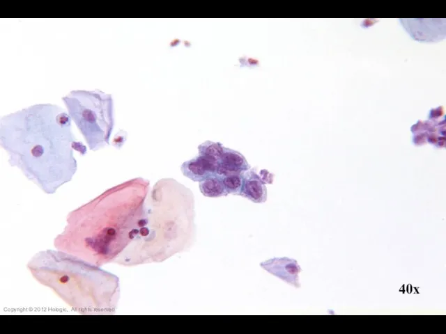
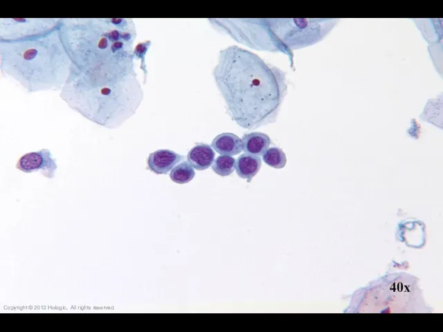
Слайд 4Differential Diagnoses
Endocervical Adenocarcinoma vs. Poorly Differentiated Squamous Cell Carcinoma (SCC)
Endocervical Adenocarcinoma
Differential Diagnoses
Endocervical Adenocarcinoma vs. Poorly Differentiated Squamous Cell Carcinoma (SCC)
Endocervical Adenocarcinoma
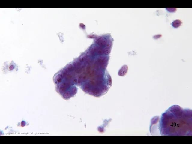
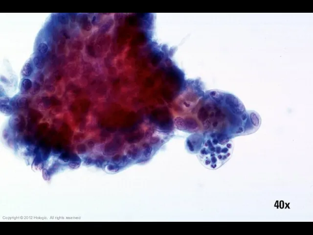
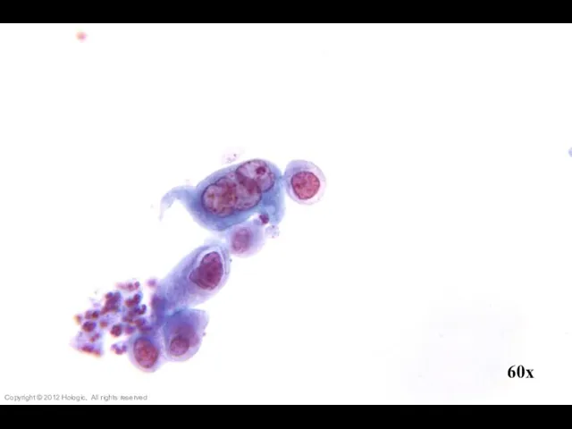
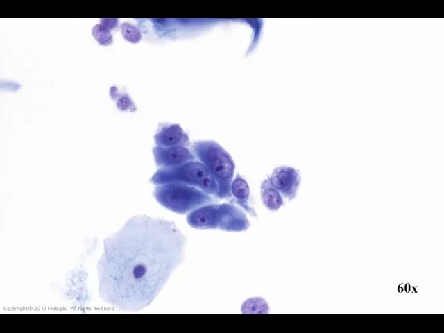
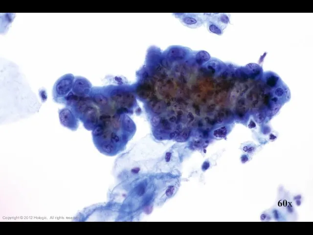
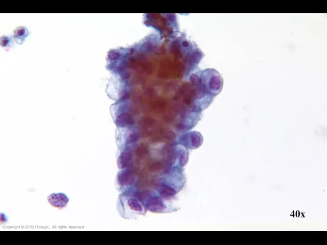
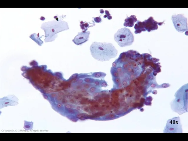
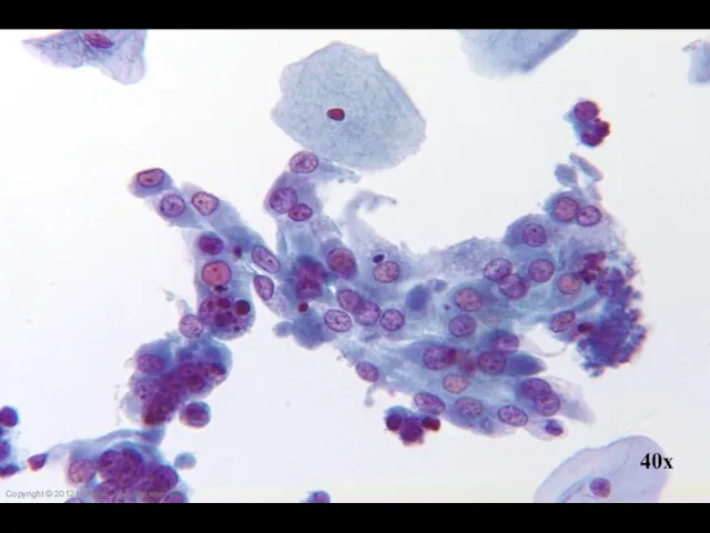
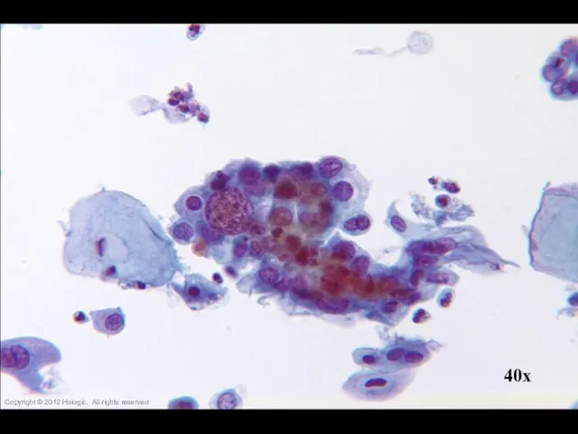
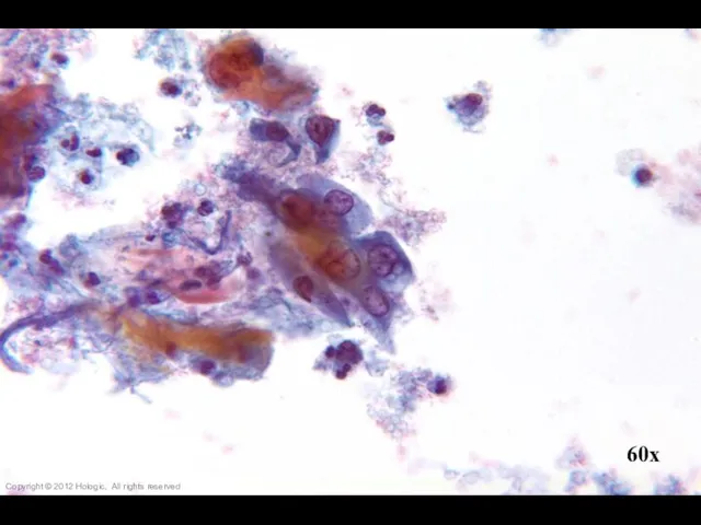
Слайд 5Poorly Differentiated Squamous Cell Carcinoma vs. Endocervical Adenocarcinoma
Poorly Differentiated SCC
2D sheets and
Poorly Differentiated Squamous Cell Carcinoma vs. Endocervical Adenocarcinoma
Poorly Differentiated SCC
2D sheets and
Слайд 6Copyright © 2012 Hologic, All rights reserved
Copyright © 2012 Hologic, All rights reserved
Слайд 720x
Copyright © 2012 Hologic, All rights reserved
20x
Copyright © 2012 Hologic, All rights reserved

Слайд 840x
Copyright © 2012 Hologic, All rights reserved
40x
Copyright © 2012 Hologic, All rights reserved
Слайд 9Copyright © 2012 Hologic, All rights reserved
Copyright © 2012 Hologic, All rights reserved
Слайд 1060x
Copyright © 2012 Hologic, All rights reserved
60x
Copyright © 2012 Hologic, All rights reserved
Слайд 1160x
Copyright © 2012 Hologic, All rights reserved
60x
Copyright © 2012 Hologic, All rights reserved
Слайд 12Copyright © 2012 Hologic, All rights reserved
Copyright © 2012 Hologic, All rights reserved
Слайд 1360x
Copyright © 2012 Hologic, All rights reserved
60x
Copyright © 2012 Hologic, All rights reserved
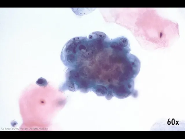
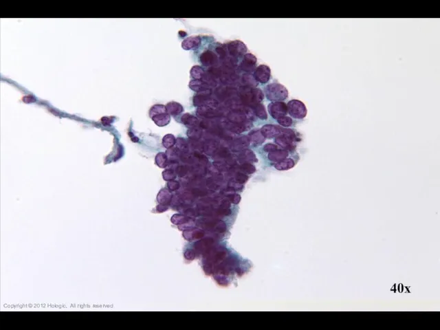
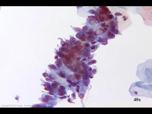
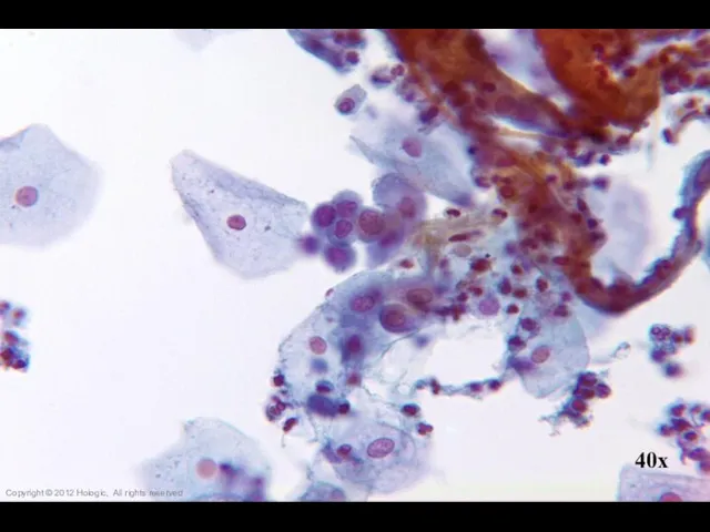
Слайд 14Endocervical Adenocarcinoma vs. Endometrial Adenocarcinoma
Endocervical Adenocarcinoma
Abundant abnormal material
directly scraped
Well preserved material
Cells and
Endocervical Adenocarcinoma vs. Endometrial Adenocarcinoma
Endocervical Adenocarcinoma
Abundant abnormal material
directly scraped
Well preserved material
Cells and
Слайд 1520x
Copyright © 2012 Hologic, All rights reserved
20x
Copyright © 2012 Hologic, All rights reserved
Слайд 1660x
Copyright © 2012 Hologic, All rights reserved
60x
Copyright © 2012 Hologic, All rights reserved
Слайд 1740x
Copyright © 2012 Hologic, All rights reserved
40x
Copyright © 2012 Hologic, All rights reserved
Слайд 1860x
Copyright © 2012 Hologic, All rights reserved
60x
Copyright © 2012 Hologic, All rights reserved
Слайд 1940x
Copyright © 2012 Hologic, All rights reserved
40x
Copyright © 2012 Hologic, All rights reserved
Слайд 2060x
Copyright © 2012 Hologic, All rights reserved
60x
Copyright © 2012 Hologic, All rights reserved

Слайд 21Copyright © 2012 Hologic, All rights reserved
Copyright © 2012 Hologic, All rights reserved
Слайд 22Copyright © 2012 Hologic, All rights reserved
Copyright © 2012 Hologic, All rights reserved
Слайд 2360x
Copyright © 2012 Hologic, All rights reserved
60x
Copyright © 2012 Hologic, All rights reserved
Слайд 2440x
Copyright © 2012 Hologic, All rights reserved
40x
Copyright © 2012 Hologic, All rights reserved
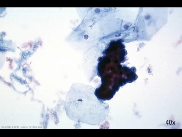
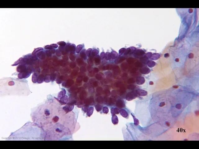
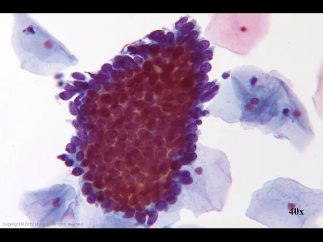
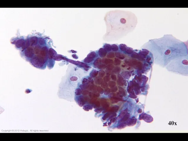
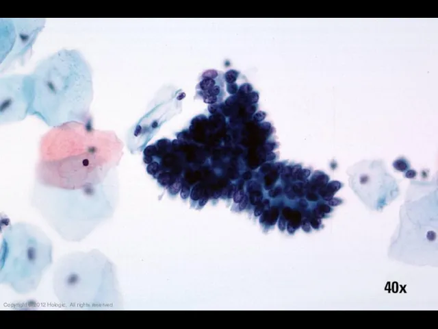
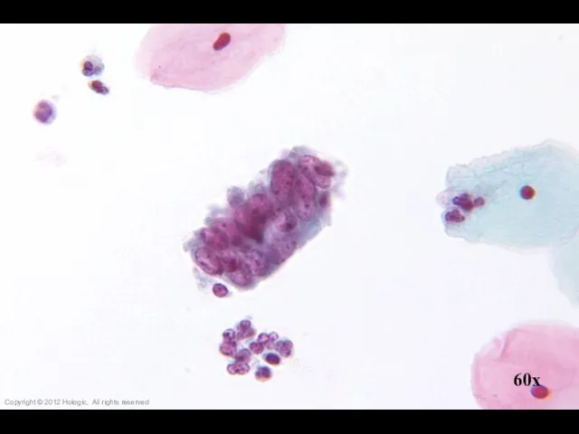
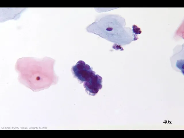
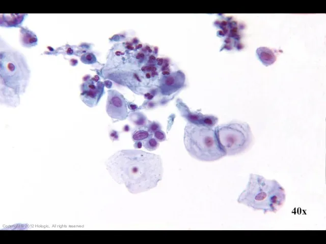
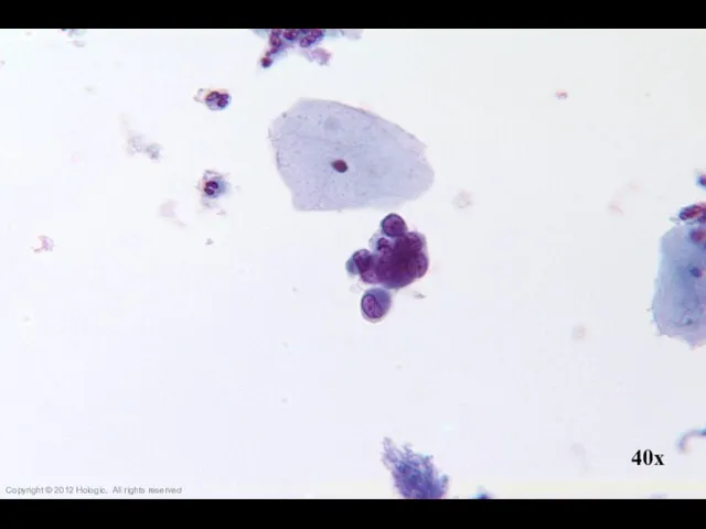
Слайд 25Endometrial Adenocarcinoma vs. Small Cell SCC
Endometrial Adenocarcinoma
Isolated abnormal groups
cell shedding
Occasional clusters, fewer
Endometrial Adenocarcinoma vs. Small Cell SCC
Endometrial Adenocarcinoma
Isolated abnormal groups
cell shedding
Occasional clusters, fewer
Слайд 2640x
Copyright © 2012 Hologic, All rights reserved
40x
Copyright © 2012 Hologic, All rights reserved
Слайд 2740x
Copyright © 2012 Hologic, All rights reserved
40x
Copyright © 2012 Hologic, All rights reserved
Слайд 2860x
Copyright © 2012 Hologic, All rights reserved
60x
Copyright © 2012 Hologic, All rights reserved
Слайд 2940x
Copyright © 2012 Hologic, All rights reserved
40x
Copyright © 2012 Hologic, All rights reserved
Слайд 3040x
Copyright © 2012 Hologic, All rights reserved
40x
Copyright © 2012 Hologic, All rights reserved
Слайд 3120x
Copyright © 2012 Hologic, All rights reserved
20x
Copyright © 2012 Hologic, All rights reserved
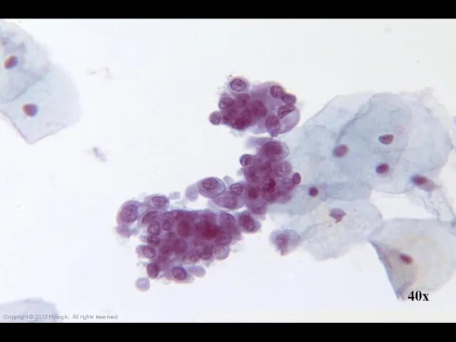
Слайд 32Adenocarcinoma In Situ vs. Tubal Metaplasia
Adenocarcinoma In Situ
Crowded sheets & strips
Feathering &
Adenocarcinoma In Situ vs. Tubal Metaplasia
Adenocarcinoma In Situ
Crowded sheets & strips
Feathering &
Слайд 3340x
Copyright © 2012 Hologic, All rights reserved
40x
Copyright © 2012 Hologic, All rights reserved
Слайд 3440x
Copyright © 2012 Hologic, All rights reserved
40x
Copyright © 2012 Hologic, All rights reserved
Слайд 3540x
Copyright © 2012 Hologic, All rights reserved
40x
Copyright © 2012 Hologic, All rights reserved
Слайд 36Copyright © 2012 Hologic, All rights reserved
Copyright © 2012 Hologic, All rights reserved
Слайд 3760x
Copyright © 2012 Hologic, All rights reserved
60x
Copyright © 2012 Hologic, All rights reserved
Слайд 3840x
Copyright © 2012 Hologic, All rights reserved
40x
Copyright © 2012 Hologic, All rights reserved
Слайд 3940x
Copyright © 2012 Hologic, All rights reserved
40x
Copyright © 2012 Hologic, All rights reserved
Слайд 4040x
Copyright © 2012 Hologic, All rights reserved
40x
Copyright © 2012 Hologic, All rights reserved
Слайд 4140x
Copyright © 2012 Hologic, All rights reserved
40x
Copyright © 2012 Hologic, All rights reserved
Слайд 4240x
Copyright © 2012 Hologic, All rights reserved
40x
Copyright © 2012 Hologic, All rights reserved
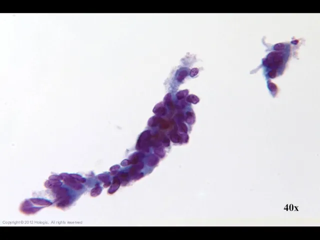
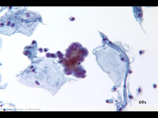
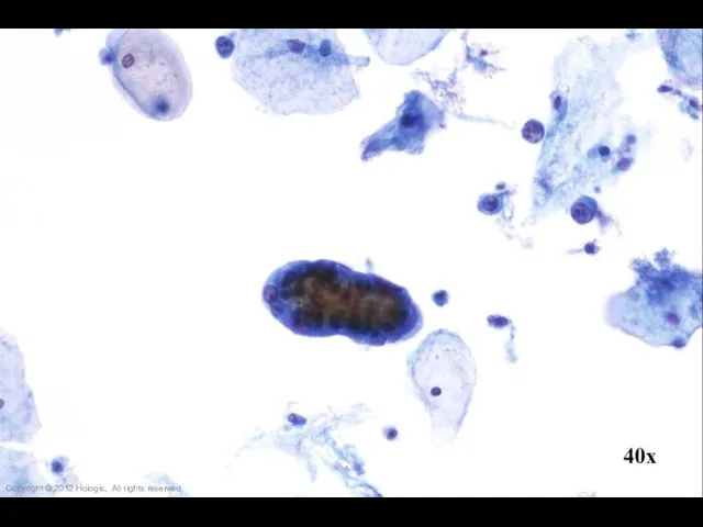
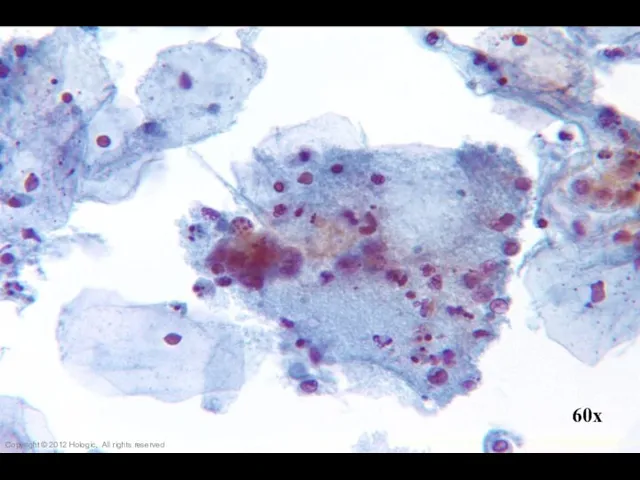
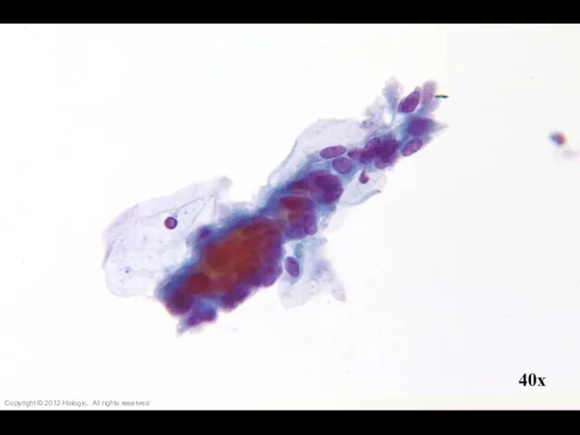
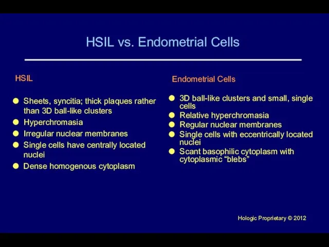
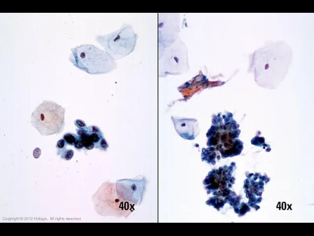
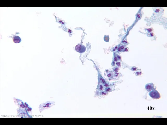
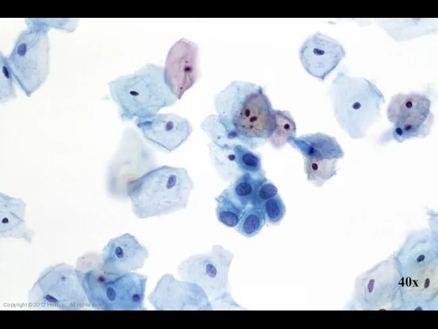
Слайд 43HSIL vs. Endometrial Cells
HSIL
Sheets, syncitia; thick plaques rather than 3D ball-like clusters
Hyperchromasia
Irregular
HSIL vs. Endometrial Cells
HSIL
Sheets, syncitia; thick plaques rather than 3D ball-like clusters
Hyperchromasia
Irregular
Слайд 44Copyright © 2012 Hologic, All rights reserved
Copyright © 2012 Hologic, All rights reserved
Слайд 4540x
Copyright © 2012 Hologic, All rights reserved
40x
Copyright © 2012 Hologic, All rights reserved
Слайд 4640x
Copyright © 2012 Hologic, All rights reserved
40x
Copyright © 2012 Hologic, All rights reserved
Слайд 4740x
Copyright © 2012 Hologic, All rights reserved
40x
Copyright © 2012 Hologic, All rights reserved
Слайд 4840x
Copyright © 2012 Hologic, All rights reserved
40x
Copyright © 2012 Hologic, All rights reserved
Слайд 49Copyright © 2012 Hologic, All rights reserved
Copyright © 2012 Hologic, All rights reserved
Слайд 5040x
Copyright © 2012 Hologic, All rights reserved
40x
Copyright © 2012 Hologic, All rights reserved
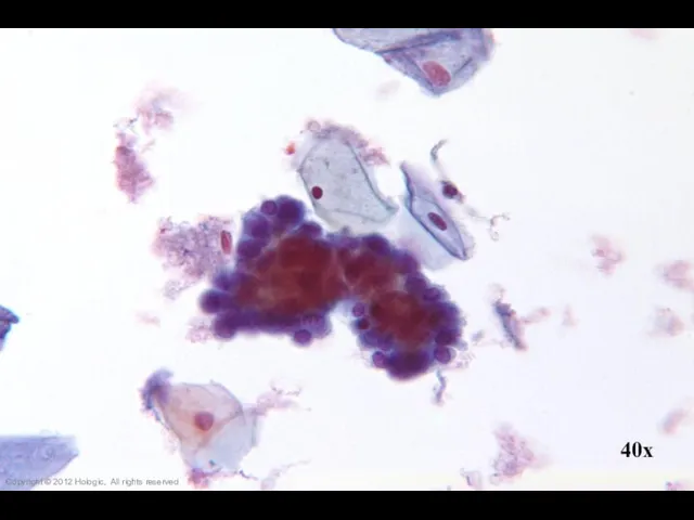
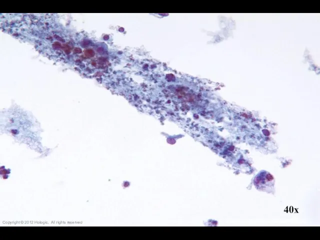
Слайд 51HSIL vs. Immature Squamous Metaplasia
HSIL
Single cells, clusters, and thickened plaques
Variable hyperchromasia
Irregular nuclear
HSIL vs. Immature Squamous Metaplasia
HSIL
Single cells, clusters, and thickened plaques
Variable hyperchromasia
Irregular nuclear
Слайд 5260x
Copyright © 2012 Hologic, All rights reserved
60x
Copyright © 2012 Hologic, All rights reserved
Слайд 5340x
Copyright © 2012 Hologic, All rights reserved
40x
Copyright © 2012 Hologic, All rights reserved
Слайд 5440x
40x
Copyright © 2012 Hologic, All rights reserved
40x
40x
Copyright © 2012 Hologic, All rights reserved
Слайд 5540x
40x
Copyright © 2012 Hologic, All rights reserved
40x
40x
Copyright © 2012 Hologic, All rights reserved
Слайд 5640x
Copyright © 2012 Hologic, All rights reserved
40x
Copyright © 2012 Hologic, All rights reserved
Слайд 5740x
Copyright © 2012 Hologic, All rights reserved
40x
Copyright © 2012 Hologic, All rights reserved
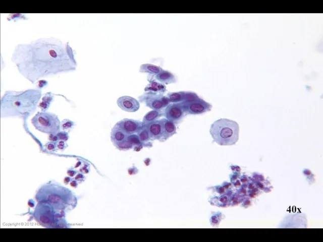
Слайд 58Repair vs. Poorly Differentiated SCC
Poorly Differentiated SCC
Poorly cohesive sheets and single cells
Irregular
Repair vs. Poorly Differentiated SCC
Poorly Differentiated SCC
Poorly cohesive sheets and single cells
Irregular
Слайд 59Copyright © 2012 Hologic, All rights reserved
Copyright © 2012 Hologic, All rights reserved
Слайд 60Copyright © 2012 Hologic, All rights reserved
Copyright © 2012 Hologic, All rights reserved
Слайд 6140x
Copyright © 2012 Hologic, All rights reserved
40x
Copyright © 2012 Hologic, All rights reserved
Слайд 6240x
Copyright © 2012 Hologic, All rights reserved
40x
Copyright © 2012 Hologic, All rights reserved
Слайд 6340x
Copyright © 2012 Hologic, All rights reserved
40x
Copyright © 2012 Hologic, All rights reserved
Слайд 6460x
Copyright © 2012 Hologic, All rights reserved
60x
Copyright © 2012 Hologic, All rights reserved
Слайд 65Trademark Statement
CytoLyt, Hologic, PreservCyt, ThinPrep, and UroCyte are registered trademarks of
Trademark Statement
CytoLyt, Hologic, PreservCyt, ThinPrep, and UroCyte are registered trademarks of
 Обзор доклинических данных, подтверждающих секретомоторную активность Синупрета экстракта(BNO 1016*/BNO 1011**)
 Профессиональные знания для врачей и техников. Учебный центр LOGOS
 О качестве оказания медицинской помощи населению
 Неблагородные металлы
 First aid
 О проведении вакцинации против коронавирусной инфекции Сovid-19 на территории Челябинской области
 Каноны этических требований к моральному облику врача
 Городская детская поликлиника, г. Петрозаводск
 Стажировки для кибернетиков
 Всемирный день иммунитета
 Качество мяса
 Оказание медицинской помощи при анатомически и клинически узком тазе
 Иммунодефицитные состояния у детей
 Первичный и гематогенный туберкулез. Патологическая анатомия
 Состав лекарственных препаратов и медицинских изделий входящих в неотложную медицинскую помощь
 Первая помощь при кровотечениях
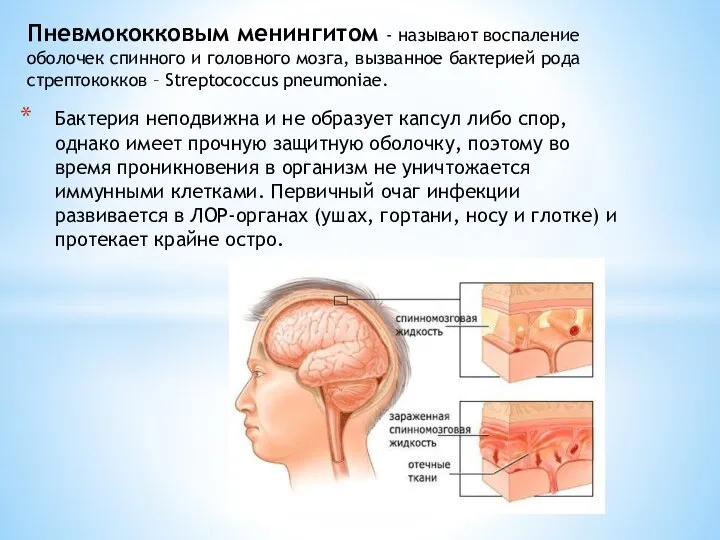 Пневмококковый менингит
 Регрессиялық талдаудың негізгі әдістерін қолданып биологиялық және медициналық мазмұны
 Симптомсыз бактериурия
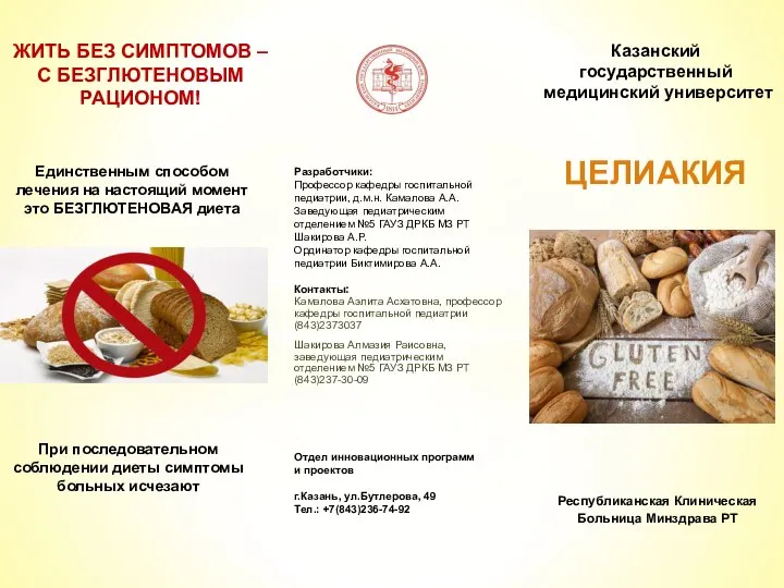 Жить без симптомов – с безглютеновым рационом. Целиакия
 Хирургическая техника одно- и двусторонней трансплантации легких
 Мышление у лиц с первичной речевой патологией
 Здоровая еда - еда против COVID-19
 Профилактика простатита и дисфункций половой сферы Dim-prost Programm
 Кровеносная система. Кровь (7 класс)
 Лекарственные средства, влияющие на функцию матки
 Клинический случай. Генерализованный атеросклероз. Эверсионная каротидная эндартерэктомия
 Оцінка професійних ризиків інфікування працівників галузі охорони здоров'я в Україні