- Heart anatomy

Содержание
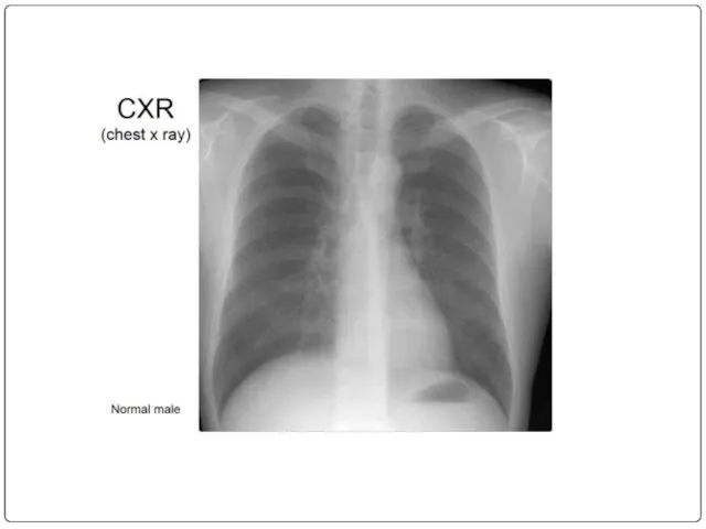
- 2. Plan: Size, Location, and Orientation Coverings Heart Wall Chambers Chest x ray
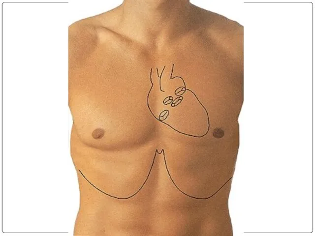
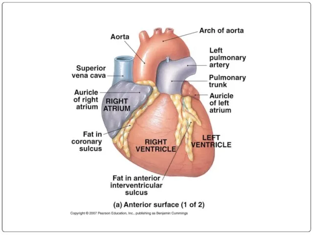
- 5. Heart Anatomy Size, Location, and Orientation Enclosed in the mediastinum Base (posteriorsuperior portion) and Apex (inferioranterior
- 6. Heart Anatomy Coverings Pericardium protects the heart anchors the heart to surrounding structures such as the
- 7. Heart Anatomy Coverings pericardial cavity contains a film of serous fluid pericarditis: inflammation of the pericardium
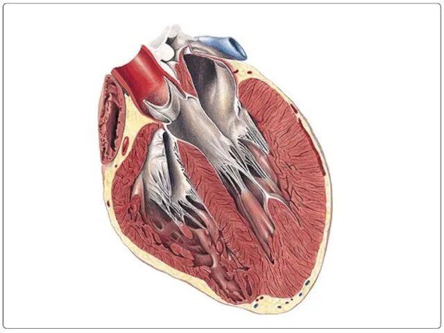
- 8. Heart Anatomy Heart Wall Epicardium Myocardium bulk of the heart consisting mainly of cardiac muscle
- 9. Heart Anatomy Heart Wall Endocardium simple squamous epithelium and a thin CT layer that lines the
- 11. Heart Anatomy Chambers Atria Features small, thin-walled chambers Functions receiving chambers for blood returning to the
- 12. Heart Anatomy Chambers Atria Receive blood from right side Superior and Inferior Vena Cava Coronary Sinus
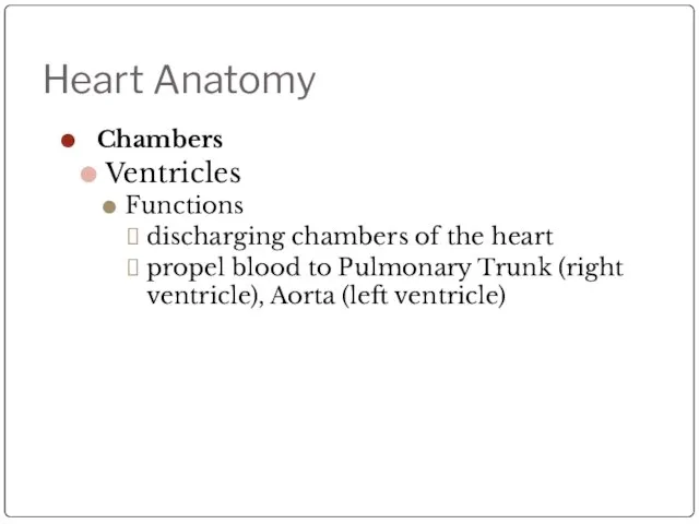
- 13. Heart Anatomy Chambers Ventricles Features make up most of the mass of the heart the walls
- 14. Heart Anatomy Chambers Ventricles Functions discharging chambers of the heart propel blood to Pulmonary Trunk (right
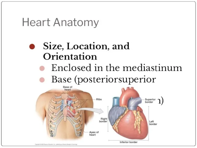
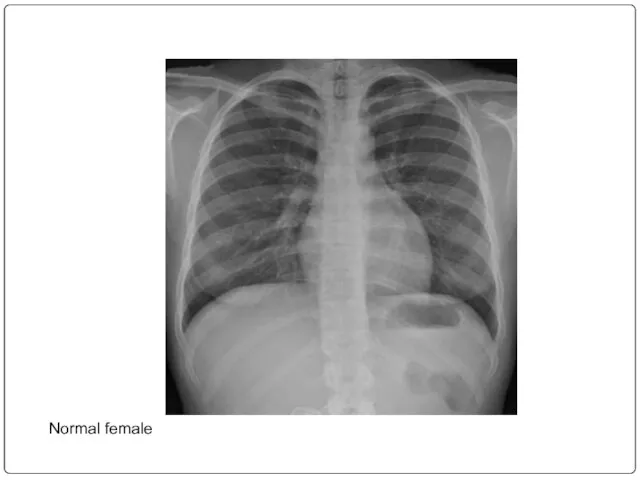
- 17. Normal female
- 19. Скачать презентацию
Слайд 5Heart Anatomy
Size, Location, and Orientation
Enclosed in the mediastinum
Base (posteriorsuperior
Heart Anatomy
Size, Location, and Orientation
Enclosed in the mediastinum
Base (posteriorsuperior
Слайд 6Heart Anatomy
Coverings
Pericardium
protects the heart
anchors the heart to surrounding
Heart Anatomy
Coverings
Pericardium
protects the heart
anchors the heart to surrounding
Слайд 7Heart Anatomy
Coverings
pericardial cavity contains a film of serous fluid
pericarditis: inflammation
Heart Anatomy
Coverings
pericardial cavity contains a film of serous fluid
pericarditis: inflammation
Слайд 8Heart Anatomy
Heart Wall
Epicardium
Myocardium
bulk of the heart consisting mainly of
Heart Anatomy
Heart Wall
Epicardium
Myocardium
bulk of the heart consisting mainly of
Слайд 9Heart Anatomy
Heart Wall
Endocardium
simple squamous epithelium and a thin CT layer
Heart Anatomy
Heart Wall
Endocardium
simple squamous epithelium and a thin CT layer
Слайд 11Heart Anatomy
Chambers
Atria
Features
small, thin-walled chambers
Functions
receiving chambers for blood returning
Heart Anatomy
Chambers
Atria
Features
small, thin-walled chambers
Functions
receiving chambers for blood returning
Слайд 12Heart Anatomy
Chambers
Atria
Receive blood from
right side
Superior and Inferior Vena Cava
Heart Anatomy
Chambers
Atria
Receive blood from
right side
Superior and Inferior Vena Cava
Слайд 13Heart Anatomy
Chambers
Ventricles
Features
make up most of the mass of the heart
Heart Anatomy
Chambers
Ventricles
Features
make up most of the mass of the heart
Слайд 14Heart Anatomy
Chambers
Ventricles
Functions
discharging chambers of the heart
propel blood
Heart Anatomy
Chambers
Ventricles
Functions
discharging chambers of the heart
propel blood

Слайд 17Normal female
Normal female
 Судебный этикет как составляющая культуры уголовно-процессуальной деятельности. Нормативные основы судебного этикета
 Оружие массового поражения
 Модельный ряд грузовых автомобилей Mercedes-Benz
 Энергетика: вчера, сегодня, завтра
 ГИИС ЭБ. Запрос на аннулирование
 Элементы интернет-маркетинга и их взаимодействие
 1. ОСНОВНЫЕ ПОНЯТИЯ Компьютерный исполнитель – это виртуальный объект, действующий в виртуальной среде обитания. Примеры: –Чертеж
 Beerfest
 Красота осени
 Социально-психологическая служба в школе
 Как принимать управленческие решения
 Денежные переводы в Республике Таджикистан
 Темы в Drupal 6 Что нового, и чем оно грозит
 Память в камне
 Помещение для открытия магазина Белорусская косметика
 Огневая подготовка. Версия 3
 Горный Дагестан
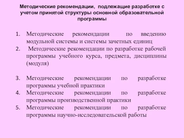 Методические рекомендации по введению модульной системы и системы зачетных единиц Методические рекомендации по разработке рабо
 Внутренний мир болезни
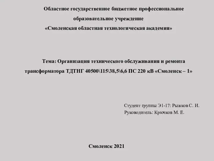 Техническое обслуживание и ремонт трансформатора ТДТНГ 40500\115\38,5\6,6 кВ Смоленск – 1
 Кинестетик
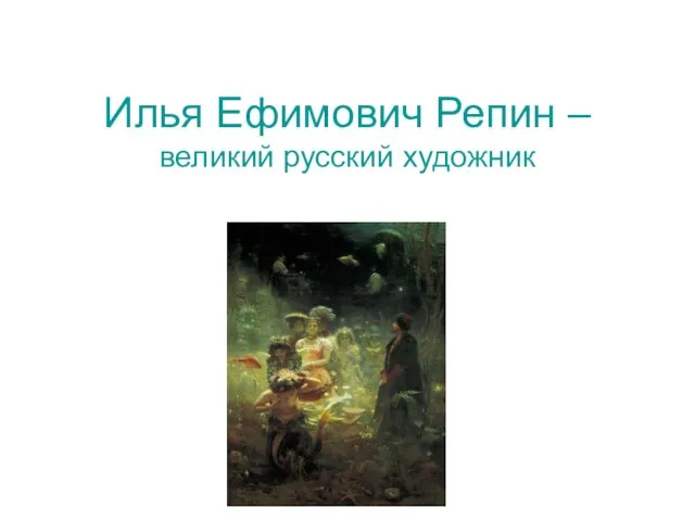 Илья Ефимович Репин –великий русский художник
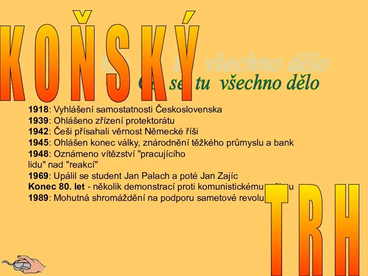 Václavské náměstí
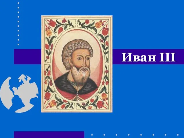 Презентация на тему Иван третий (4 класс)
 11 причин инвестировать в Свердловскую область
 Технология установки врезного замка
 Религия и мораль
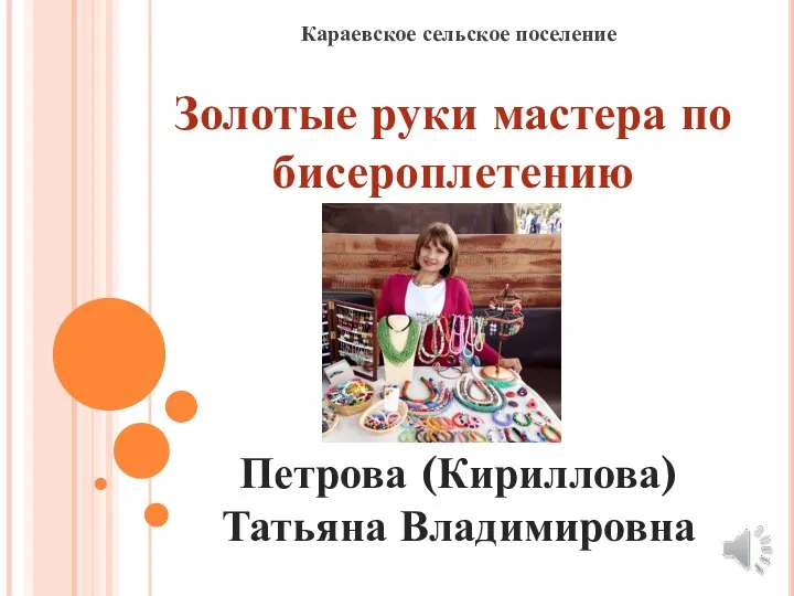 Караевское сельское поселение. Золотые руки мастера по бисероплетению